

TRPS1 is a Highly Sensitive Marker for Breast Cancer: A Tissue Microarray Study Evaluating More Than 19,000 Tumors From 152 Different Tumor Entities
- PMID: 38647255
- PMCID: PMC11093513
- DOI: 10.1097/PAS.0000000000002213
TRPS1 is a Highly Sensitive Marker for Breast Cancer: A Tissue Microarray Study Evaluating More Than 19,000 Tumors From 152 Different Tumor Entities
Abstract
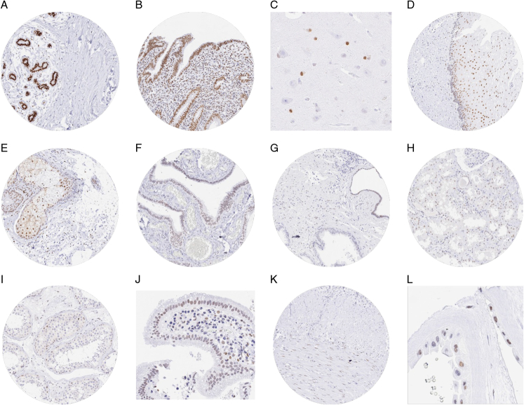
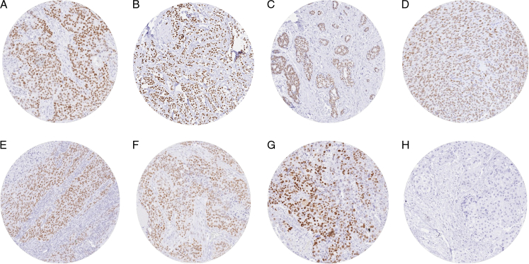
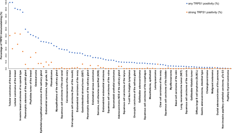
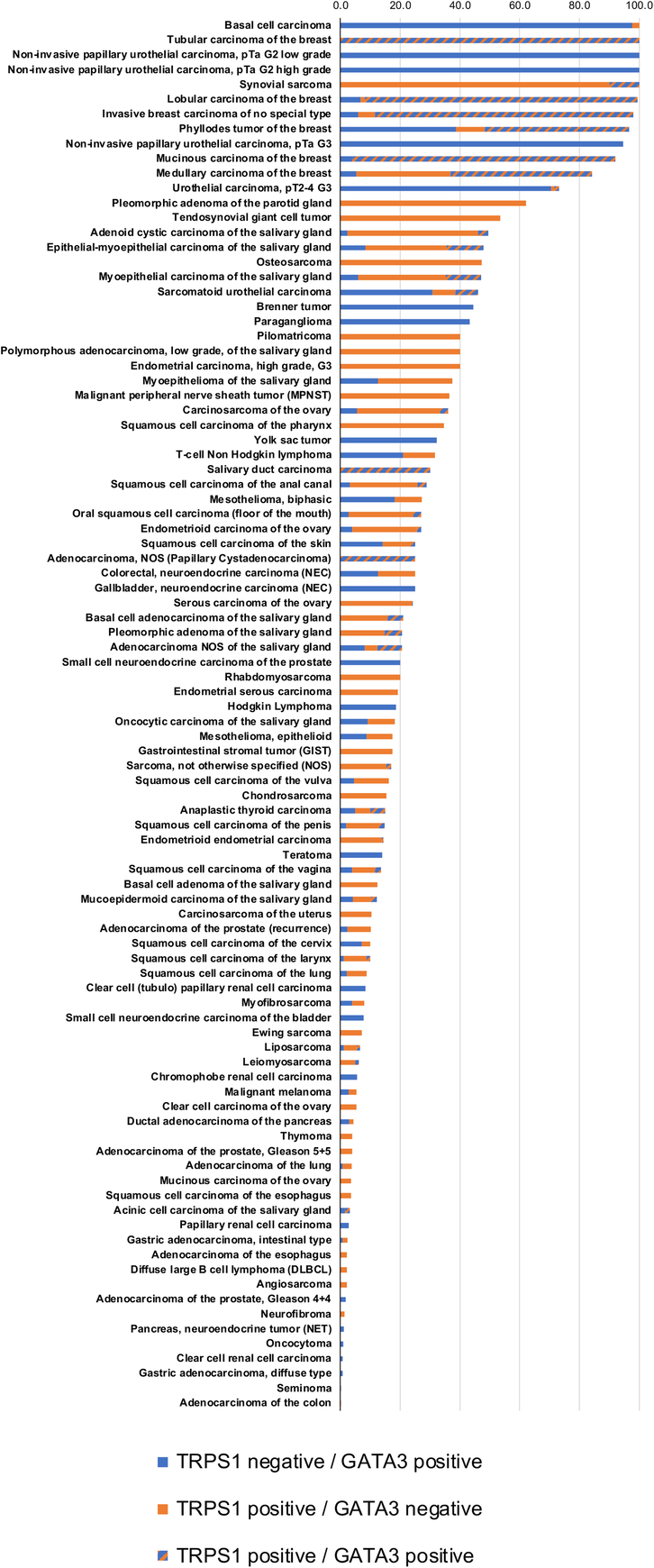
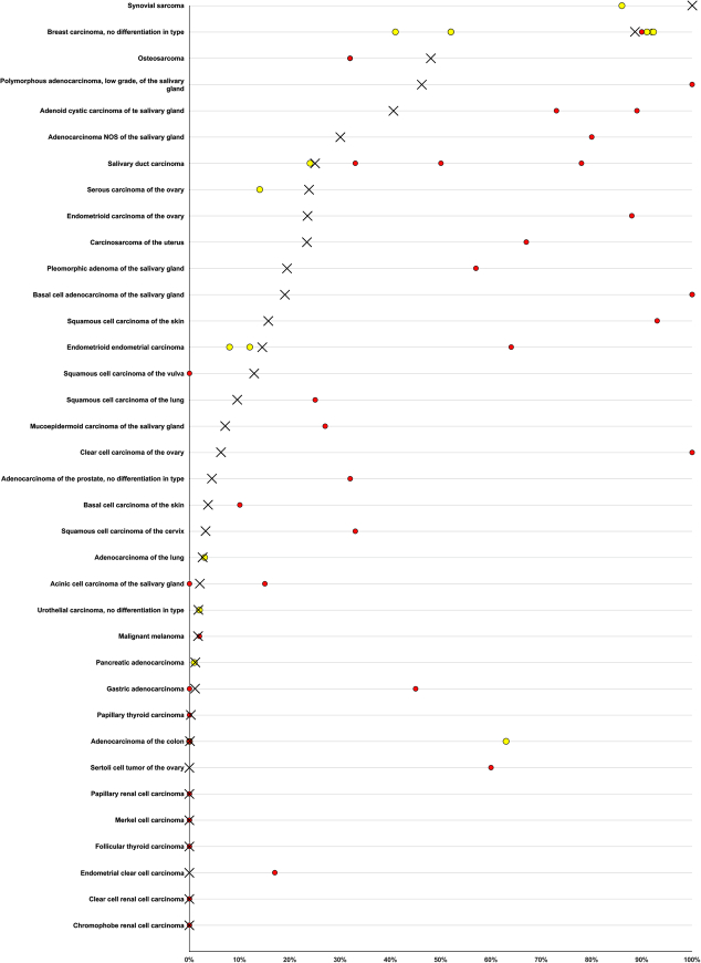
Trichorhinophalangeal syndrome 1 (TRPS1) is a nuclear protein highly expressed in breast epithelial cells. TRPS1 immunohistochemistry (IHC) has been suggested as a breast cancer marker. To determine the diagnostic and prognostic utility of TRPS1 IHC, tissue microarrays containing 19,201 samples from 152 different tumor types and subtypes were analyzed. GATA3 IHC was performed in a previous study. TRPS1 staining was seen in 86 of 152 tumor categories with 36 containing at least one strongly positive case. TRPS1 staining predominated in various types of breast carcinomas (51%-100%), soft tissue tumors (up to 100%), salivary gland tumors (up to 46%), squamous cell carcinomas (up to 35%), and gynecological cancers (up to 40%). TRPS1 positivity occurred in 1.8% of 1083 urothelial neoplasms. In invasive breast carcinoma of no special type, low TRPS1 expression was linked to high grade ( P = 0.0547), high pT ( P < 0.0001), nodal metastasis ( P = 0.0571), loss of estrogen receptor and progesterone receptor expression ( P < 0.0001 each), and triple-negative status ( P < 0.0001) but was unrelated to patient survival ( P = 0.8016). In squamous cell carcinomas from 11 different sites, low TRPS1 expression was unrelated to tumor phenotype. Positivity for both TRPS1 and GATA3 occurred in 47.4% to 100% of breast cancers, up to 30% of salivary gland tumors, and 29 (0.3%) of 9835 tumors from 134 other cancer entities. TRPS1 IHC has high utility for the identification of cancers of breast (or salivary gland) origin, especially in combination with GATA3. The virtual absence of TRPS1 positivity in urothelial neoplasms is useful for the distinction of GATA3-positive urothelial carcinoma from breast cancer.
Copyright © 2024 The Author(s). Published by Wolters Kluwer Health, Inc.
Conflict of interest statement
Conflicts of Interest and Source of Funding: The rabbit recombinant TRPS1 antibody, clone MSVA-512R was provided from MS Validated Antibodies GmbH (owned by a family member of G.S.). The company MS Validated Antibodies is owned by a relative of G.S. For the remaining authors none were declared.
Figures
References
-
- Yang L, Gong X, Wang J, et al. Functional mechanisms of TRPS1 in disease progression and its potential role in personalized medicine. Pathol Res Pract. 2022;237:154022. - PubMed
-
- Kanno S, Gui T, Itoh S, et al. Aberrant expression of the P2 promoter-specific transcript Runx1 in epiphyseal cartilage of Trps1-null mice. Exp Mol Pathol. 2011;90:143–148. - PubMed
-
- Ergoren MC, Akcan N, Manara E, et al. Characterization of a novel frameshift mutation within the TRPS1 gene causing trichorhinophalangeal syndrome type 1 in a kindred cypriot family. Appl Immunohistochem Mol Morphol. 2022;30:635–639. - PubMed
-
- Huang JZ, Chen M, Zeng M, et al. Down-regulation of TRPS1 stimulates epithelial-mesenchymal transition and metastasis through repression of FOXA1. J Pathol. 2016;239:186–196. - PubMed
MeSH terms
Substances
LinkOut - more resources
Full Text Sources
Medical
Research Materials
