. 2024 Apr 22;20(4):e1012186.
doi: 10.1371/journal.ppat.1012186. eCollection 2024 Apr.

Beyond the VSG layer: Exploring the role of intrinsic disorder in the invariant surface glycoproteins of African trypanosomes

Affiliations

Beyond the VSG layer: Exploring the role of intrinsic disorder in the invariant surface glycoproteins of African trypanosomes

Hagen Sülzen et al. PLoS Pathog. .

Abstract

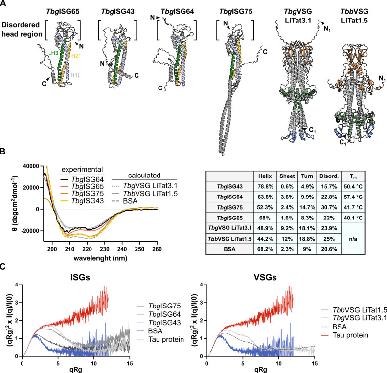
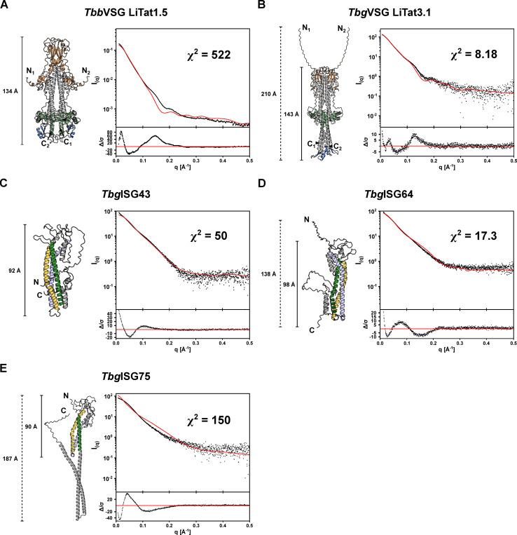
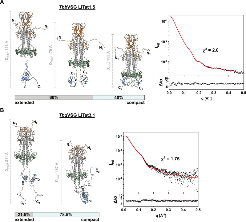
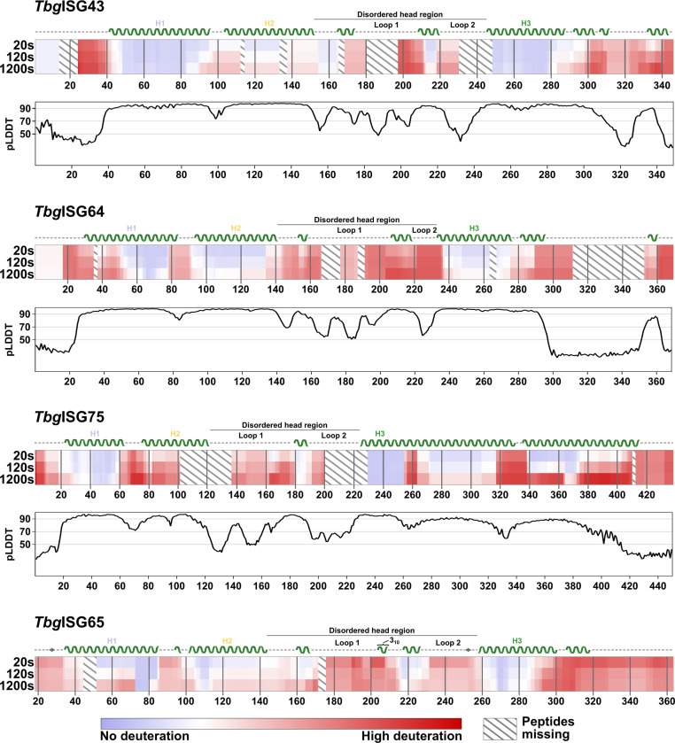
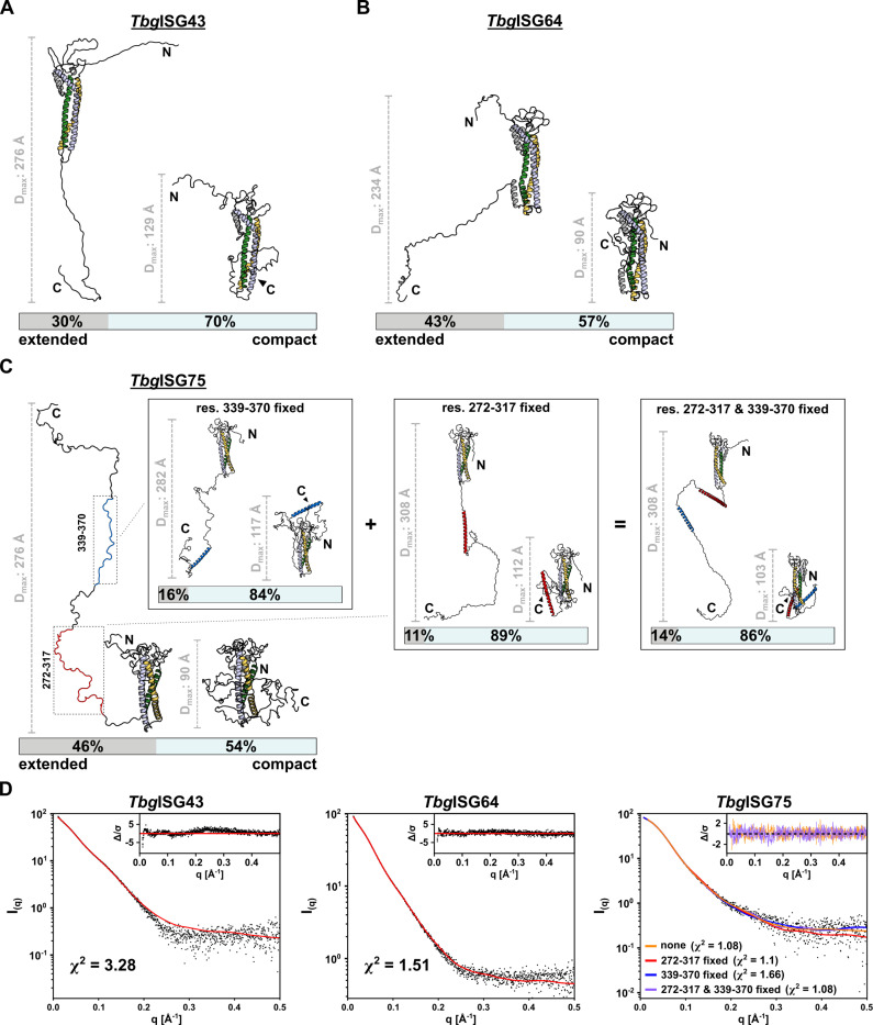
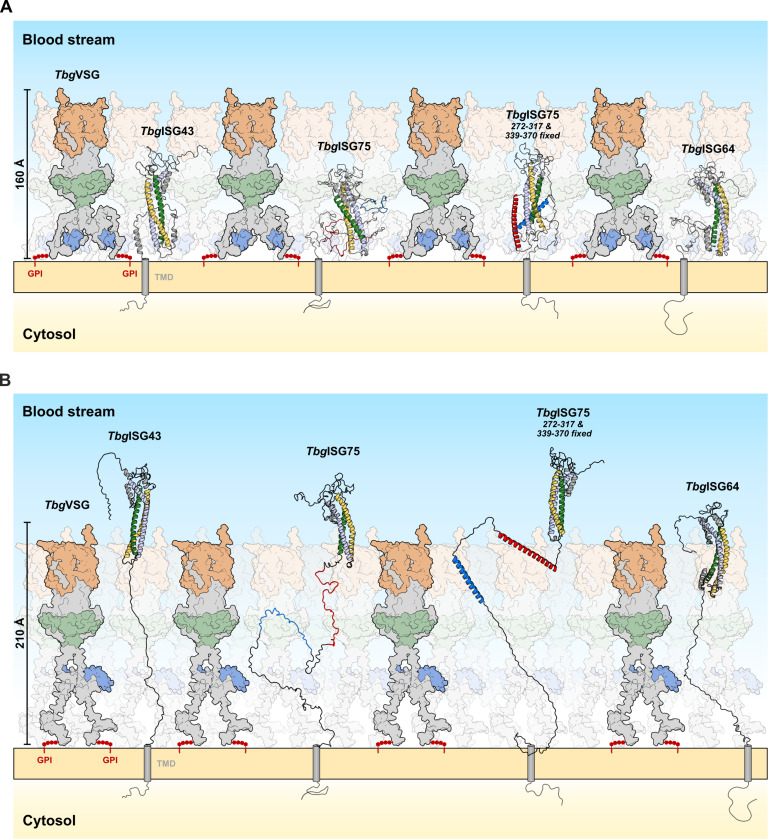
In the bloodstream of mammalian hosts, African trypanosomes face the challenge of protecting their invariant surface receptors from immune detection. This crucial role is fulfilled by a dense, glycosylated protein layer composed of variant surface glycoproteins (VSGs), which undergo antigenic variation and provide a physical barrier that shields the underlying invariant surface glycoproteins (ISGs). The protective shield's limited permeability comes at the cost of restricted access to the extracellular host environment, raising questions regarding the specific function of the ISG repertoire. In this study, we employ an integrative structural biology approach to show that intrinsically disordered membrane-proximal regions are a common feature of members of the ISG super-family, conferring the ability to switch between compact and elongated conformers. While the folded, membrane-distal ectodomain is buried within the VSG layer for compact conformers, their elongated counterparts would enable the extension beyond it. This dynamic behavior enables ISGs to maintain a low immunogenic footprint while still allowing them to engage with the host environment when necessary. Our findings add further evidence to a dynamic molecular organization of trypanosome surface antigens wherein intrinsic disorder underpins the characteristics of a highly flexible ISG proteome to circumvent the constraints imposed by the VSG coat.


Conflict of interest statement

The authors have declared that no competing interests exist.

Figures

Fig 1
Fig 1. The VSG and ISG solution structures exhibit a high degree of flexibility.
(A) The predicted AlphaFold2 models of TbgISG43, TbgISG64, TbgISG75, TbgVSG LiTat3.1 and TbbVSG LiTat1.5 illustrated in cartoon representation, depicted next to the hybrid structure of TbgISG6518-363 (PDBDEV_00000201). The three helices in the TbgISGs constituting the canonical three helix bundle have been colored uniformly and labelled on TbgISG65, arrows indicate the direction of the peptide chain from N- to C-terminus. Top and bottom lobe as well as C-terminal domains of the VSGs are highlighted in orange, green and blue, respectively. For representation purposes, the N-termini of TbgISG64 and TbgVSG have been shortened, deleted residues are indicated with a line break (full length models are shown in Fig 2). (B) CD spectra for TbgISGs (experimental), VSGs (calculated from AlphaFold2 models) and BSA (calculated from PDB 4F5S). Secondary structure analyses (performed using CONTINLL [48, 49]) for the respective spectra and TbgISG melting temperatures (Tm) are summarized in the table. All CD spectra exhibit minima at 208 and 222 nm, characteristic for a largely alpha-helical fold. Similarly, all proteins have a high content of turns and disordered regions. (C) Dimensionless Kratky plots for TbgISGs (left) and TbbVSG LiTat1.5 and TbgVSG LiTat3.1 (right). Comparison of the plots for Tbg and Tbb proteins to reference samples for strictly folded (BSA, blue) and completely disordered proteins (Tau protein, red) demonstrate that all measured proteins contain significant fractions of both ordered and disordered components.
Fig 2
Fig 2. The VSG and ISG single-conformer AlphaFold2 models do not account for the experimentally observed solution behavior.
The theoretical scattering curves of the unaltered AlphaFold2 models for TbbVSG LiTat1.5 (A), TbgVSG LiTat3.1 (B), TbgISG43 (C), TbgISG64 (D) and TbgISG75 (E) were calculated (red curves) and compared to the experimental SAXS data (black dots). The fit residuals are displayed as insets below the scattering curves. The respective AlphaFold2 models used for the calculations are shown (colored using the same scheme as in Fig 1).
Fig 3
Fig 3. Conformational ensemble modelling reveals structural flexibility of VSGs in solution.
(A) The solution scattering of TbbVSG LiTat1.5 is best described by a conformational ensemble composed of 5 models. Three representative conformations are shown, alongside a scale bar indicating the Dmax of the individual conformer. (B) The SAXS data of TbgVSG LiTat3.1 is best described by a conformational ensemble composed of 2 models. Both conformations are depicted next to a scale bar indicating the Dmax of the individual conformer. In both panels, the experimental scattering data (black dots) and calculated ensemble scattering curves (red line) are shown. The residuals of the fit are shown below.
Fig 4
Fig 4. HDX-MS reveals intrinsically disordered regions in TbgISGs.
Chiclet plots showing the relative deuteration across the sequences of TbgISGs at 20, 120 and 1200 s of incubation. The amount of relative deuteration is indicated by color gradients, ranging from blue (no deuteration) to red (high deuteration). Peptides absent in the analysis are represented by diagonal lines. For TbgISG75, TbgISG64 and TbgISG43, the secondary structure, as predicted by AlphaFold2, is indicated above the corresponding chiclet plot. For TbgISG65, the secondary structure of the integrative TbgISG65 hybrid structure deposited to the PDBDev (PDBDEV_00000201) is shown [7]. Disordered regions are represented by a dashed line, beta sheets as arrows and alpha helices as helices. The three helices constituting the canonical three-helical bundle are labelled (H1 to H3) and colored as in previous figures. The 310-helix in ISG65 is indicated with a label. The disordered loops forming the disordered, membrane-distal head region of the ISGs are labeled accordingly. Underneath the chiclet plots of TbgISG43, TbgISG64 and TbgISG75, the per-residue pLDDT scores of the respective AlphaFold2 models are plotted. Thresholds for high (>90), medium (>70) and low (<50) modelling confidence are indicated with grey lines.
Fig 5
Fig 5. Conformational ensemble modelling reveals structural flexibility of ISGs in solution.
Representative ensembles of (A) TbgISG43, (B) TbgISG64 and (C) TbgISG75 for the experimental SAXS data, calculated using BILBOMD. The smallest ensemble producing a reasonable improvement in goodness of fit of the theoretical scattering curve to the experimental data was chosen. Models constituting the selected ensemble are shown with their respective weights. For each model, the Dmax is displayed with a representative scalebar. For ISG75 in (C) four ensembles are shown, corresponding to ensemble calculation without HDX-MS restraints, restraints applied to residues Arg272-Arg317 (highlighted in red) [left insert], residues Lys339-Ala370 (highlighted in blue) [middle insert] and both [right insert]. (D) Experimental scattering data are shown, overlaid with the theoretical scattering curves of the selected ensembles and the respective χ2 of the fit. Residuals are shown as insets. For ISG75, the residuals for the ensemble without restraints for modelling of the CTD (orange) and with restraints in both res. 272–317 and res. 339–370 (purple) are shown.
Fig 6
Fig 6. An updated model of the Trypanosoma surface coat–Functional implications of ISG conformational flexibility within the VSG umbrella model.
(A) In the retracted form, ISGs reside within the VSG coat (represented here by TbgVSG LiTat3.1), leaving only their disordered, loop-rich head domains accessible to molecules in the host’s blood stream, while concealing all other epitopes under the protective VSG umbrella. (B) In the extended conformation, both ISG43 and ISG75 protrude well beyond the boundaries of the VSG layer, even when the latter is in its extended conformation. Whilst in this model ISG64 would not be able to fully extend beyond the maximally extended VSG umbrella, it still protrudes beyond the VSGs CTD, thereby implying accessibility to potential ligands. The GPI anchors of the VSGs are depicted in red, while transmembrane domains of ISGs are shown as gray cylinders. The N- and C-termini of the protein models obtained from SAXS modeling were manually modified for this figure to allow for membrane anchoring via the C-terminal linker without altering the Dmax of the models.

Similar articles

Cited by

References

    1. Salmon D, Vanwalleghem G, Morias Y, Denoeud J, Krumbholz C, Lhommé F, et al.. Adenylate cyclases of Trypanosoma brucei inhibit the innate immune response of the host. Science (New York, NY). 2012;337: 463–466. doi: 10.1126/science.1222753 - DOI - PubMed
    1. Salmon D, Bachmaier S, Krumbholz C, Kador M, Gossmann JA, Uzureau P, et al.. Cytokinesis of Trypanosoma brucei bloodstream forms depends on expression of adenylyl cyclases of the ESAG4 or ESAG4-like subfamily. 2012;84: 225–242. doi: 10.1111/j.1365-2958.2012.08013.x - DOI - PubMed
    1. Salmon D. Adenylate Cyclases of Trypanosoma brucei, Environmental Sensors and Controllers of Host Innate Immune Response. Pathogens. 2018;7: 48. doi: 10.3390/pathogens7020048 - DOI - PMC - PubMed
    1. SEED JR, SECHELSKI OB, LOOMIS MR. A Survey for a Trypanocidal Factor in Primate Sera. J Protozool. 1990;37: 393–400. doi: 10.1111/j.1550-7408.1990.tb01163.x - DOI - PubMed
    1. Rifkin MR. Identification of the trypanocidal factor in normal human serum: high density lipoprotein. Proc Natl Acad Sci. 1978;75: 3450–3454. doi: 10.1073/pnas.75.7.3450 - DOI - PMC - PubMed

Publication types

MeSH terms

Substances