. 2024 May 7;121(19):e2313568121.
doi: 10.1073/pnas.2313568121. Epub 2024 Apr 22.

Impact of repeated blast exposure on active-duty United States Special Operations Forces

Affiliations

Impact of repeated blast exposure on active-duty United States Special Operations Forces

Natalie Gilmore et al. Proc Natl Acad Sci U S A. .

Abstract

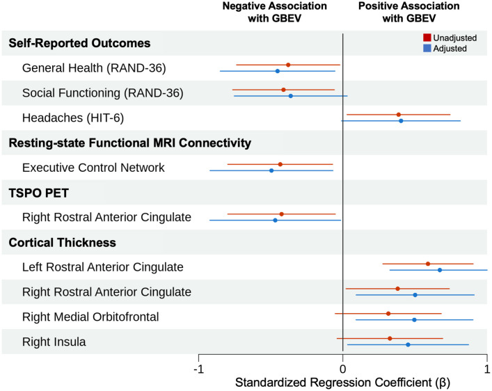
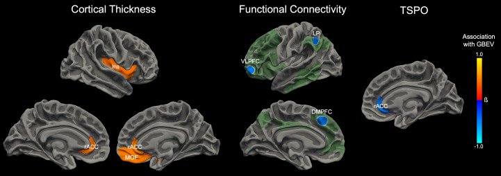
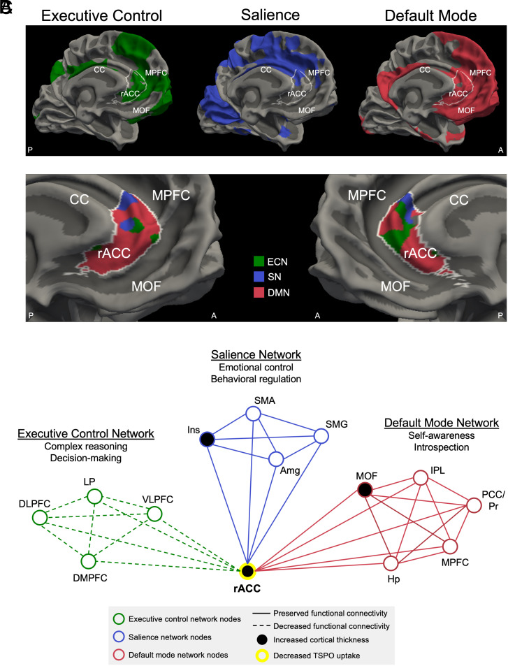
United States (US) Special Operations Forces (SOF) are frequently exposed to explosive blasts in training and combat, but the effects of repeated blast exposure (RBE) on SOF brain health are incompletely understood. Furthermore, there is no diagnostic test to detect brain injury from RBE. As a result, SOF personnel may experience cognitive, physical, and psychological symptoms for which the cause is never identified, and they may return to training or combat during a period of brain vulnerability. In 30 active-duty US SOF, we assessed the relationship between cumulative blast exposure and cognitive performance, psychological health, physical symptoms, blood proteomics, and neuroimaging measures (Connectome structural and diffusion MRI, 7 Tesla functional MRI, [11C]PBR28 translocator protein [TSPO] positron emission tomography [PET]-MRI, and [18F]MK6240 tau PET-MRI), adjusting for age, combat exposure, and blunt head trauma. Higher blast exposure was associated with increased cortical thickness in the left rostral anterior cingulate cortex (rACC), a finding that remained significant after multiple comparison correction. In uncorrected analyses, higher blast exposure was associated with worse health-related quality of life, decreased functional connectivity in the executive control network, decreased TSPO signal in the right rACC, and increased cortical thickness in the right rACC, right insula, and right medial orbitofrontal cortex-nodes of the executive control, salience, and default mode networks. These observations suggest that the rACC may be susceptible to blast overpressure and that a multimodal, network-based diagnostic approach has the potential to detect brain injury associated with RBE in active-duty SOF.

Keywords: Special Operations Forces; blast overpressure; traumatic brain injury.


Conflict of interest statement

Competing interests statement:The authors declare no competing interest.

Figures

Fig. 1.
Fig. 1.
Blast exposure associations with psychological health, physical symptoms, and neuroimaging measures. We display all measures that showed an association with GBEV in univariate (i.e., unadjusted) or multivariable (i.e., adjusted) analyses. Filled circles represent standardized regression coefficients (β). Solid lines represent 95% CIs. In multivariable analyses, higher GBEV was associated with lower scores on the general health subscale of the RAND-36 Measure of Health-Related Quality of Life, decreased functional connectivity in the executive control network, decreased TSPO signal in the right rACC, and increased cortical thickness in the left rACC, right rACC, right medial orbitofrontal cortex, and right insula. The association between higher GBEV and increased cortical thickness in the left rACC survived correction for multiple comparisons.
Fig. 2.
Fig. 2.
Associations between blast exposure and neuroimaging measures. Cumulative blast exposure, measured by an interview-based GBEV, was associated with alterations in T1-weighted measures of cortical thickness (Left), 7T resting-state fMRI measures of functional connectivity (Middle), and PET-MRI measures of TSPO (Right). For each modality, the anatomic regions that showed a significant association with GBEV in the adjusted regression models (controlling for age, combat exposure, and blows to the head) are superimposed on the surface of the brain. Orange colors indicate a positive association with GBEV, whereas blue colors represent a negative association with GBEV. The scalar bar color is weighted by the standardized regression coefficient (β). For the functional connectivity analysis, the seed nodes used to assess executive control network connectivity are delineated by white outlines. The extended executive control network derived from the seed nodes is shown in semitransparent green for visualization purposes. The executive control network shown in green represents the mean connectivity derived from the entire cohort. For each neuroimaging measure—cortical thickness, functional connectivity, and TSPO—the associations with GBEV converged on the rACC (outlined in purple). Abbreviations: DMPFC = dorsomedial prefrontal cortex; Ins = insula; MOF = medial orbitofrontal cortex; LP = lateral parietal lobe; VLPFC = ventrolateral prefrontal cortex.
Fig. 3.
Fig. 3.
Brain network alterations associated with cumulative blast exposure. (A) The executive control network (ECN, green), salience network (SN, blue), and default mode network (DMN, red) are shown on the medial surface of the left cerebral hemisphere. Each network represents the mean connectivity derived from the entire cohort of 28 participants who had usable 7T resting-state fMRI data. All three networks connect with the rACC, outlined in white, indicating that the rACC is a hub node with functional connectivity to the ECN, SN, and DMN. (B) rACC connectivity with the ECN, SN, and DMN is shown on the medial surface of the left and right cerebral hemispheres. rACC vertices on the cortical surface are color-coded according to which network has the highest level of functional connectivity with the rACC at that anatomic location. (C) Structural, functional, and neuroimmune alterations are shown in schematic form within the ECN, SN, and DMN. These network alterations converge on the rACC hub node. Abbreviations: A = anterior; Amg = amygdala; CC = corpus callosum; DLPFC = dorsolateral prefrontal cortex; DMPFC = dorsomedial prefrontal cortex; Hp = hippocampus; Ins = insula; IPL = inferior parietal lobule; LP = lateral parietal lobe; MOF = medial orbitofrontal cortex; MPFC = medial prefrontal cortex; P = posterior; PCC/Pr = posterior cingulate cortex/precuneus; SMA = supplementary motor area; SMG = supramarginal gyrus; TSPO = translocator protein; VLPFC = ventrolateral prefrontal cortex. Of note, the Raichle atlas nodes, which were used as seed regions to generate each functional network (as shown in SI Appendix, Fig. S2), are a subset of the ECN, SN, and DMN nodes shown in the schematic in (C).

References

    1. Garcia A., et al. , Health conditions among Special Operations Forces versus conventional military service members: A VA TBI model systems study. J. Head Trauma Rehabil. 37, E292–E298 (2022). - PubMed
    1. Modica L. C. M., Egnoto M. J., Statz J. K., Carr W., Ahlers S. T., Development of a blast exposure estimator from a department of defense-wide survey study on military service members. J. Neurotrauma 38, 1654–1661 (2021). - PubMed
    1. Edlow B. L., et al. , Optimizing brain health of United States Special Operations Forces. J. Spec. Oper. Med. 23, 47–56 (2023). - PubMed
    1. Rosenfeld J. V., et al. , Blast-related traumatic brain injury. Lancet Neurol. 12, 882–893 (2013). - PubMed
    1. Mac Donald C. L., et al. , Detection of blast-related traumatic brain injury in U.S. military personnel. N. Engl. J. Med. 364, 2091–2100 (2011). - PMC - PubMed

Publication types