. 2024 Apr 22;22(4):e3002585.
doi: 10.1371/journal.pbio.3002585. eCollection 2024 Apr.

Mrj is a chaperone of the Hsp40 family that regulates Orb2 oligomerization and long-term memory in Drosophila

Affiliations

Mrj is a chaperone of the Hsp40 family that regulates Orb2 oligomerization and long-term memory in Drosophila

Meghal Desai et al. PLoS Biol. .

Abstract

Orb2 the Drosophila homolog of cytoplasmic polyadenylation element binding (CPEB) protein forms prion-like oligomers. These oligomers consist of Orb2A and Orb2B isoforms and their formation is dependent on the oligomerization of the Orb2A isoform. Drosophila with a mutation diminishing Orb2A's prion-like oligomerization forms long-term memory but fails to maintain it over time. Since this prion-like oligomerization of Orb2A plays a crucial role in the maintenance of memory, here, we aim to find what regulates this oligomerization. In an immunoprecipitation-based screen, we identify interactors of Orb2A in the Hsp40 and Hsp70 families of proteins. Among these, we find an Hsp40 family protein Mrj as a regulator of the conversion of Orb2A to its prion-like form. Mrj interacts with Hsp70 proteins and acts as a chaperone by interfering with the aggregation of pathogenic Huntingtin. Unlike its mammalian homolog, we find Drosophila Mrj is neither an essential gene nor causes any gross neurodevelopmental defect. We observe a loss of Mrj results in a reduction in Orb2 oligomers. Further, Mrj knockout exhibits a deficit in long-term memory and our observations suggest Mrj is needed in mushroom body neurons for the regulation of long-term memory. Our work implicates a chaperone Mrj in mechanisms of memory regulation through controlling the oligomerization of Orb2A and its association with the translating ribosomes.


Conflict of interest statement

The authors have declared that no competing interests exist.

Figures

Fig 1
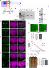
Fig 1. Identification of Mrj as a regulator of Orb2A’s prion-like oligomerization.
(A) Schematic of immunoprecipitation assay to identify Hsp40 interactors of Orb2A. Individual constructs coding for members of the Hsp40 family with HA tag were cotransfected in S2 cells with Orb2A construct and immunoprecipitation was performed with anti Orb2 antibody. The immunoprecipitate was probed with an anti-HA antibody to detect if the Hsp40 proteins were interacting with Orb2A. (B) From 37 proteins of the Hsp40 family, the immunoprecipitation (IP) screen identified CG9828, CG4164, Mrj, Tpr2, DroJ2, and CG7130 as interactors. Lys is the input lysate, Ctl IP is the control IP with beads only and Orb2 IP is the IP with anti-Orb2 antibody. (C) Schematic of immunoprecipitation assay to identify Hsp70 interactors of Orb2A. Individual constructs coding for members of the Hsp70 family with Flag tag were cotransfected in S2 cells with Orb2A construct and immunoprecipitation was performed with anti Orb2 antibody. The immunoprecipitate was probed with an anti-Flag antibody to detect if the Hsp70 proteins were interacting with Orb2A. (D) All 4 proteins of the Hsp70 family screened in the immunoprecipitation experiment, Hsp70Aa, Hsc70-1, Hsc70Cb, and Hsc70-4 were found to be interacting with Orb2A. (E) Schematic of the yeast based screen. A Sup35 knockout strain rescued by a chimeric construct expressing Orb2A’s prion-like domain tagged with the C-terminal domain of Sup35 is transformed with galactose inducible Hsp40 and Hsp70 constructs. A premature stop codon in the Ade 1–4 gene is used as a reporter. The screen consists of inducing the chaperones with galactose in the prion negative strain and screening for their ability to change the color to white and grow in adenine-deficient media. (F) Galactose induction of individual chaperones of both Hsp40 and Hsp70 family of proteins in prion negative Orb2APrD-Sup35C strain, caused only Mrj to convert the prion negative state of the cells to prion positive state, as evidenced by the change in the colony color to white in YPD media and causing it to now grow in adenine. (G) Schematic of SDD-AGE assay to quantitate the change in Orb2A-GFP oligomerization in presence of Mrj. Sf9 cells were infected with viruses for Orb2A alone and Orb2A with Mrj. The lysate from these cells was centrifuged and the resulting pellet was resuspended in an SDS-containing buffer, subjected to SDD-AGE, and further probed with anti-Orb2 antibody. (H) Representative SDD-AGE blots showing increased levels of Orb2A oligomers in presence of Mrj. (I) Quantitation of Orb2A oligomers in presence and absence of Mrj. Data is represented as a relative fold change for Orb2A in presence of Mrj as compared to without Mrj. Data is represented as mean ± SEM and significance is tested using two-tailed Student’s unpaired t test. (J) Recombinant GST-Mrj bound to Glutathione beads on incubation with purified recombinant Orb2A-His from E. coli could pulldown Orb2A. The same blot is probed first with an anti-Orb2 antibody followed by probing with an anti-Mrj antibody. The band marked with * is the GST-Mrj protein and the band below it is possibly a breakdown product from the former. GST protein bound to Glutathione beads was used as a negative control. (K) Recombinant GST-Mrj bound to Glutathione beads on incubation with purified recombinant Orb2A-GFP-His from Sf9 cells could pulldown Orb2A-GFP. The same blot is probed first with an anti-Orb2 antibody followed by probing with an anti-Mrj antibody. The band marked with * is the GST-Mrj protein and the band below it is possibly a breakdown product from the former. GST protein bound to Glutathione beads was used as a negative control. The data underlying this figure are available at: https://figshare.com/s/f5d913a0a289339ee16b.
Fig 2
Fig 2. Drosophila Mrj behaves like its mammalian homolog as a chaperone preventing Huntingtin aggregation.
(A) Schematic of immunoprecipitation assay to check the possibility of self-oligomerization of Mrj. Two Mrj constructs, Mrj-HA and Mrj-RFP were cotransfected in S2 cells. The cells were lysed and immunoprecipitation was done with anti-RFP (RFPTrap beads). The immunoprecipitate was next probed with anti-HA antibody. (B) Left panel shows a western blot of immunoprecipitated Mrj-RFP probed with anti-HA antibody indicating the presence of Mrj-HA in the immunoprecipitate, suggesting an interaction between Mrj-RFP and Mrj-HA. The right panel shows the same blot probed with an anti-RFP antibody to confirm the pulldown of Mrj-RFP. (C) Schematic of immunoprecipitation assay to identify Hsp70 interactors of Mrj. Flag-tagged Hsp70 constructs were cotransfected with Mrj-RFP. Lysate from these cells was used for immunoprecipitation with anti-RFP beads, and the immunoprecipitate was probed with anti-Flag antibody. (D) In the IP experiment, of the 4 Hsp70’s tested here, Hsp70Aa and Hsc70-4 show their presence in the immunoprecipitate suggesting of their interaction with Mrj. (E) Schematic of immunoprecipitation assay to check if Hsp70Aa and Hsc70-4 interacts with HPD motif mutated Mrj. Flag-tagged Hsp70 constructs were cotransfected with Mrj H31Q-RFP. Lysate from these cells was used for immunoprecipitation with anti-RFP beads, and the immunoprecipitate was probed with anti-Flag antibody. (F) In the IP experiment on probing with anti-Flag antibody both Hsp70Aa and Hsc70-4 could not be detected in the immunoprecipitate. The same blots when probed with an anti-RFP antibody, confirmed the pulldown of Mrj H31Q-RFP. (G) Representative confocal image of a single S2 cell coexpressing Mrj-RFP and Httexon1-Q103-GFP shows colocalization of Mrj with Htt aggregates. The scale bar is 10 microns. (H) Representative images of Httexon1Q103-GFP cells coexpressing with CG7133-HA, Mrj-RFP, and Mrj-HA suggest a decrease in the Htt aggregates in presence of Mrj. Scale bars are 5 microns. (I) Quantitation of the percentage of HttQ103-exon1GFP expressing cells with aggregates in presence of CG7133-HA, Mrj-RFP, and Mrj-HA suggests a significant decrease of Htt aggregates in presence of both Mrj-RFP and Mrj-HA. The Mrj-HA construct is more efficient in decreasing Htt aggregation compared to Mrj-RFP. Data is represented as mean ± SEM and significance is tested with two-tailed Student’s paired t test. (J) Western blot of lysates from S2 cells coexpressing Httexon1Q103-GFP with Mrj and CG7133 shows similar amounts of Htt (monomer size marked with *) in SDS-PAGE. Right panel shows the same lysates probed with anti-Tubulin antibody. (K) Quantitation of the Htt bands from SDS-PAGE shows no significant difference. Data is represented as mean ± SEM and significance is tested with two-tailed Student’s unpaired t test. (L) SDD-AGE from S2 cell lysate coexpressing Httexon1Q103-GFP along with CG7133 and Mrj showed a decreased amount of Htt oligomers in presence of Mrj. (M) Quantitation of SDD-AGE shows a significantly decreased levels of Htt oligomers in presence of Mrj. Data is represented as mean ± SEM and significance is tested with two-tailed Student’s unpaired t test. The data underlying this figure are available at: https://figshare.com/s/f5d913a0a289339ee16b.
Fig 3
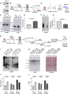
Fig 3. Drosophila Mrj is not an essential gene unlike its mammalian homolog.
(A) Schematic of the genomic organization of the Mrj gene and the knockin of the Gal4-loxP-3XP3-RFP-loxP cassette in the locus to make Mrj knockout. The black arrows represent the PCR primers for confirming the Mrj knockout, and the green and purple arrows represent the PCR primers for up and down PCR to check the knockin of the cassette in the Mrj locus. (B) Confirmation of Mrj knockout using genomic DNA PCR with the knockout, up and down PCR primers. The red * marks the amplified band for Mrj in the wild type (+/+) and its absence in the Mrj knockout (-/-). GAPDH PCR amplification (blue **) was used as the loading control in the PCR. (C) Confirmation of Mrj knockout using western blot using an anti-Mrj antibody. Anti-α-Tubulin antibody was used as the loading control. (D) Immunostaining of wild type (+/+) and Mrj knockout (-/-) with anti-FasII antibody and Phalloidin shows no gross difference in the overall morphology of the mushroom body and the brain. Scale bar is 20 micron. (E) Immunostaining of wild type (+/+) and Mrj knockout (-/-) with anti-Ref(2)P antibody and Phalloidin and (F) with anti-Ubiquitin antibody and Phalloidin shows no gross difference between the two sets. (G) Phalloidin staining of muscles from third instar larvae of wild type (+/+) and Mrj knockout (-/-) shows no gross difference. (H) Quantitation of the average speed of wild type (+/+) and Mrj knockout (-/-) shows no significant difference between the two. Data is represented as mean ± SEM and Mann–Whitney U test is used to check significance. (I) Kaplan–Meier survivor curve shows no significant difference in the life span of wild type (Red) and Mrj knockout (-/-) (Blue). Significance was tested with log-rank Mantel–Cox test. (J) Representative image of silver-stained gel of soluble and insoluble protein fractions, from wild type (+/+) and Mrj knockout (-/-) fly heads. (K) Quantitation of soluble to insoluble protein ratio from wild-type (+/+) and Mrj knockout (-/-) flies showed no significant difference. Data is represented as mean ± SEM and a two-tailed Student’s unpaired t test is used to check significance. The data underlying this figure are available at: https://figshare.com/s/f5d913a0a289339ee16b.
Fig 4
Fig 4. Mrj interacts with both the Orb2 isoforms.
(A) Representative image of S2 cell co-expressing Orb2A-GFP and Mrj-RFP showing colocalization of Mrj with Orb2A aggregates. (B) Representative image of S2 cell co-expressing Orb2A325-GFP (RNA-binding domain deleted construct) and Mrj-RFP showing colocalization of Mrj with Orb2A325 aggregates. (C) Representative image of S2 cell co-expressing Orb2AΔ162-GFP (prion-like domain deleted construct) and Mrj-RFP showing no colocalization of Mrj with Orb2AΔ162. (D) Representative image of S2 cell co-expressing Orb2B-GFP and Mrj-RFP showing colocalization of Mrj with Orb2B aggregates. (E) Representative image of S2 cell co-expressing Orb2B478-GFP and Mrj-RFP showing colocalization of Mrj with Orb2B478 aggregates. (F) Schematic of immunoprecipitation assay to check for interaction between Mrj-RFP and Orb2A-GFP, Orb2A325-GFP, Orb2AΔ162-GFP, Orb2B-GFP, and Orb2B478-GFP. (G) Immunoprecipitated Mrj-RFP pulls down Orb2A-GFP. (H) Immunoprecipitatated Mrj-RFP pulls down Orb2A325 suggesting the interaction between Mrj and Orb2A is independent of its RNA-binding domain. (I) No interaction detected between Orb2AΔ162-GFP and Mrj-RFP in immunoprecipitation experiment suggesting the interaction of Orb2A with Mrj is dependent on the prion-like domain of Orb2A. (J) Immunoprecipitated Mrj-RFP pulls down Orb2B-GFP. (K) Immunoprecipitated Mrj-RFP pulls down Orb2B478-GFP suggesting the interaction of Mrj with Orb2B is independent of its RNA-binding domain. (L) Schematic of immunoprecipitation assay to check for interaction between Mrj-HA and Orb2B. (M) Immunoprecipitation experiment showed interaction between Orb2B and Mrj-HA. (N) Schematic of immunoprecipitation assay to check for interaction between endogenous Mrj and Orb2. (O) Probing the SDS-PAGE of Mrj immunoprecipitate with anti-Orb2 antibody detects the presence of monomeric Orb2B. (P) Probing the SDD-AGE of Mrj immunoprecipitate with anti Orb2 antibody shows the presence of Orb2 oligomers. The data underlying this figure are available at: https://figshare.com/s/f5d913a0a289339ee16b.
Fig 5
Fig 5. Mrj knockout shows reduced Orb2 oligomers.
(A) Western blot of fly head extracts from wild type and Mrj KO shows similar amounts of Orb2B in both of them. α-Tubulin is used as a loading control here. (B) Schematic of the process of preparing the soluble and insoluble fractions from fly head extract to check for differential distribution of Orb2B. (C) Western blot of soluble and insoluble fractions from wild-type and Mrj KO fly heads show a reduced amount of Orb2B in the insoluble fraction of Mrj knockout. α-Tubulin was used as the loading control. (D) Quantitation of relative Orb2B level ratio in soluble to insoluble fractions show a significant increase in Mrj KO flies as compared to control wild-type flies. Data is represented as mean ± SEM and significance is tested using two-tailed Student’s paired t test. (E) Schematic of the Orb2 pulldown experiment followed by SDD-AGE to check for Orb2 oligomers. (F) Representative SDD-AGE blot shows the reduced amount of Orb2 oligomers in Mrj knockout. (G) Quantitation of Orb2 oligomers in Mrj knockout and wild type. Data is represented as mean ± SEM and two-tailed Student’s unpaired t test is used to check significance. (H) Schematic of sequential detergent-based extraction using TritonX-100, 0.1% SDS, and 2% SDS containing buffers to check for differential distribution of Orb2 in wild type and Mrj knockout. (I) Probing the SDD-AGE of different fractions from wild-type and Mrj knockout fly heads show, an increased presence of Orb2 in the TritonX-100 extracted fraction from Mrj knockout in comparison to the wild type. However, for both the 0.1% and 2% SDS extracted fractions the Orb2 oligomer levels were found to be reduced in the Mrj knockout. (J) Quantitation of the SDD-AGE shows increased levels of Orb2 oligomer in the TritonX-100 soluble fraction in Mrj knockout and decreased levels in the 0.1% and 2% SDS soluble fractions. Data is represented as mean ± SEM and two-tailed Student’s unpaired t test is used to check significance. (K) Probing the SDS-PAGE of the same extracted fractions with anti-Orb2 antibody shows a similar trend of increased amounts of Orb2B monomers in the TritonX-100 extracted fraction and decreased amounts of Orb2B monomers in the 0.1% and 2% SDS extracted fractions from Mrj knockout in comparison to wild type. For the 2% SDS extracted fraction, 25% of the total eluate was run to avoid saturation of the signal. The right panel shows the same blot stained with Ponceau stain. (L) Quantitation of the SDS-PAGE shows increased levels of Orb2B in the TritonX-100 soluble fraction in Mrj knockout and decreased levels in the 0.1% and 2% SDS soluble fractions. Data is represented as mean ± SEM and two-tailed Student’s unpaired t test is used to check significance. The data underlying this figure are available at: https://figshare.com/s/f5d913a0a289339ee16b.
Fig 6
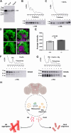
Fig 6. Mrj regulates long-term memory.
(A) Schematic of male courtship suppression-based memory assay. (B) Mrj knockout flies show significant memory deficit in comparison to the wild-type flies from 16 h onwards. Data are represented as mean ± SEM and Mann–Whitney U test is done to test for significance. (C) Representative image of the expression pattern of Mrj KO Gal4 visualized using the reporter CD8GFP (green) and counterstained with anti-FasII antibody (magenta) shows the expression of GFP in mushroom body neurons. Scale bar is 20 micron. (D) Confirmation of knockdown of Mrj by pan-neuronal expression of an Mrj RNAi line using a western blot with anti-Mrj antibody. Anti-α-Tubulin antibody was used as the loading control. (E) Representative image of the expression pattern of 201Y Gal4 visualized using the reporter CD8GFP. (F) Knockdown of Mrj in specific mushroom body neurons using 201Y Gal4 causes a significant memory deficit in comparison to the control from 16 h onwards. Data are represented as mean ± SEM and Mann–Whitney U test is done to test for significance. The data underlying this figure are available at: https://figshare.com/s/f5d913a0a289339ee16b.
Fig 7
Fig 7. Mrj interacts with ribosomes and helps in regulating Orb2A’s association with polysomes.
(A) Immunoprecipitated Rpl18-Flag which is incorporated in ribosomes pulls down Mrj, suggesting a possible association of Mrj with ribosomes. (B) Upper panel shows a representative polysome profile of lysate obtained from cells expressing Mrj-HA. The numbers in X-axis represent the fraction number. The lower panel is a western blot of individual fractions, showing the presence of Mrj in polysome fractions. (C) Upper panel shows a representative polysome profile of EDTA-treated lysate obtained from cells expressing Mrj-HA. The numbers in X-axis represent the fraction number. The lower panel is a western blot of individual fractions, showing the shift of Mrj to heavier fractions in comparison to non-EDTA treated lysate. (D) Representative images of mushroom body Kenyon cells immunostained with anti-Puromycin antibody to detect newly translated proteins. Magenta shows the counterstain with Phalloidin. (E) Quantitation of anti-Puromycin staining intensity shows no significant difference between the wild type (+/+) and Mrj knockout (-/-). Data is represented as mean ± SEM and significance is tested using two-tailed Student’s unpaired t test. (F) Upper panel shows a representative polysome profile of lysate obtained from cells expressing Orb2A-HA. The numbers in X-axis represent the fraction number. The lower panel shows a western blot detecting Orb2A in individual fractions. (G) Upper panel shows a representative polysome profile of lysate obtained from cells co-expressing Orb2A-HA and Mrj-HA. The numbers in X-axis represent the fraction number. The lower panel shows a western blot detecting Orb2A and Mrj in individual fractions. In comparison to only Orb2A, in presence of Mrj, more Orb2A is detected in polysome fractions. (H) Schematic of the model originating from this work. We hypothesize association of Mrj with Ribosomes is probably stabilizing it and regulating its oligomerization. Mrj acts as an inhibitor of aggregation of pathogenic Huntingtin (Htt) and positively regulates the formation of Orb2 oligomers. The data underlying this figure are available at: https://figshare.com/s/f5d913a0a289339ee16b.

Similar articles

Cited by

References

    1. Bliss TV, Lomo T. Long-lasting potentiation of synaptic transmission in the dentate area of the anaesthetized rabbit following stimulation of the perforant path. J Physiol. 1973;232:331–356. doi: 10.1113/jphysiol.1973.sp010273 - DOI - PMC - PubMed
    1. Harris KM. Structural LTP: from synaptogenesis to regulated synapse enlargement and clustering. Curr Opin Neurobiol. 2020;63:189–197. doi: 10.1016/j.conb.2020.04.009 - DOI - PMC - PubMed
    1. Nicoll RA. A Brief History of Long-Term Potentiation. Neuron. 2017;93:281–290. doi: 10.1016/j.neuron.2016.12.015 - DOI - PubMed
    1. Agranoff BW. Memory and protein synthesis. Sci Am. 1967;216:115–122. doi: 10.1038/scientificamerican0667-115 - DOI - PubMed
    1. Agranoff BW, Davis RE, Casola L, Lim R. Actinomycin D Blocks Formation of Memory of Shock-Avoidance in Goldfish. Science. 1967;158:1600–1601. doi: 10.1126/science.158.3808.1600 - DOI - PubMed

Publication types

MeSH terms

LinkOut - more resources