. 2024 Jun;1870(5):167193.
doi: 10.1016/j.bbadis.2024.167193. Epub 2024 Apr 20.

The IRE1α-XBP1 arm of the unfolded protein response is a host factor activated in SARS-CoV-2 infection

Affiliations

The IRE1α-XBP1 arm of the unfolded protein response is a host factor activated in SARS-CoV-2 infection

Jose Javier Fernández et al. Biochim Biophys Acta Mol Basis Dis. 2024 Jun.

Abstract

SARS-CoV-2 infection can cause severe pneumonia, wherein exacerbated inflammation plays a major role. This is reminiscent of the process commonly termed cytokine storm, a condition dependent on a disproportionated production of cytokines. This state involves the activation of the innate immune response by viral patterns and coincides with the biosynthesis of the biomass required for viral replication, which may overwhelm the capacity of the endoplasmic reticulum and drive the unfolded protein response (UPR). The UPR is a signal transduction pathway composed of three branches that is initiated by a set of sensors: inositol-requiring protein 1 (IRE1), protein kinase RNA-like ER kinase (PERK), and activating transcription factor 6 (ATF6). These sensors control adaptive processes, including the transcriptional regulation of proinflammatory cytokines. Based on this background, the role of the UPR in SARS-CoV-2 replication and the ensuing inflammatory response was investigated using in vivo and in vitro models of infection. Mice and Syrian hamsters infected with SARS-CoV-2 showed a sole activation of the Ire1α-Xbp1 arm of the UPR associated with a robust production of proinflammatory cytokines. Human lung epithelial cells showed the dependence of viral replication on the expression of UPR-target proteins branching on the IRE1α-XBP1 arm and to a lower extent on the PERK route. Likewise, activation of the IRE1α-XBP1 branch by Spike (S) proteins from different variants of concern was a uniform finding. These results show that the IRE1α-XBP1 system enhances viral replication and cytokine expression and may represent a potential therapeutic target in SARS-CoV-2 severe pneumonia.

Keywords: COVID-19; Cytokines; Fluvoxamine; Pneumonia; TLR; Transcription factors; Unfolded protein response; Variants of concern; Viral sepsis.


Conflict of interest statement

Declaration of competing interest The A.G.S. laboratory has received research support from GSK, Pfizer, Senhwa Biosciences, Kenall Manufacturing, Blade Therapeutics, Avimex, Johnson & Johnson, Dynavax, 7Hills Pharma, Pharmamar, ImmunityBio, Accurius, Nanocomposix, Hexamer, N-fold LLC, Model Medicines, Atea Pharma, Applied Biological Laboratories and Merck, outside of the reported work. A.G.S. has consulting agreements for the following companies involving cash and/or stock: Castlevax, Amovir, Vivaldi Biosciences, Contrafect, 7Hills Pharma, Avimex, Pagoda, Accurius, Esperovax, Farmak, Applied Biological Laboratories, Pharmamar, CureLab Oncology, CureLab Veterinary, Synairgen, Paratus and Pfizer, outside of the reported work. A.G.S. has been an invited speaker in meeting events organized by Seqirus, Janssen, Abbott and Astrazeneca. A.G.S is inventor on patents and patent applications on the use of antivirals and vaccines for the treatment and prevention of virus infections and cancer, owned by the Icahn School of Medicine at Mount Sinai, New York, outside of the reported work. A.M. is the creator of Omics Bioinformatics S.L. and owns all the stocks of this company. The M.S. laboratory has received unrelated research funding in sponsored research agreements from 7Hills Pharma, ArgenX N.V., Moderna and Phio Pharmaceuticals, which has no competing interest with this work. The article reflects the views of the authors and does not represent the views or policies of the FDA. All other authors declare that they have no conflict of interest.

Figures

Fig. 1.
Fig. 1.
SARS-CoV-2 activates the Ire1α-Xbp1 arm of the UPR in the lungs of K18-hACE2 mice after infection with 1 × 10⁴ PFU of the ancestral SARS-CoV-2 strain WA1/2020. (A) Analysis of Xbp1 by RT-PCR and resolution in agarose gel of the amplicons of three independent animals per condition (left panel) and quantification of sXbp1(right panel). (B) Western blot of sXbp1 and Chop in Mock and infected mice at 2 and 5 dpi. (C) Analysis of the mRNA of sXbp1, Hspa5, Atf4, and Ddit3. (D) Assay of viral titers measured by TCID50 on days 2 and 5 in lung tissue. (E) Weight loss monitored in animals up to 10 days after infection. (F) Percentage of survival. Data are presented as mean ± SEM. *p < 0.05, **p < 0.01. Unpaired Student’s t-test.
Fig. 2.
Fig. 2.
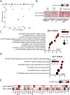
Bioinformatic analysis of the lung tissue of K18-hACE2 mice during SARS-CoV-2 infection. (A) Principal component analysis showing distinct clustering of each group of animals. (B) Heat map of UPR genes comparing lung tissue in Mock mice with mice infected with 1 × 10⁴ PFU of ancestral SARS-CoV-2 strain lineage B at 3 and 7 dpi. (C-D) GO enriched pathways analysis related to ER function at 3 and 7 dpi. Data are represented as bubble color with FDR < 0.05, and the number of genes within the GO pathway are represented as bubble size. (E) Heat map of cytokines comparing mock with 3 and 7 dpi. DEG analysis was carried out by DESeq2, and data are represented as Log2 fold change.
Fig. 3.
Fig. 3.
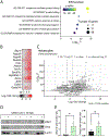
SARS-CoV-2 infection drives activation of the Ire1α-Xbp1 arm of the UPR in the lungs of Syrian hamsters. (A) GO enriched pathways analysis and differential biological processes related to ER function at 6 dpi. (B) Heat map of UPR target genes comparing Mock with Syrian hamsters infected with 5 × 10⁵ PFU of rSARS-CoV-2 WT. Lungs were collected at 2, 4, and 6 dpi. (C) Volcano plot of UPR genes at 6 dpi. Significant genes are represented with a cut-point of 1 in Log2 fold change and −Log10P. The differential expression analysis shown was carried out using the DESeq2 program. The significant genes object of study are represented using the library EnhancedVolcano v1.14.0 of R v4.2.0 (Bioconductor). (D-E) Western blot analysis of sXbp1 and Chop proteins at 6 dpi and densitometric quantification.
Fig. 4.
Fig. 4.
Lungs of Syrian hamster infected with SARS-CoV-2 show increased levels of cytokines. (A) GO enriched pathways analysis and differential biological processes related to cytokines at 6 dpi. (B) Volcano plot of the cytokines showing significant changes at 2, 4, and 6 dpi.
Fig. 5.
Fig. 5.
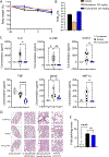
Effect of fluvoxamine in 129S1 mice infected with MA-SARS-CoV-2. (A) 129S1 mice were infected with MA-SARS-CoV-2 at 104 PFU and daily treated with 150 mg/kg s.c. fluvoxamine. Body weight was monitored throughout the experiment. (B) Viral titers measured by TCID50. Mice treated with 100 mg/kg of remdesivir were used as an antiviral positive control. (C) Blood cytokine levels from 129S1 mice were assayed at 3 dpi using the MD44 Multiplex assay. Data are presented as mean ± SEM. *p < 0.05, **p < 0.01, ***p < 0.005, ****p < 0.001. Ordinary one-way ANOVA. (D) H&E-stained sections of a lung from a 10-week-old female 129S1 mice at 3 dpi. (E) Total pathology score was examined by implementing a semi-quantitative, 5-point grading scheme including four different histopathological parameters. Results only showed differences between uninfected, and vehicle and fluvoxamine-treated groups.
Fig. 6.
Fig. 6.
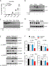
Role of sXBP1, CHOP, and GADD34 during SARS-CoV-2 infection in A549-ACE2 human epithelial cells. (A) Viral load measured by RT-qPCR at different times after infection. (B-D) sXBP1, CHOP, and GADD34 protein expression. (E) 20 nM of siRNA were used to knockdown DDIT3, GADD34, and XBP1 24 h prior to infection with SARS-CoV-2 at 1 MOI for 16 h. After this time, protein extracts were collected for the immunodetection of S protein, DDIT3, GADD34, and sXBP1. (F-G) DDIT3, GADD34, and XBP1 were knocked down 24 h before infection with SARS-CoV-2 under different MOI and timepoint conditions. Viral replication was assayed by immunostaining of N protein. The percentage of infection was quantified as (Infectedcells/TotalcellsBackground)×100, and the DMSO control was then set to 100 % infection for analysis. Data are presented as mean ± SEM of three biological replicates. *p < 0.05, **p < 0.01, ***p < 0.005, ****p < 0.001. Ordinary two-way ANOVA.
Fig. 7.
Fig. 7.
Effect of different SARS-CoV-2 VOCs on the UPR. (A) HEK293 T cells were transfected with plasmids encoding SARS-CoV-2 S protein VOCs and harvested at 36 hpt for Western blot analysis of S protein, sXBP1, HERPUD1, and GAPDH proteins. Tunicamycin (TM) was used as a positive control for UPR activation. (B) Densitometric scanning of sXBP1 protein expression normalized to GAPDH in HEK293 T cells transfected and treated as in (A). (C) Analysis of Xbp1 splicing by RT-PCR and agarose gel electrophoresis of HEK293 T cells treated as in (A). (D) A549-ACE2/TMPRSS2 cells were infected with SARS-CoV-2 VOCs at 0.1 MOI and incubated in the presence and absence of 10 μM KIRA8. Cell extracts were collected after 16 hpi for Western blot analysis of S protein, HERPUD1, and GAPDH. (E) Densitometric scanning of S protein. *p < 0.05. One sample t-test. ***p < 0.001. (F) Analysis of Xbp1 splicing by RT-PCR and resolution in agarose gel of cells infected with different SARS-CoV-2 VOCs at 0.1 MOI for 16 hpi and treated in the presence and absence of 10 μM KIRA8. (G) Quantitation of viral RNA in A549-ACE2/TMPRSS2 infected cells. N gene was assayed for viral load quantitation and RPL19 as a cell housekeeping gene. The IRE1α inhibitor KIRA8 was added immediately after the virus absorption period and maintained in the medium until cell harvesting 16 h later. (H) Viral titers measured by TCID50. Immunoblots are representative of three independent biological replicates. Data are presented as mean ± SEM. *p < 0.05. ***p < 0. 001. Paired Student’s t-test.

References

    1. V’kovski P, Kratzel A, Steiner S, Stalder H, Thiel V, Coronavirus biology and replication: implications for SARS-CoV-2, Nat. Rev. Microbiol. 19 (2021) 155–170, 10.1038/s41579-020-00468-6. - DOI - PMC - PubMed
    1. Lmanza A, Carlesso A, Chintha C, Creedican S, Doultsinos D, Leuzzi B, Luís A, McCarthy N, Montibeller L, More S, Endoplasmic reticulum stress signalling–from basic mechanisms to clinical applications, FEBS J. 286 (2019) 241–278, 10.1111/febs.14608. - DOI - PMC - PubMed
    1. Grootjans J, Kaser A, Kaufman RJ, Blumbergn RS, The unfolded protein response in immunity and inflammation, Nat. Rev. Immunol. 16 (2016) 469–484, 10.1038/nri.2016.62. - DOI - PMC - PubMed
    1. Iwakoshi NN, Lee AH, Glimcher LH, The X-box binding protein-1 transcription factor is required for plasma cell differentiation and the unfolded protein response, Immunol. Rev. 194 (2003) 29–38, 10.1034/j.1600-065x.2003.00057.x. - DOI - PubMed
    1. Martinon F, Chen X, Lee AH, Glimcher LH, TLR activation of the transcription factor XBP1 regulates innate immune responses in macrophages, Nat. Immunol. 11 (2010) 411–418, 10.1038/ni.1857. - DOI - PMC - PubMed

Publication types

MeSH terms