. 2024 Jun:370:110-123.
doi: 10.1016/j.jconrel.2024.04.029. Epub 2024 Apr 23.

Cadherin-11 targeted cell-specific liposomes enabled skin fibrosis treatment by inducing apoptosis

Affiliations

Cadherin-11 targeted cell-specific liposomes enabled skin fibrosis treatment by inducing apoptosis

Himanshu N Bhatt et al. J Control Release. 2024 Jun.

Abstract

Continuous and aberrant activation of myofibroblasts is the hallmark of pathological fibrosis (e.g., abnormal wound healing). The deposition of excessive extracellular matrix (ECM) components alters or increases the stiffness of tissue and primarily accounts for multiple organ dysfunctions. Among various proteins, Cadherin-11 (CDH11) has been reported to be overexpressed on myofibroblasts in fibrotic tissues. Anti-apoptotic proteins such as (B cell lymphoma-2) (BCL-2) are also upregulated on myofibroblasts. Therefore, we hypothesize that CDH11 could be a targeted domain for cell-specific drug delivery and targeted inhibition of BCL-2 to ameliorate the development of fibrosis in the skin. To prove our hypothesis, we have developed liposomes (LPS) conjugated with CDH11 neutralizing antibody (antiCDH11) to target cell surface CDH11 and loaded these LPS with a BCL-2 inhibitor, Navitoclax (NAVI), to induce apoptosis of CDH11 expressing fibroblasts. The developed LPS were evaluated for physicochemical characterization, stability, in vitro therapeutic efficacy using dermal fibroblasts, and in vivo therapeutic efficacy in bleomycin-induced skin fibrosis model in mice. The findings from in vitro and in vivo studies confirmed that selectivity of LPS was improved towards CDH11 expressing myofibroblasts, thereby improving therapeutic efficacy with no indication of adverse effects. Hence, this novel research work represents a versatile LPS strategy that exhibits promising potential for treating skin fibrosis.

Keywords: Cadherin 11; Dermal fibrosis; Lipid nanoparticle; Myofibroblast; Scleroderma.


Conflict of interest statement

Declaration of competing interest The authors report no conflict of interest.

Figures

Fig. 1.
Fig. 1.
Preparation and characterization of LPS. (A) Scheme of preparation of NAVI-LPS and antiCDH11-NAVI-LPS. mPEG2000-DSPE and DSPE-PEG2000-Mal were used to prepare NAVI-LPS and antiCDH11-NAVI-LPS, respectively, in the lipid phase. (B) Representative TEM image of antiCDH11-NAVI-LPS. Scale bars, 100 nm. (C) Size measurements of NAVI-LPS and antiCDH11-NAVI-LPS. (D) in vitro, release testing of NAVI-LPS and antiCDH11-NAVI-LPS in PBS of pH 7.4, and data are presented as mean ± SD, where n = 3.
Fig. 2.
Fig. 2.
Cellular internalization of LPS in dermal fibroblasts. (A) Cellular uptake of FITC-LPS and antiCDH11-FITC-LPS in TGF-β1 (10 ng/mL) activated dermal fibroblasts using confocal microscopy. Green, blue, yellow, and red fluorescence indicates LPS, nucleus, COL1α1, and α-SMA, respectively. Scale bars, 50 μm. (B and C) Quantification analysis of FITC fluorescence in COL1α1+ and α-SMA+ area in dermal fibroblasts treated with FITC-LPS and antiCDH11-FITC-LPS (n = 4). (D) Cellular uptake of FITC-LPS and antiCDH11-FITC-LPS in non-activated and TGF-β1 (10 ng/mL) activated dermal fibroblasts measured by flow cytometer at 2 h. Significant differences were evaluated using a one-way ANOVA (B & C). The data are presented as means ± SD, n = 4. *P < 0.05, ***P < 0.001. (For interpretation of the references to colour in this figure legend, the reader is referred to the web version of this article.)
Fig. 3.
Fig. 3.
In vitro therapeutic efficacy in dermal fibroblasts. (A) Representative images of inhibition of fibrotic proteins such as CDH11, BCL-2, COL1α1, and α-SMA immunostaining in TGF-β1 (10 ng/mL) activated dermal fibroblasts treated with PBS, NAVI, NAVI-LPS, and antiCDH11-NAVI-LPS. Scale bars, 20 μm. (B to E) Quantification analysis of expression of CDH11, BCL-2, COL1α1, and α-SMA in TGF-β1 (10 ng/mL) activated dermal fibroblasts treated with PBS, NAVI, NAVI-LPS, and antiCDH11-NAVI-LPS (n = 4). The data are presented as means ± SD, n = 4. (F) RT-PCR measurement of CDH11 RNA level in non-activated, TGF-β1 (10 ng/ml) activated dermal fibroblasts, TGF-β1 (10 ng/mL) activated dermal fibroblasts treated with NAVI, NAVI-LPS, and antiCDH11-NAVI-LPS. The data are presented as means ± SD, n = 3. Significant differences were evaluated using a one-way ANOVA (B to F). *P < 0.05, ***P < 0.001, ****P < 0.0001, n.s., not significant, P > 0.05.
Fig. 4.
Fig. 4.
Trans-well migration assay. (A) Schematic presentation of transwell migration assay. (B and C) Representative images and quantification of fibroblasts migrated from the upper to the lower chamber after treatment of PBS, PBS + TGF-β1, NAVI, NAVI-LPS, and antiCDH11-NAVI-LPS (n = 4). Data are presented as means ± SD. *P < 0.05, **P < 0.01, ****P < 0.0001, n.s., not significant, P > 0.05. Fig. 4A was created from Biorender.com.
Fig. 5.
Fig. 5.
Biodistribution of LPS in mice model of skin fibrosis. (A) Time-dependent NIR fluorescence images of fibrotic mice following IV administration of DiR-LPS and antiCDH11-DiR-LPS (DiR dose: 1 mg/kg). (B) Ex-vivo imaging of major organs and skin excised after 48 h post-injection. (C) The fluorescence intensity of LPS in the fibrotic region of the skin was recorded as total photon counts in fibrotic mice kept in the dorsal position during real-time imaging in the IVIS chamber. (D) Mean fluorescent intensity of DiR in organs after 48 h post-injection of LPS. Data are presented as mean ± SD, where n = 3. Li-Liver; Sk-Skin; K-Kidney; Lu-Lungs; Sp-Spleen; H-Heart.
Fig. 6.
Fig. 6.
In vivo therapeutic validation of LPS in a skin fibrosis mouse model. (A) Schematics of in vivo experimental design. (B) Representative micrographs of H&E staining. Scale bars, 100 μm. (C) Representative micrographs of Masson’s trichrome stain. Scale bars, 100 μm. (D) The dermal thickness of fibrotic skin. Five determinations per high-powered field from 5 mice/group. (E) Measurement of hydroxyproline content in mouse skin tissues (n = 5). (F) Western blot assay of CDH11, COL1α1, α-SMA, and BCL-2 expression in skin tissue homogenates collected from different groups. (G to J) Quantification of western blot bands of (G) CDH11 (H) BCL-2 (I) COL1α1, (J) α-SMA in homogenates of skin tissue collected from different treatment groups. GAPDH was used as a housekeeping protein. Significant differences were evaluated using a one-way ANOVA (D & E, G to J). Data are presented as means ± SD. *P < 0.05, **P < 0.01, ****P < 0.0001, n.s., not significant, P > 0.05.
Fig. 7.
Fig. 7.
Immunofluorescence analysis in fibrotic skin after treatments. (A) Representative immunofluorescence images of CDH11, BCL-2, BCL-xL, COL1α1, α-SMA, and TUNEL from mouse fibrotic skin sections treated with NAVI, NAVI-LPS, and antiCDH11-NAVI-LPS (5 mg/kg of NAVI). Scale bars, 200 μm. (B to G) Quantitative analysis of immunofluorescence staining in terms of expression of CDH11 (B), BCL-2 (C), BCL-xL (D), COL1α1 (E), α-SMA (F), and TUNEL+ area (G). The significant difference was evaluated using a one-way ANOVA (B to G). The data are presented as means ± SD, n = 5. *P < 0.05, **P < 0.01, ****P < 0.0001, n.s., not significant, P > 0.05.
Fig. 8.
Fig. 8.
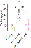
Immunogenicity study. In vivo TNF-α levels in serum following intravenous administration (five doses) with Blank-LPS and antiCDH11-LPS in C57BL/6 mice. Data are presented as mean ± SD (n = 5). The significant difference was evaluated using a one-way ANOVA. *P < 0.05, P > 0.05, n.s., not significant.

References

    1. Pathogenesis of Systemic Sclerosis: Recent Insights of Molecular and Cellular Mechanisms and Therapeutic Opportunities - John Varga, Maria Trojanowska, Masataka Kuwana, 2017, (n.d.). 10.5301/jsrd.5000249 (accessed November 29, 2023). - DOI
    1. Emerging targets of disease-modifying therapy for systemic sclerosis Nat. Rev. Rheumatol., (n.d.). https://www.nature.com/articles/s41584-019-0184-z (accessed November 29, 2023). - PubMed
    1. Scleroderma Facts, n.d. https://scleroderma.org/wp-content/uploads/2022/02/SIP-2022-Updated-7_5_....
    1. Abraham DJ, Varga J, Scleroderma: from cell and molecular mechanisms to disease models, Trends Immunol. 26 (2005) 587–595, 10.1016/j.it.2005.09.004. - DOI - PubMed
    1. Arno AI, Gauglitz GG, Barret JP, Jeschke MG, New molecular medicine-based scar management strategies, Burns 40 (2014) 539–551, 10.1016/j.burns.2013.11.010. - DOI - PMC - PubMed

MeSH terms

LinkOut - more resources