. 2024 Apr 22;15(1):3415.
doi: 10.1038/s41467-024-47689-4.

Tyrosine phosphorylation of CARM1 promotes its enzymatic activity and alters its target specificity

Affiliations

Tyrosine phosphorylation of CARM1 promotes its enzymatic activity and alters its target specificity

Hidehiro Itonaga et al. Nat Commun. .

Abstract

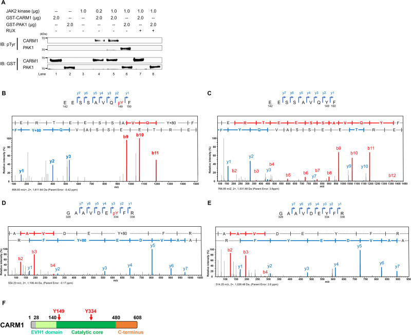
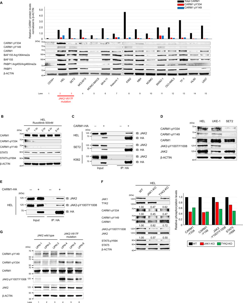
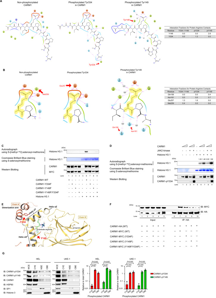
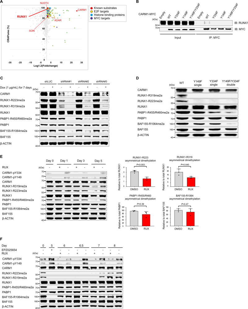
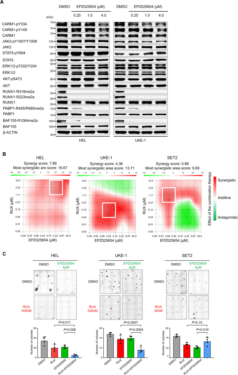
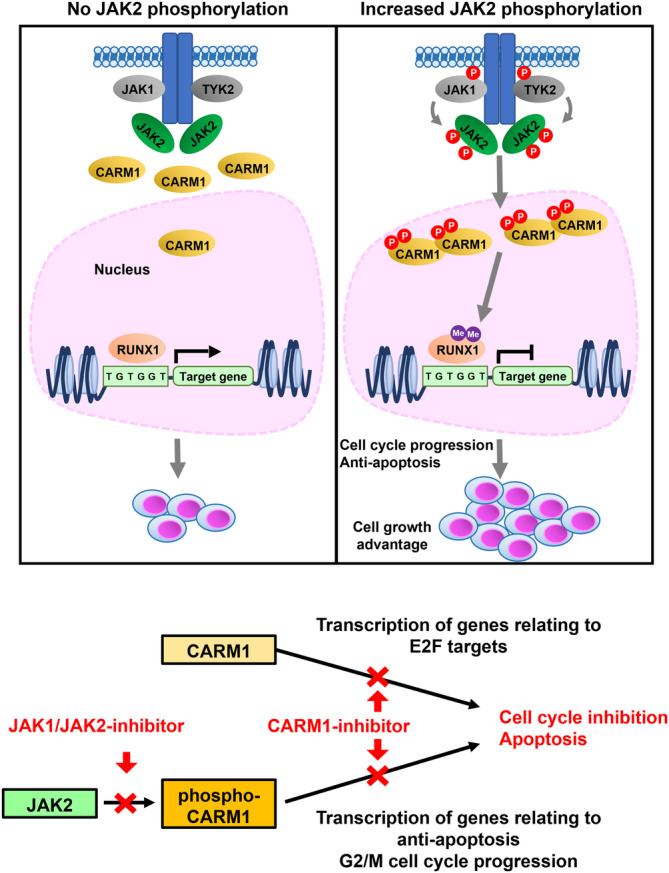
An important epigenetic component of tyrosine kinase signaling is the phosphorylation of histones, and epigenetic readers, writers, and erasers. Phosphorylation of protein arginine methyltransferases (PRMTs), have been shown to enhance and impair their enzymatic activity. In this study, we show that the hyperactivation of Janus kinase 2 (JAK2) by the V617F mutation phosphorylates tyrosine residues (Y149 and Y334) in coactivator-associated arginine methyltransferase 1 (CARM1), an important target in hematologic malignancies, increasing its methyltransferase activity and altering its target specificity. While non-phosphorylatable CARM1 methylates some established substrates (e.g. BAF155 and PABP1), only phospho-CARM1 methylates the RUNX1 transcription factor, on R223 and R319. Furthermore, cells expressing non-phosphorylatable CARM1 have impaired cell-cycle progression and increased apoptosis, compared to cells expressing phosphorylatable, wild-type CARM1, with reduced expression of genes associated with G2/M cell cycle progression and anti-apoptosis. The presence of the JAK2-V617F mutant kinase renders acute myeloid leukemia (AML) cells less sensitive to CARM1 inhibition, and we show that the dual targeting of JAK2 and CARM1 is more effective than monotherapy in AML cells expressing phospho-CARM1. Thus, the phosphorylation of CARM1 by hyperactivated JAK2 regulates its methyltransferase activity, helps select its substrates, and is required for the maximal proliferation of malignant myeloid cells.


Conflict of interest statement

The authors declare no competing interests.

Figures

Fig. 1
Fig. 1. JAK2 phosphorylates CARM1 in vitro.
A JAK2 phosphorylates CARM1 in an in vitro kinase assay, in which active JAK2 kinase and recombinant CARM1 proteins were used; PAK1 was used as a positive control. The amounts of protein in the reaction are indicated. The phosphorylation of CARM1 was completely abolished by the JAK1/JAK2 inhibitor (ruxolitinib, RUX; 500 nM) (lanes 7 and 8). The experiment was repeated independently twice. B Peptide fragments in the mass spectrometry analysis were generated from proteolytic cleavage of CARM1 following in vitro kinase assays in the presence of active JAK2 kinase. Tyrosine-149 (Y149) along with the series of y- and b-ions, including the phosphorylated residue, is shown as the phosphorylated peptide (EESSAVQpYF). C Peptide fragments in the mass spectrometry analysis were generated from proteolytic cleavage of CARM1 following in vitro kinase assays in the absence of active JAK2 kinase. The peptide fragment around Y149 residue (EESSAVQYF) is shown without phosphorylation of tyrosine, indicating no gain in molecular weight of 80 Da (i.e. the weight of PO4). D Peptide fragments in the mass spectrometry analysis were generated from proteolytic cleavage of CARM1 following in vitro kinase assays in the presence of active JAK2 kinase. Tyrosine-334 (Y334) along with the series of y- and b-ions, including the phosphorylated residue, is shown as the phosphorylated peptide (GAAVDEpYFR). (E) Peptide fragments in the mass spectrometry analysis were generated from proteolytic cleavage of CARM1 following in vitro kinase assays in the absence of active JAK2 kinase. The peptide fragment around Y334 residue (GAAVDEYFR) is shown without phosphorylation of tyrosine, indicating no gain in molecular weight of 80 Da. F The regions containing amino acid residues Y149 and Y334 are located within the core catalytic domain (residue 140-480) of CARM1. Residues 28-140 in CARM1 are highly homologous to a family of Drosophila-enbaled/vasodilator-stimulated phosphoprotein homology 1 (EVH1) domains, which specifically bind to target proline-rich sequences with low affinity and high specificity.
Fig. 2
Fig. 2. JAK2-V617F promotes the tyrosine phosphorylation of CARM1 in myeloid leukemia cells.
A The expression of CARM1 protein and Y149/Y334 phosphorylated CARM1 protein was assessed in 14 myeloid leukemia cell lines and human CD34+ cord blood cells, by immunoblotting analysis. B Phosphorylation of Y149 and Y334 of CARM1 in HEL cells is abolished following treatment with the JAK1/JAK2 inhibitor (RUX at low concentration to avoid severe apoptosis; 500 nM), as is phosphorylation of tyrosine-694 in STAT5. C Immunoprecipitation was performed using HEL, SET2, and K562 cells that express HA-tagged CARM1, with an anti-HA antibody. Immunoblotting with anti-HA and anti-JAK2 antibodies revealed the interaction between JAK2 and CARM1. D HEL and UKE-1 cells harboring homozygous JAK2-V617F mutations had phosphorylated CARM1-Y149/Y334 and (auto) phosphorylated JAK2-Y1007/Y1008, while SET2 cells harboring heterozygous JAK2-V617F mutation did not. E Proteins were immunoprecipitated from HEL cell extracts that express HA-tagged CARM1, using an anti-HA antibody; immunoblotting was then performed using an anti-HA antibody, anti-phosphorylated JAK2 rabbit antibody, or anti-JAK2 mouse antibody. F Extracts from JAK1 or TYK2 knockout HEL cells were immunoblotted using phosphospecific anti-CARM1, JAK2, and STAT5 antibodies. G Phospho-CARM1 and phospho-JAK2 were evaluated in mononuclear cells isolated from patients with the following myeloid neoplasms: UPN-1 (unique patient number-1), chronic myeloid leukemia blast phase; UPN-2, acute myeloid leukemia with mutated NPM1; UPN-3, acute myeloid leukemia not otherwise specified; UPN-4, essential thrombocythemia; UPN-5, primary myelofibrosis; and UPN-6, polycythemia vera. All experiments were repeated at least two times independently. A n = 3, BG n = 2.
Fig. 3
Fig. 3. Biochemical regulation of CARM1 enzymatic activity by tyrosine phosphorylation.
A Based on the crystal structures of CARM1, CARM1-Y334 phosphorylation (middle) increased the interaction of the region containing Y334 itself with the substrate compared with non-phosphorylated Y334 (left). CARM1-Y149 phosphorylation (right) also increased the binding of the region containing Y149 itself with the substrate. B CARM1-Y334 and -Y149 phosphorylation impairs the loss of binding between CARM1 methionine-259 (Met259) and substrate. The decreased Met259 binding increases the interaction of glutamic acid-257 (Glu257) and glutamic acid-266 (Glu266) with substrates (middle) in the presence of Y334 phosphorylation, compared to non-phosphorylated Y334 (left). Furthermore, the loss of methionine-259 binding increases the interaction of glutamine-158 (Gln158) and aspartic acid-161 (Asn161) with substrates in the presence of Y149 phosphorylation (right). C Immunoprecipitation was performed using an anti-MYC antibody and 293 T cells that express MYC-tagged WT or non-phosphorylatable CARM1. The mutant CARM1 Y149F, Y334F, and Y149F/Y334F proteins show reduced methyltransferase activity for histone H3.1, compared to wild-type (WT) CARM1, in in vitro methylation assay. CARM1 and MYC protein lanes are shown to demonstrate equal loading (Top). Autoradiograph of the methylated 3H-histone H3.1 (Middle). Coomassie staining shows histone H3.1 used in the assay (Bottom). Western blotting shows the relative amount of CARM1 and MYC. D Recombinant CARM1 protein (produced in E.coli), histone H3.1, and 14C-SAM were incubated to perform an in vitro methylation assay. CARM1 was also incubated with JAK2 kinase, leading to its phosphorylation on Y334 in lanes 3, 4, 7, and 8. Phosphorylated CARM1 shows increased methyltransferase activity for histone H3.1 in in vitro methylation assay (Top). Autoradiograph of the methylated 3H-histone H3.1 (Middle). Coomassie staining shows histone H3.1 used in the assay (Bottom). Western blotting shows the relative amount of CARM1 and phosphorylated CARM1. E CARM1-Y149 and -Y334 localize at dimerization arm and helix αX, respectively. These residues lie close to the dimerization interface in the modeled CARM1 structure. F Co-immunoprecipitation of HA- and MYC-tagged CARM1 from 293 T cell extracts transiently transfected with plasmid expressing HA-tagged WT and MYC-tagged WT or mutant CARM1. HA-tagged WT CARM1 was immunoprecipitated from cell extracts with anti-HA antibodies, and then the coimmunoprecipitated MYC-tagged CARM1 was probed with anti-MYC antibodies. The levels of MYC-tagged CARM1 Y149F and Y149F/Y334F from the HA immunoprecipitates were lower than those of MYC-tagged CARM1 WT. G Subcellular fractionations of HEL cells and UKE-1 cells were immunoblotted using anti-total CARM1, CARM1-pY334, and -pY149 antibodies; cytoplasmic extraction, CYE; nuclear soluble extraction, NSE; and chromatin-bound extraction, CBE. The left lane represents the expression levels of the indicated proteins of whole-cell lysates (WCE). The bar graph on the right represents the ratio of cytoplasmic, nuclear, or chromatin-binding CARM1-pY334 and -pY149 to total cytoplasmic, nuclear, or chromatin-binding CARM1, respectively (bands inside the boxes). Data represent the mean ± SD. n = 3, unpaired two-tailed Student’s t-test. C, D, F All experiments were repeated two times independently.
Fig. 4
Fig. 4. Identification of RUNX1 as CARM1-interacting proteins by Proximity BioID proteomics.
A Scatter plot comparing mean-fold change for CARM1-BirA* fusion vs. BirA* alone with abundance in published negative control AP-MS datasets (%CRAPome). Green dots represent proteins (i) with a cutoff frequency of ≥80% CRAPome and an average spectral count fold change ≥1.2 or (ii) with a cutoff frequency of <80% CRAPome but the average spectral count fold change ≥3.0. Known substrates of CARM1 are indicated as red, and E2F-targets, histone binding proteins, and MYC-targets are shown in yellow, blue, and violet, respectively. See also supplementary data 1 (HEL cells, n = 1) and 2 (K562 cells, n = 1). B Proteins were immunoprecipitated from HEL cell extracts that express MYC-tagged CARM1 (WT and non-phosphorylatable mutants), using an anti-MYC antibody; immunoblotting was then performed using an anti-MYC antibody and anti-RUNX1 mouse antibody. C Doxycycline-inducible short hairpin RNAs (shRNAs) directed against CARM1 decreased CARM1 protein levels and the ADMA levels of RUNX1-R223 and -R319 as well as well-established targets, such as PABP1-R455/R460 and BAF155-R1064. D Clustered regularly interspaced short palindromic repeat (CRISPR)/CRISPR-associated protein-9 (Cas9)-mediated non-phosphorylatable CARM1 mutants decreased the ADMA levels of RUNX1-R223 and -R319 as well as PABP1 and BAF155. E Expression of total and asymmetry dimethylated RUNX1, PABP1, and BAF155 were assessed in HEL cells treated with RUX 250 nM or DMSO control for 5 days. Fresh media with RUX or DMSO was added on days 0, 2, and 4. Quantification of the ADMA levels of RUNX1, BAF155, and PABP1 at 5 days after RUX treatment are shown in the right panels. Data represent the mean ± SD. n = 3, one-way ANOVA. The relative cell viability at day 5 was 85% for cells treated with RUX, compared to those cultured with DMSO (P = 0.005)(n = 3, biological replicates). F The levels of ADMA RUNX1, BAF155, and PABP1 were measured in HEL cells treated with RUX (or DMSO) after EPZ025654 treatment for 5 days followed by a wash-out phase lasting up to 3 days (labeled as day 8). The relative cell viability at day 8 was 86.8% for cells treated with RUX compared to those cultured with DMSO (P = 0.006)(n = 3 biological replicates). The experiments were repeated at least two times independently. B, D n = 2, (C) n = 3.
Fig. 5
Fig. 5. Functional analysis of non-phosphorylatable mutant CARM1.
A Cell proliferation assays of non-phosphorylatable CARM1 mutant knock-in HEL cells, where cell numbers were measured using a cell-counting apparatus. Y149F/Y334F (n = 3) vs. WT type (n = 3); day 3 (p < 0.001), day 5 (p = 0.003), day 7 (p = 0.001). B The flow cytometry analysis of BrdU-stained HEL cells expressing non-phosphorylatable CARM1 mutants. Mean fractions ± s.d. in sub G1, G0/G1, S, and G2/M populations. n = 3. S population (p = 0.015) in Y149F vs. WT; S (p = 0.011) and G2/M populations (p = 0.009) in Y334F vs. WT; S (p = 0.008) and G2/M populations (p = 0.002) in Y149F/Y334F vs. WT. C Heatmap shows the differentially expressed coding genes at 2-fold cut-off, representing replicates of HEL cells expressing CARM1 WT or two independent cells expressing CAMR1-Y149F/Y334F double mutation (Y149F/Y334F-1 and Y149F/Y334F-2). D Gene ontology analysis of significant downregulated genes in HEL cells expressing CARM1-Y149F/Y334F compared to CARM1-WT. E Heatmaps of FDR (q < 0.25) values from GSEA of hallmark gene set collections. F Representative GSEA plot depicting the downregulation of G2/M checkpoint and apoptosis/anti-apoptosis pathways. G Volcano plot representing gene expression changes triggered by CARM1-Y149F/Y334F mutation knock-in in HEL cells. Genes associated with apoptosis/anti-apoptosis, G2/M checkpoints, stemness in hematopoietic stem cells, and RUNX1-target are shown in red, yellow, blue, and violet, respectively. The red dots indicate upregulated genes in HEL cells expressing CARM1-Y149F/Y334F, whereas the blue dots indicate downregulated genes. P values correspond to a two-sided Wilcoxon rank-sum test with Bonferroni correction. H Representative GSEA plot depicting the downregulation of “hematopoietic stem cell up” signature. I qRT-PCR analysis showing BMI-1 and CD34 in HEL cells expressing CARM1 WT (n = 3), Y149F (n = 6), Y334F (n = 6), and Y149F/Y334F mutation (n = 6). Mean and SD are expressed as a percentage of HPRT-1 expression. J qRT-PCR analysis showing ID2 and MIR144 in HEL cells expressing CARM1 WT (n = 3), Y149F (n = 6), Y334F (n = 6), and Y149F/Y334F mutation (n = 6). Mean and SD are expressed as a percentage of HPRT-1 expression. n = 3. K Heat map of total R319-RUNX1 or asymmetrically dimethylated R319-RUNX1 binding tag intensity by ChIP-seq analysis for HEL cells expressing CARM1 WT, Y149F, Y334F, or Y149F/Y334F mutant proteins. L ChIP-seq analyses were performed to assess total RUNX1 and asymmetrically dimethylated R319-RUNX1 chromatin binding. Target occupancies at the ID2 gene are shown in IGV genome browser tracks. All error bars represent the mean ± SD. P values were determined by two-tailed Student’s t-test (A, B) and one-way ANOVA followed by Dunnett’s post hoc test (I, J). *p < 0.05, **p < 0.01, ***p < 0.001.
Fig. 6
Fig. 6. Inhibition of CARM1 targets cells harboring phosphorylated CARM1 mediated by JAK2-V617F mutant.
A Western blot assessment of phosphorylation in JAK2, STAT5, ERK, and AKT, and asymmetric demethylated arginine in RUNX1, BAF155, and PABP1 in HEL and UKE-1 cells treated with 5 days with increasing concentrations of EPZ025654 (μM). These experiments were repeated independently twice. B Excess over Bliss plots (Bliss method) showing synergistic effects between EPZ025654 and RUX were visualized in the calculated 2D synergy maps. Red and green areas represent synergistic (synergy score >+10), addictive (synergy score 0- + 10), and antagonistic effect (<−10), respectively. In 2D synergy maps, white rectangles show the maximum synergy area in each cell. C The colony formation of HEL, UKE-1, and SET2 cells treated with DMSO (control), RUX, EPZ025654, or a combination of RUX and EPZ025654. The concentration of RUX was applied based on the IC50 values for each cell line. Representative pictures of colonies on semi-solid methylcellulose media are shown on the upper panels. Quantification of the number of colonies at 14 days after plating are shown in the lower panels. Data represent the mean ± SD. n = 4, one-way ANOVA.
Fig. 7
Fig. 7. A schematic model showing JAK2-CARM1 axis.
JAK2-V617F mutant kinase, when activated by JAK2, JAK1, or TYK2, strongly phosphorylates CARM1-Y149 and -Y334, increasing its methyltransferase activity and the asymmetrical dimethylation of its substrates, including histone 3 and RUNX1. CARM1 phosphorylation promotes cell-cycle progression and inhibits apoptosis, and regulates the genes associated with stemness (BMI-1).

References

    1. Larsen SC, et al. Proteome-wide analysis of arginine monomethylation reveals widespread occurrence in human cells. Sci. Signal. 2016;9:rs9. doi: 10.1126/scisignal.aaf7329. - DOI - PubMed
    1. Bedford MT, Clarke SG. Protein arginine methylation in mammals: who, what, and why. Mol. Cell. 2009;33:1–13. doi: 10.1016/j.molcel.2008.12.013. - DOI - PMC - PubMed
    1. Greenblatt SM, Liu F, Nimer SD. Arginine methyltransferases in normal and malignant hematopoiesis. Exp. Hematol. 2016;44:435–441. doi: 10.1016/j.exphem.2016.03.009. - DOI - PubMed
    1. Yang Y, Bedford M. Protein arginine methyltransferases and cancer. Nat. Rev Cancer. 2013;13:37–50. doi: 10.1038/nrc3409. - DOI - PubMed
    1. Gao WW, et al. Arginine methylation of HSP70 regulates retinoid acid-mediated RARβ2 gene activation. Proc. Natl Acad. Sci. USA. 2015;112:E3327–E3336. doi: 10.1073/pnas.1509658112. - DOI - PMC - PubMed

Publication types

MeSH terms

Substances