. 2024 Apr 22;7(1):486.
doi: 10.1038/s42003-024-06130-8.

A viral assembly inhibitor blocks SARS-CoV-2 replication in airway epithelial cells

Affiliations

A viral assembly inhibitor blocks SARS-CoV-2 replication in airway epithelial cells

Li Du et al. Commun Biol. .

Abstract

The ongoing evolution of SARS-CoV-2 to evade vaccines and therapeutics underlines the need for innovative therapies with high genetic barriers to resistance. Therefore, there is pronounced interest in identifying new pharmacological targets in the SARS-CoV-2 viral life cycle. The small molecule PAV-104, identified through a cell-free protein synthesis and assembly screen, was recently shown to target host protein assembly machinery in a manner specific to viral assembly. In this study, we investigate the capacity of PAV-104 to inhibit SARS-CoV-2 replication in human airway epithelial cells (AECs). We show that PAV-104 inhibits >99% of infection with diverse SARS-CoV-2 variants in immortalized AECs, and in primary human AECs cultured at the air-liquid interface (ALI) to represent the lung microenvironment in vivo. Our data demonstrate that PAV-104 inhibits SARS-CoV-2 production without affecting viral entry, mRNA transcription, or protein synthesis. PAV-104 interacts with SARS-CoV-2 nucleocapsid (N) and interferes with its oligomerization, blocking particle assembly. Transcriptomic analysis reveals that PAV-104 reverses SARS-CoV-2 induction of the type-I interferon response and the maturation of nucleoprotein signaling pathway known to support coronavirus replication. Our findings suggest that PAV-104 is a promising therapeutic candidate for COVID-19 with a mechanism of action that is distinct from existing clinical management approaches.


Conflict of interest statement

S.S., A.F.L., M.M., S.F.Y. and K.P. are employees of Prosetta Biosciences. V.R.L. is the CEO of Prosetta Biosciences, which manufactures PAV-104 presented in the manuscript. All other authors declare no competing interests.

Figures
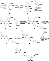
Fig. 1
Fig. 1. Synthesis and molecular structure of PAV-104.
To a solution of aldehyde1 (10 g, 65.79 mmol, 1.0 eq) in toluene was added 2,4-dimethoxybenzyl amine 2 (10.99 g, 65.79 mmol, 1.0 eq), and the reaction mixture was heated at 80 °C for 24 h. Solvent was removed, and the residue was taken in MeOH and cooled using an ice bath. Then sodium borohydride (4.97 g, 131.58 mmol, 2.0 eq) was added slowly, and the reaction mixture was stirred at room temperature for 12 h. Solvent was removed, and the residue was taken in ethyl acetate and then sat. NaHCO3 was added and stirred for 1 h. The organic layer was separated, dried (MgSO4), and the solvent was removed to give amine 3, which was used in the next step without further purification. To a solution of the crude amine 3 (5.0 g, 19.1 mmol, 1.0 eq) in DMF (25 mL) was added acid 4 (3.17 g, 19.1 mmol, 1.0 eq), HATU (8.7 g, 22.92 mmol, 1.2 eq,), and DIEA (12.32 g, 95.5 mmol, 5.0 eq) and the reaction mixture was stirred at room temperature for 12 h. The reaction mixture was then diluted with ethyl acetate (EtOAc) and washed with 10% aqueous HCl (1×), sat. NaHCO3 (1×) and water (3×). Organic layer was collected, dried (MgSO4), and evaporated to give a residue, which was taken in MeOH, and then K2CO3 (2.64 g. 19.1 mmol, 1.0 eq) was added and stirred at room temperature for 12 h. Solvent was removed, and the residue was taken in Ethyl acetate and washed with 10% HCl (1×). Organic layer was separated, dried, and solvent was removed to give a residue, which was purified by column chromatography (EtOAc/ Hexane) to give compound 5. To a stirred solution of compound 5 (1.0 g, 2.22 mmol, 1.0 eq) and cesium carbonate (1.08 g, 3.33 mmol, 1.5 eq) in DMF (15 mL) was added methyl 4-(chloromethyl) benzoate 6 (450 mg, 2.44 mmol, 1.2 eq) and the reaction mixture was stirred at room temperature for 18 h. The reaction mixture was diluted with ethyl acetate and washed with water (3×). Organic layer was dried and concentrated to give crude product 7. The crude compound 7 was stirred in a 1:1 mixture of TFA: DCM for 12 h. Concentration followed by chromatography purification (Hexane/EtOAc) provided compound 8. To a stirred solution of compound 8 (0.84 mmol, 1.0 eq) in 3:1 mixture of THF: H2O (12 mL) was added LiOH (40 mg, 1.68 mmol, 2.0 eq) and the reaction mixture was stirred at 65 °C for 12 h. The reaction mixture was evaporated under vacuum to give a residue, which was stirred in a mixture of 10% aqueous HCl and ethyl acetate for 30 min. Organic layer was collected, washed (H2O, 1×), dried, and concentrated to give crude acid 9. To a solution of the amine 10 (68 mg, 0.552 mmol, 1.2 eq) in DMF (25 mL) were added acid 9 (200 mg, 0.46 mmol, 1.0 eq), HATU (210 mg, 0.552 mmol, 1.2 eq,) and DIEA (0.300 mg, 2.3 mmol, 5.0 eq). The reaction mixture was stirred at room temperature for 12 h. The reaction mixture was then diluted with EtOAc and washed with 10% aqueous HCl (1×), sat. NaHCO3 (1×) and water (3×). The organic layer was collected, dried (MgSO4), and evaporated to give a residue, which was purified by column chromatography (EtOAc/ Hexane) to give PAV-104. PAV-104 was dissolved in DMSO.
Fig. 2
Fig. 2. PAV-104 decreases virus production in SARS-CoV-2-infected Calu-3 cells.
a MTT assay was performed on Calu-3 cells to examine the cellular toxicity of PAV-104. Relative cell viability was displayed based on the PAV-104-untreated control (set at 100%). The concentration of 100 nM of PAV-104 is represented by the black arrow. The red arrow represents the CC50 value (1306 nM) of PAV-104. b Anti-SARS-CoV-2 activity of PAV-104 in Calu-3 cells was measured by RT-qPCR targeting the N genes. Cells were pretreated with PAV-104 at the indicated concentrations for 1 h, followed by infection with SARS-CoV-2 (USA-WA1/2020, MOI = 0.01) for 24 h in the presence of PAV-104. RNA isolation and RT-qPCR assay were performed 24 h post infection. Data were nomalized to the DMSO negative control. c The SARS-CoV-2 titer (TCID50) was measured after treatment with varying doses of PAV-104 as described in b. d Immunofluorescence staining of Calu-3 cells with DAPI (blue) was performed at 72 h post infection. Cells were pretreated with PAV-104 at the indicated concentrations, followed by infection with SARS-CoV-2 virus. Scale bar, 500 μm. e Quantification of SARS-CoV-2 (USA-WA1/2020, MOI = 0.01) infected Calu-3 (FITC-positive) cells (shown in d). Data are representative of the results of three independent experiments (n = 3 biologically independent samples, mean ± standard error of mean (SEM)). Statistical significance was analyzed by t test. p ≤ 0.05 [*], p ≤ 0.01 [**], p ≤ 0.001 [***], p ≤ 0.0001 [****].
Fig. 3
Fig. 3. PAV-104 inhibits SARS-CoV-2 replication in Calu-3 cells more potently than remdesivir.
a Reduction of SARS-CoV-2 replication by PAV-104 and remdesivir in Calu-3 cells, as determined by RT-qPCR targeting the N gene. Calu-3 cells were pretreated with DMSO, PAV-104, or remdesivir for one hour, then infected with SARS-CoV-2 at an MOI of 0.001. Cells were collected at 48 hpi. b Percent inhibition of SARS-Cov-2 replication by PAV-104 and remdesivir in Calu-3 cells, as determined by RT-qPCR (PAV-104: EC50 = 1.725 nM, EC90 = 24.5 nM; remdesivir: EC50 = 7.9 nM, EC90 = 219.9 nM). c Reduction of SARS-CoV-2 replication by PAV-104 and remdesivir in Calu-3 cells, as determined by infectious viral titer in the supernatant at 48 hpi. d Percent inhibition of SARS-CoV-2 replication by PAV-104 and remdesivir in Calu-3 cells, as determined by infectious viral titer (PAV-104: EC50 = 0.5 nM, EC90 = 10.3 nM; remdesivir: EC50 = 0.65 nM, EC90 = 19.3 nM). Data are representative of the results of three independent experiments (n = 3 biologically independent samples, mean ± SEM). Statistical significance was determined by t test. p ≤ 0.05 [*], p ≤ 0.01 [**], p ≤ 0.001 [***], p ≤ 0.0001 [****].
Fig. 4
Fig. 4. PAV-104 inhibits the replication of SARS-CoV-2 variants in human primary airway epithelial cells.
a Antiviral activity of PAV-104 against SARS-CoV-2 in primary AECs, as determined by RT-qPCR. ALI-cultured primary AECs were pre-incubated with DMSO or PAV-104 at indicated concentrations for one hour and were then infected with heat-inactivated virus and SARS-CoV-2 (lineage P.1, MOI = 0.1) at the apical and basal compartment for two hours. Cells were then washed and supplemented with fresh media containing DMSO or PAV-104. Cells were collected for RNA isolation and RT-qPCR at 36 hpi. Each color represents data from one donor. b Antiviral activity of PAV-104 against SARS-CoV-2 variants (Delta and Omicron) in primary AECs, as determined by RT-qPCR. Each dot represents data from one donor. Data are representative of the results of three independent experiments (n = 3 biologically independent samples, mean ± SEM). Statistical significance was analyzed by paired t tests. p ≤ 0.05 [*], p ≤ 0.01 [**], p ≤ 0.001 [***], p ≤ 0.0001 [****].
Fig. 5
Fig. 5. PAV-104 inhibits SARS-CoV-2 replication at a post-entry step of the viral life cycle.
a Relative infectivity of SARS-2-S pseudotyped virus and VSV-G pseudotyped virus in HEK-293T cells overexpressing the ACE2 and TMPRRS2 receptors (HEK293T-ACE2-TMPRSS2) treated with PAV-104 at the indicated concentrations. HEK293T-ACE2-TMPRSS2 cells were exposed to PAV-104 for 1 h and then infected with SARS-2-S pseudotyped virus or VSV-G pseudotyped virus. Pseudotyped viral entry was analyzed by luciferase activity 24 hpi. Positive serum predetermined to possess anti-SARS-CoV-2 neutralizing activity was used as a positive control. Luciferase signals obtained in the absence of PAV-104 were used for normalization. n = 4 biologically independent samples. b Schematic timeline of PAV-104 treatment in Calu-3 cells. Calu-3 cells were incubated with PAV-104 or infected with SARS-CoV-2 at indicated time points as the diagram shows. c Virus production (measured as viral titer) in Calu-3 cells treated with PAV-104 at indicated doses and time points. n = 3 biologically independent samples. d Virus production (measured as viral N gene expression by RT-qPCR) in primary AECs treated with PAV-104 at indicated doses and time points. Heat-inactivated SARS-Cov-2 treatment was used for normalization. n = 3 biologically independent samples. Data are representative of the results as mean ± SEM. Statistical significance was analyzed by t test or paired t test. p ≤ 0.05 [*], p ≤ 0.01 [**], p ≤ 0.001 [***], p ≤ 0.0001 [****].
Fig. 6
Fig. 6. PAV-104 blocks SARS-CoV-2 virus-like particle assembly/budding.
a Western blot analysis of structural protein expression in cell lysates and ultracentrifuged pellets. HEK293T cells were transfected with plasmids encoding the proteins indicated at the top. Western blots were performed with the primary antibodies indicated on the left of the blots. Anti-β-actin antibody was used as a loading control. b, c Relative quantification of the indicated protein from western blot (a). β-actin was used as a loading control for cell lysates and pellets. The loading control was measured on the same blot (after stripping) alongside the other proteins in the experiment. d Quantification of SARS-CoV-2 VLPs by nanoparticle tracking analysis. HEK293T cells were transfected with plasmids encoding the proteins indicated at the top. VLPs containing nanoparticles in the ultracentrifuged pellets from cell culture supernatants were diluted to a concentration in the range of 107–109/ml and examined using a NanoSight NS300 (NanoSight, Ltd) equipped with a 405 nm laser. n = 5 biologically independent samples. Data are representative of the results as mean ± SEM. Statistical significance was analyzed by t test. p ≤ 0.05 [*], p ≤ 0.01 [**], p ≤ 0.001 [***], p ≤ 0.0001 [****].
Fig. 7
Fig. 7. PAV-104 specifically inhibits SARS-CoV-2 at a late life cycle stage.
a Nucleocapsid mRNA level of single-cycle virus ΔS-VRP in Calu-3 cells with or without PAV-104 treatment, measured by RT-qPCR. b Nucleocapsid (N) protein level of single-cycle virus ΔS-VRP in Calu-3 cells with or without PAV-104 treatment, measured by western blot. c Relative quantification of N density from western blot (b). GAPDH was used as a loading control. The loading control was re-probed on the same blot alongside the other proteins in the experiment. d Quantification of viral particles by nanoparticle tracking analysis. Data are representative of the results of three independent experiments (n = 3 biologically independent samples, mean ± SEM). Statistical significance was determined by t test. p ≤ 0.05 [*], p ≤ 0.01 [**], p ≤ 0.001 [***], p ≤ 0.0001 [****].
Fig. 8
Fig. 8. PAV-104 interacts with N and interferes with N oligomerization.
a Quantitation of resin-bound N band density detected by western blot. DRAC experiments were performed on the PAV-104 resin column in triplicate and control resin column in singlicate from cell extracts prepared from Calu-3 cells that were uninfected (Un-Inf) or infected with SARS-CoV-2 Delta variant (Delta) or SARS-CoV-2 Omicron variant (Omicron). Material bound to the PAV-104 resin was run on gels and western blot for SARS-CoV-2 N. n = 3 biologically independent samples. The quantitation of each fraction was normalized to the total N protein quantity. Data are representative of the results as mean ± SEM. Statistical significance was analyzed by t test. p ≤ 0.05 [*], p ≤ 0.01 [**], p ≤ 0.001 [***], p ≤ 0.0001 [****]. b Quantitation of SARS-CoV-2 N in each fraction. Cell extracts from N-transfected cells in the presence or absence of PAV-104 were sedimented in a 10–40% glycerol gradient at 135,000 × g for 20 h. Twenty-two fractions were collected, and protein content analyzed using a commercial SARS-CoV-2 N protein sandwich ELISA kit (duplicate). n = 2 biologically independent samples.
Fig. 9
Fig. 9. Impact of SARS-CoV-2 infection and PAV-104 treatment on the transcriptome of primary AECs.
ac Volcano plots showing the proportion of differentially expressed genes (DEGs) in the setting of SARS-CoV-2 infection (MOI = 0.1) (SARS-CoV-2 infection vs Control (SC)) (a), SARS-CoV-2 infection in the presence of PAV-104 (SARS-CoV-2 infection+PAV-104 vs Control (PC)), and SARS-CoV-2 infection in the presence of PAV-104 vs SARS-CoV-2 infection (PS). DEGs (FDR < 0.05) with log2(fold change) >0.5 are indicated in red. DEGs (FDR < 0.05) with log2(fold change) < −0.5 are indicated in blue. The absolute value of Log2(fold change) <0.5 and non-significant DEGs are indicated in gray. d Sample coverage tracks from the QIAGEN genome browser depicting SARS-CoV-2 assembly. Mapped read counts of Control, SARS-CoV-2 infection, and SARS-CoV-2 infection in the presence of PAV-104 (SARS-CoV-2 infection+PAV-104) are 0 to 3, 0 to 392,760, and 0 to 1790, respectively. e Scatter plot highlighting SARS-CoV-2 infection-regulated genes reversed by PAV-104. The X axis indicates the log2 fold change between the SARS-CoV-2 infection+PAV-104 treatment group and control. The Y axis indicates that log2 fold change between SARS-CoV-2 infection and control. Red points denote genes significantly induced by SARS-CoV-2 infection and significantly reversed by PAV-104 treatment. f Top enriched REACTOME pathways in response to SARS-CoV-2 infection or PAV-104 treatment identified using gene set enrichment analysis (GSEA). The orange and blue-colored bars in the bar chart indicate predicted pathway activation or predicted inhibition, respectively, based on enrichment score. Y represents FDR < 0.25.

Update of

Similar articles

Cited by

  • SARS-CoV-2 Omicron variations reveal mechanisms controlling cell entry dynamics and antibody neutralization.
    Qing E, Salgado J, Wilcox A, Gallagher T. Qing E, et al. PLoS Pathog. 2024 Dec 2;20(12):e1012757. doi: 10.1371/journal.ppat.1012757. eCollection 2024 Dec. PLoS Pathog. 2024. PMID: 39621785 Free PMC article.
  • MMPred: a tool to predict peptide mimicry events in MHC class II recognition.
    Guerri F, Junet V, Farrés J, Daura X. Guerri F, et al. Front Genet. 2024 Dec 10;15:1500684. doi: 10.3389/fgene.2024.1500684. eCollection 2024. Front Genet. 2024. PMID: 39722794 Free PMC article.
  • SARS-CoV-2 Assembly: Gaining Infectivity and Beyond.
    Katiyar H, Arduini A, Li Y, Liang C. Katiyar H, et al. Viruses. 2024 Oct 22;16(11):1648. doi: 10.3390/v16111648. Viruses. 2024. PMID: 39599763 Free PMC article. Review.
  • Small molecule protein assembly modulators with pan-cancer therapeutic efficacy.
    Lingappa AF, Akintunde O, Samueli E, Ewald C, Michon M, Ziari N, Lu M, Yu SF, Froehlich M, Le PU, Fernandez Y, Mallesh S, Lin J, Kitaygorodskyy A, Solas D, Reed JC, Lingappa JR, Müller-Schiffmann A, Korth C, Prasad D, Nalca A, Aston E, Fabbri B, Anand SK, Campi TW, Petrouski E, Dey D, Andrews DW, Rubenstein JL, Lingappa VR. Lingappa AF, et al. Open Biol. 2024 Dec;14(12):240210. doi: 10.1098/rsob.240210. Epub 2024 Dec 18. Open Biol. 2024. PMID: 39689856 Free PMC article.
  • A pan-respiratory antiviral chemotype targeting a transient host multi-protein complex.
    Michon M, Müller-Schiffmann A, Lingappa AF, Yu SF, Du L, Deiter F, Broce S, Mallesh S, Crabtree J, Lingappa UF, Macieik A, Müller L, Ostermann PN, Andrée M, Adams O, Schaal H, Hogan RJ, Tripp RA, Appaiah U, Anand SK, Campi TW, Ford MJ, Reed JC, Lin J, Akintunde O, Copeland K, Nichols C, Petrouski E, Moreira AR, Jiang IT, DeYarman N, Brown I, Lau S, Segal I, Goldsmith D, Hong S, Asundi V, Briggs EM, Phyo NS, Froehlich M, Onisko B, Matlack K, Dey D, Lingappa JR, Prasad DM, Kitaygorodskyy A, Solas D, Boushey H, Greenland J, Pillai S, Lo MK, Montgomery JM, Spiropoulou CF, Korth C, Selvarajah S, Paulvannan K, Lingappa VR. Michon M, et al. Open Biol. 2024 Jun;14(6):230363. doi: 10.1098/rsob.230363. Epub 2024 Jun 19. Open Biol. 2024. PMID: 38889796 Free PMC article.

References

    1. Hu B, Guo H, Zhou P, Shi Z-L. Characteristics of SARS-CoV-2 and COVID-19. Nat. Rev. Microbiol. 2021;19:141–154. doi: 10.1038/s41579-020-00459-7. - DOI - PMC - PubMed
    1. Razonable RR, et al. Casirivimab–Imdevimab treatment is associated with reduced rates of hospitalization among high-risk patients with mild to moderate coronavirus disease-19. eClinicalMedicine. 2021;40:101102. doi: 10.1016/j.eclinm.2021.101102. - DOI - PMC - PubMed
    1. Thilagar BP, et al. Anti-spike monoclonal antibody therapy in pregnant women with mild-to-moderate coronavirus disease 2019 (COVID-19) Obstet. Gynecol. 2022;139:616–618. doi: 10.1097/AOG.0000000000004700. - DOI - PMC - PubMed
    1. Chen RE, et al. In vivo monoclonal antibody efficacy against SARS-CoV-2 variant strains. Nature. 2021;596:103–108. doi: 10.1038/s41586-021-03720-y. - DOI - PMC - PubMed
    1. Beigel JH, et al. Remdesivir for the treatment of Covid-19 - final report. N. Engl. J. Med. 2020;383:1813–1826. doi: 10.1056/NEJMoa2007764. - DOI - PMC - PubMed

Publication types

MeSH terms

Substances

Supplementary concepts