. 2024 Apr 22;7(1):484.
doi: 10.1038/s42003-024-06174-w.

Single-cell T-cell receptor repertoire profiling in dogs

Affiliations

Single-cell T-cell receptor repertoire profiling in dogs

My H Hoang et al. Commun Biol. .

Abstract

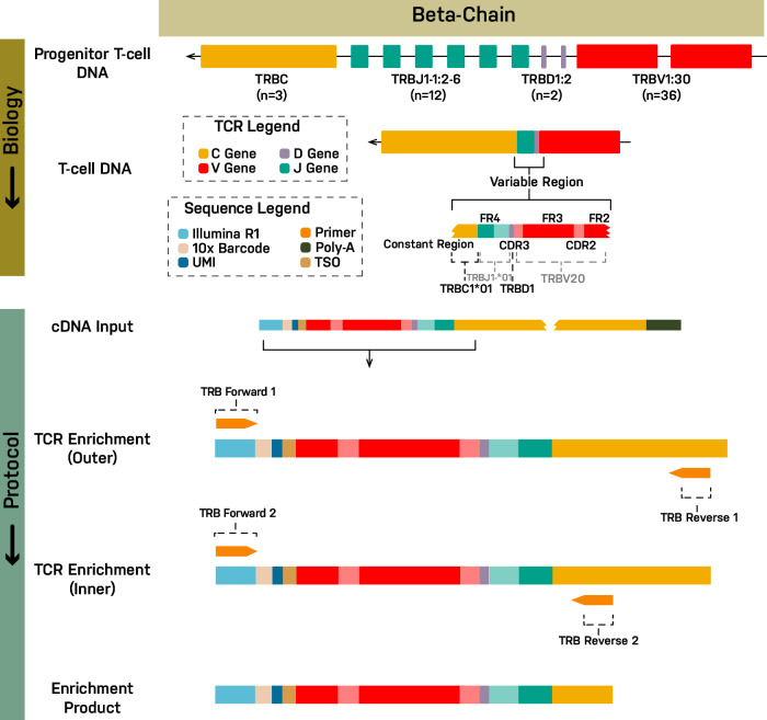
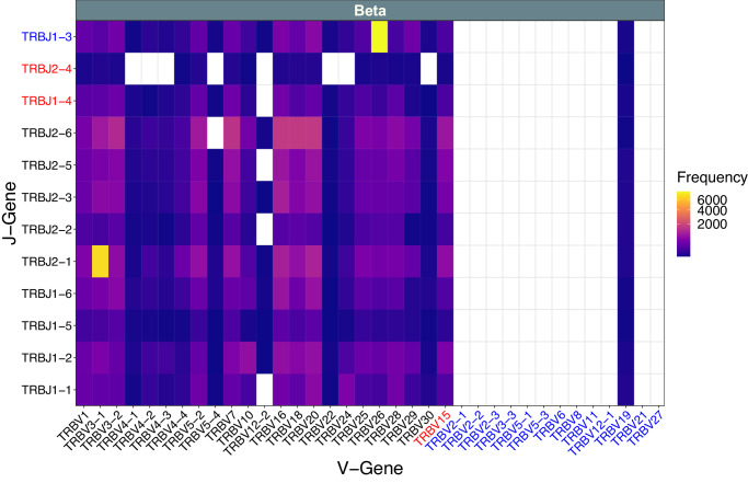
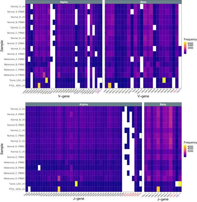
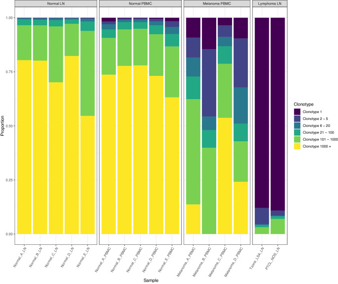
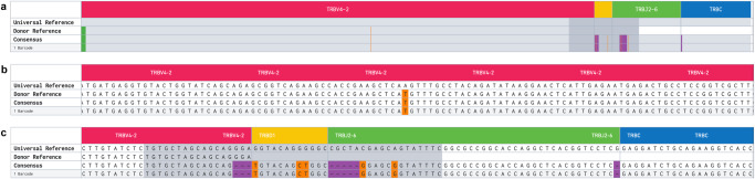
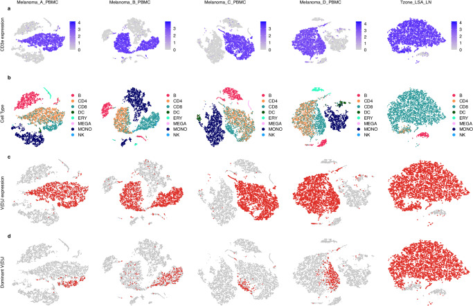
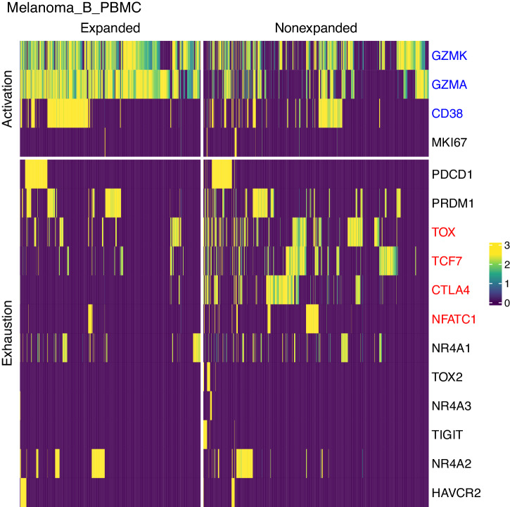
Spontaneous cancers in companion dogs are robust models of human disease. Tracking tumor-specific immune responses in these models requires reagents to perform species-specific single cell T cell receptor sequencing (scTCRseq). scTCRseq and integration with scRNA data have not been demonstrated on companion dogs with cancer. Here, five healthy dogs, two dogs with T cell lymphoma and four dogs with melanoma are selected to demonstrate applicability of scTCRseq in a cancer immunotherapy setting. Single-cell suspensions of PBMCs or lymph node aspirates are profiled using scRNA and dog-specific scTCRseq primers. In total, 77,809 V(D)J-expressing cells are detected, with an average of 3498 (348 - 5,971) unique clonotypes identified per sample. In total, 29/34, 40/40, 22/22 and 9/9 known functional TRAV, TRAJ, TRBV and TRBJ gene segments are observed respectively. Pseudogene or otherwise defective gene segments are also detected supporting re-annotation of several as functional. Healthy dogs exhibit highly diverse repertoires, T cell lymphomas exhibit clonal repertoires, and vaccine-treated melanoma dogs are dominated by a small number of highly abundant clonotypes. scRNA libraries define large clusters of V(D)J-expressing CD8+ and CD4 + T cells. Dominant clonotypes observed in melanoma PBMCs are predominantly CD8 + T cells, with activated phenotypes, suggesting possible anti-tumor T cell populations.


Conflict of interest statement

The authors declare no competing interests.

Figures

Fig. 1
Fig. 1. TCR β chain enrichment strategy for use with 10x scRNA sequencing.
The TCR V(D)J enrichment strategy is depicted using the β chain for illustration (see Supplementary Fig. 1 for α chain). At top, the genomic un-rearranged TRB locus is shown (Note: TRB is located on the negative strand in dogs). During T cell development from progenitor T cells to mature T cells, individual V, D, J and C gene segments are rearranged by somatic recombination to produce a functional TRB locus. Transcription and splicing produce a pre-mRNA and then mRNA for the complete V(D)JC transcript sequence (not shown). In the modified 10x protocol, mRNA (including TRB mRNA) is converted to cDNA. TRB cDNA is then amplified using a nested PCR design. The forward primers from the 10x protocol were left unchanged (v2 protocol shown). In the first cycle, the forward primer (TRB Forward 1) primes off the Illumina read 1 (R1) sequencing adapter that is incorporated during generation of cDNA. In the second cycle, the identical forward primer (TRB Forward 2) again primes off the R1 sequence. The first reverse primer (TRB Reverse 1, Outer) primes off the constant (C) region gene segment. The second reverse primer (TRB Reverse 2, Inner) similarly primes off the C region but at an inner, 5’ position relative to the outer primer. The β chain primer design was based off a dog TCR β rearranged partial mRNA (GenBank: HE653957.1) which was extended to include (from 3’ to 5’) the R1 adapter, 10x cell barcode, UMI, TSO, V, D, J, and C gene segments. The constructed cDNA sequence was then used as input to primer3plus (4.0), with forward primers provided as described above, and a target region for reverse primer specified in the C region. The product of the first (outer) design was used as input for the second (inner) design.
Fig. 2
Fig. 2. Aggregate VJ Gene Segment Usage for dog T-cell receptor β chain.
The VJ gene combinations identified in samples across all dogs (n = 16; normal LN, normal PBMC, melanoma PBMC, lymphoma LN) are plotted along with their observed cell barcode counts (Frequency) for the TRB chain. The corresponding VJ gene usage for the TRA chain is shown in Supplementary Fig. 5. Text in black indicates a functional annotation according to IMGT; blue indicates a pseudogene; red indicates a gene segment that has an ORF but also a defect in the splicing sites, recombination signals and/or regulatory elements or other features disqualifying a functional annotation.
Fig. 3
Fig. 3. TRA/TRB V and J gene segment usage by sample.
The observed cell barcode counts for the TRA chain (Alpha) and TRB chain (Beta) for V-gene and J-gene segments are shown for each sample. Note, gene segments not detected in any sample are not shown. For observation of the complete set of known gene segments (aggregated across all samples) see Fig. 2 and Supplementary Fig. 5. Text in black indicates a functional annotation according to IMGT; blue indicates a pseudogene; red indicates gene segment that has an ORF but also a defect in the splicing sites, recombination signals and/or regulatory elements or other features disqualifying a functional annotation. Note, in rare cases where multiple alleles are expressed, the same cell/barcode can be counted toward multiple gene segments of the same class or even the same gene segment.
Fig. 4
Fig. 4. Single cell clonotype distribution for TRA and TRB chains for all samples.
The proportion of total barcodes, for all clonotypes, is shown for the lymph node aspirates (Normal_LN) and PBMCs (Normal_PBMC) from five healthy dogs, PBMCs from four dogs with melanoma (Melanoma_PBMC) and lymph node aspirates from two dogs with T cell lymphoma (Lymphoma_LN). Proportion was estimated by calculating the fraction of cells in each bin (Clonotype 1, Clonotype 2–5, etc where the clonotypes are sorted in descending order of cell counts). The healthy normal samples are characterized by highly diverse clonotypes, with even the most frequent clonotype observed in only a very small proportion of cells. The melanoma PBMC cases are characterized by a small number of dominant clonotypes with higher frequency. The T cell lymphoma cases are characterized by one dominant clonotype in each case.
Fig. 5
Fig. 5. Example individual clonotype.
Individual cell-specific TRA and TRB clonotypes are resolved to the nucleotide level. Illustrated here is a single such TRB clonotype from Melanoma_B (CDR3: CASSSVQLAERYF). a A specific VDJ recombination with complete TRBV4-2, TRBD1, and TRBJ2-6 and a portion of TRBC is depicted. The Universal Reference (based on IMGT V(D)JC reference sequences) is shown in the first row. Germline variants in the analyzed sample, relative to the Universal Reference, are depicted in the Donor Reference line. The germline/donor sequence is inferred by the cellranger software, by determining shared sequence between multiple clonotypes, in different cells, that use the same gene segment. The Consensus row and subclonotype row(s) show additional variants from the Donor and Universal Reference that were presumably introduced during joining of gene segments. The consensus represents the sequence of the first exact subclonotype for a receptor chain within the clonotype. In this case, only a single subclonotype for a single barcode is shown. But, in other cases, multiple subclonotypes may be grouped together, each represented by one or more cell barcodes. Subclonotypes may have small nucleotide differences or share a TRA chain but have missing TRB chain or vice versa. Single nucleotide changes are shown in orange and small deletions shown in purple. b and c are zoomed into the base pair level to visualize the variants.
Fig. 6
Fig. 6. Gene expression based t-SNE clustering for dogs with melanoma or T zone lymphoma with cells annotated by CD3E expression, inferred cell type or V(D)J transcript detection.
t-SNE clustering based on global gene expression patterns identified a number of distinct clusters of cells from Melanoma_A, Melanoma_B, Melanoma_C, and Melanoma_D PBMCs and T zone lymphoma lymph node. Genes present in fewer than 10 cells and cells with fewer than 100 genes were filtered out of the dataset. Cells whose mitochondrial expression was in the top 5% across all cells were filtered out and DoubletFinder was used to identify and filter out expected doublets. a A T cell marker (CD3E; colored gray to blue) identifies several large clusters. b Cell types, inferred based on published expression signatures of blood cell types, identified CD4 (orange) and CD8 (teal) T cell clusters largely overlapping with the CD3E-positive clusters identified in (a) as well as large monocyte (dark blue) and B cell (red) clusters and smaller clusters of several other cell types. Cell types without an assignment or with a population of less than 1% of all cells identified were excluded. c Cells identified with V(D)J rearrangements overlap strongly with those identified as CD4/CD8 T cells in (b) or CD3E-positive shown in (a). d Cells corresponding to the single most dominant clonotype largely cluster together in the CD8 T cell clusters shown in (b).
Fig. 7
Fig. 7. Expression of T cell activation and exhaustion markers in expanded clonotypes versus non-expanded clonotypes for Melanoma_B_PBMC.
Heatmap of single cell expression values (loge(x + 1) normalized and scaled for all cells in the sample) for expanded CD8 + T cells vs non-expanded CD8 + T cells for known markers of T cell activation and exhaustion. Expanded cells are those with a clonotype frequency greater than 1%. Marker gene names are colored blue if their expression is significantly increased or red if significantly decreased in expanded vs non-expanded CD8 + T cells (adjusted p value < 0.05) for this dog sample (Melanoma_B_PBMC) (Supplementary Data 8).

Similar articles

Cited by

References

    1. LeBlanc, A. K. et al. Adjuvant Sirolimus Does Not Improve Outcome in Pet Dogs Receiving Standard-of-Care Therapy for Appendicular Osteosarcoma: A Prospective, Randomized Trial of 324 Dogs. Clin. Cancer Res. 27, 3005–3016 (2021). - PMC - PubMed
    1. Regan DP, et al. Losartan blocks osteosarcoma-elicited monocyte recruitment, and combined with the kinase inhibitor toceranib, exerts significant clinical benefit in canine metastatic osteosarcoma. Clin. Cancer Res. 2022;28:662–676. doi: 10.1158/1078-0432.CCR-21-2105. - DOI - PMC - PubMed
    1. Rebhun, R. B. et al. Inhaled recombinant human IL-15 in dogs with naturally occurring pulmonary metastases from osteosarcoma or melanoma: a phase 1 study of clinical activity and correlates of response. J. Immunother. Cancer10, e004493 (2022). - PMC - PubMed
    1. Burton JH, et al. NCI comparative oncology program testing of non-camptothecin indenoisoquinoline topoisomerase i inhibitors in naturally occurring canine lymphoma. Clin. Cancer Res. 2018;24:5830–5840. doi: 10.1158/1078-0432.CCR-18-1498. - DOI - PMC - PubMed
    1. Olin, M. R. et al. Treatment combining CD200 immune checkpoint inhibitor and tumor-Lysate vaccination after surgery for pet dogs with high-grade glioma. Cancers11, 137 (2019). - PMC - PubMed

Publication types

Substances