. 2024 Sep;31(9):1355-1367.
doi: 10.1038/s41594-024-01294-7. Epub 2024 Apr 22.

Concerted SUMO-targeted ubiquitin ligase activities of TOPORS and RNF4 are essential for stress management and cell proliferation

Affiliations

Concerted SUMO-targeted ubiquitin ligase activities of TOPORS and RNF4 are essential for stress management and cell proliferation

Julio C Y Liu et al. Nat Struct Mol Biol. 2024 Sep.

Abstract

Protein SUMOylation provides a principal driving force for cellular stress responses, including DNA-protein crosslink (DPC) repair and arsenic-induced PML body degradation. In this study, using genome-scale screens, we identified the human E3 ligase TOPORS as a key effector of SUMO-dependent DPC resolution. We demonstrate that TOPORS promotes DPC repair by functioning as a SUMO-targeted ubiquitin ligase (STUbL), combining ubiquitin ligase activity through its RING domain with poly-SUMO binding via SUMO-interacting motifs, analogous to the STUbL RNF4. Mechanistically, TOPORS is a SUMO1-selective STUbL that complements RNF4 in generating complex ubiquitin landscapes on SUMOylated targets, including DPCs and PML, stimulating efficient p97/VCP unfoldase recruitment and proteasomal degradation. Combined loss of TOPORS and RNF4 is synthetic lethal even in unstressed cells, involving defective clearance of SUMOylated proteins from chromatin accompanied by cell cycle arrest and apoptosis. Our findings establish TOPORS as a STUbL whose parallel action with RNF4 defines a general mechanistic principle in crucial cellular processes governed by direct SUMO-ubiquitin crosstalk.


Conflict of interest statement

The authors declare no competing interests.

Figures
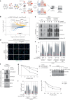
Fig. 1
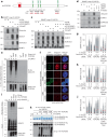
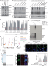
Fig. 1. Genome-wide screens reveal an essential role of TOPORS in SUMO-dependent DPC repair.
a, Workflow of FACS-based haploid genetic screens for DNMT1 abundance (created with BioRender). b, Screen for DNMT1 abundance in EdU-positive cells after co-treatment with 5-AzadC and EdU (n = 1). Positive and negative regulators of DNMT1 abundance are labeled blue and yellow, respectively (two-sided Fisher’s exact test, FDR-corrected P ≤ 0.05; non-significant genes are shown in gray). Genes scoring as significant DNMT1 regulators in a mock screen (Extended Data Fig. 1b) were filtered out, except for DNMT1. c, HAP1 cell lines released from single-round thymidine synchronization in early S phase were treated with 5-AzadC for 30 min and collected at indicated times. Chromatin-enriched fractions were immunoblotted with indicated antibodies. d, Representative images of U2OS cells transfected with indicated siRNAs, treated as in c, pre-extracted and immunostained with DNMT1 antibody. Scale bar, 10 μm. e, DNMT1 foci formation in d was analyzed by quantitative image-based cytometry (QIBC) (red bars, mean; >8,300 cells analyzed per condition). f, HeLa cells stably expressing GFP–DNMT1 were synchronized in early S phase by double thymidine block and release, treated with 5-AzadC and/or SUMOi for 30 min and subjected to GFP IP under denaturing conditions. Immobilized GFP–DNMT1 complexes were incubated with whole-cell lysate from parental HeLa cells and immunoblotted with indicated antibodies. g, Clonogenic survival of 5-AzadC-treated U2OS cells transfected with indicated siRNAs (mean ± s.e.m.; n = 3 independent experiments). h, Immunoblot analysis of U2OS cells treated with USP7i for the indicated times in presence or absence of the proteasome inhibitor MG132. Arrow indicates band corresponding to endogenous TOPORS. i, U2OS cells released from single thymidine synchronization in early S phase were treated with SUMOi and 5-AzadC for 30 min, released into medium containing USP7i or not and collected at indicated times. DNMT1 foci formation was analyzed as in d and e (red bars, mean; >20,000 cells analyzed per condition). j, Clonogenic survival of 5-AzadC-treated U2OS cells in presence or absence of USP7i (mean ± s.e.m.; n = 3 independent experiments). Data information: data are representative of five (d), three (e,f,i) and two (c,h) independent experiments with similar outcomes. Source data
Fig. 2
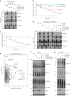
Fig. 2. TOPORS functions as a SUMO-targeted ubiquitin ligase in DPC repair.
a, Domain organization of human TOPORS and mutations introduced to generate the TOPORS *SIM mutant. b, Immunoblot analysis of U2OS cells transfected with siRNAs for a total of 72 h that were released from synchronization in early S phase, treated or not with 5-AzadC for 30 min and subjected to DNMT1 IP under denaturing conditions. c, As in b, except that cells were treated with 5-AzadC 40 h after siRNA transfection. d, As in b, except that USP7i was added where indicated. e, Immunoblot analysis of in vitro ubiquitylation reactions containing recombinant Strep–HA–TOPORS proteins (Extended Data Fig. 2c), E1 (UBA1) and E2 (UBE2D1) enzymes, FLAG–ubiquitin, ATP and Ub–VS and SUMO2–VS. f, Representative images of stable U2OS/GFP–TOPORS cell lines that were induced or not to express GFP–TOPORS proteins with doxycycline (DOX), pre-extracted and fixed and immunostained with ubiquitin conjugate-specific antibody (FK2). Scale bar, 10 μm. See Extended Data Fig. 2e for images of non-pre-extracted U2OS/GFP–TOPORS cell lines where expression of GFP–TOPORS *SIM can be seen. g, U2OS cells were sequentially transfected with siRNAs and expression plasmids (GFP–TOPORS WT and mCherry–H2B). Cells were then subjected to single-round thymidine synchronization in early S phase and, upon release from the block, treated with 5-AzadC for 30 min and pre-extracted and fixed at indicated times. After DNMT1 immunostaining, DNMT1 foci formation in transfected (mCherry-positive) cells was analyzed by quantitative image-based cytometry (QIBC) (red bars, mean; >1,030 cells analyzed per condition). h. As in g, except that cells were transfected with GFP–TOPORS *RING and mCherry–H2B plasmids (red bars, mean; >680 cells analyzed per condition). i, Immunoblot analysis of recombinant Strep–HA–TOPORS proteins incubated with poly-SUMO2 chains and subjected to Strep-Tactin pulldown. j, As in g, except that cells were transfected with GFP–TOPORS *SIM and mCherry–H2B plasmids (red bars, mean; >910 cells analyzed per condition). k, Coomassie staining (top) and immunoblot analysis using SUMO2 and HA antibodies (bottom) of in vitro STUbL reactions containing recombinant Strep–HA–TOPORS proteins, E1 (UBA1) and E2 (UBE2K) enzymes, FLAG–ubiquitin, 4×SUMO2 STUbL, Ub–VS and SUMO2–VS. Data information: data are representative of four (c) and three (b,dk) independent experiments with similar outcomes. Source data
Fig. 3
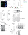
Fig. 3. TOPORS and RNF4 cooperatively drive multiple STUbL-driven responses.
a, U2OS PML-KO cells stably reconstituted with YFP-tagged PML-V (U2OS PML-KO/YFP–PML) were transfected with indicated siRNAs for 72 h and exposed to arsenic (As2O3) for 1 h. Cells were subjected to GFP IP under denaturing conditions followed by immunoblotting with indicated antibodies. b, U2OS PML-KO/YFP–PML cells transfected with indicated siRNAs were treated with arsenic and fixed at indicated timepoints. YFP–PML intensity was analyzed by quantitative image-based cytometry (QIBC) (mean ± s.e.m.; n = 3 independent experiments). c, U2OS PML-KO/YFP–PML cells were exposed to arsenic in the presence or absence of USP7i, fixed at the indicated times and analyzed as in b (mean ± s.e.m.; n = 3 independent experiments). d, As in a, except that cells were treated with arsenic 40 h after transfection with indicated siRNAs. e, U2OS PML-KO/YFP–PML cells were grown in SILAC medium and exposed to 1 µM arsenic for 2 h (heavy condition) or left untreated (light condition). PML bodies were enriched by affinity purification using GFP nanobody crosslinked to magnetic beads, and associated proteins were identified and quantified by mass spectrometry. Selected outliers with increased abundance and PML itself are indicated. A single experiment was performed. f, Immunoblot analysis of U2OS cells transfected with indicated siRNAs for 40 h. Slow-migrating, hyper-SUMOylated proteins (‘well products’) are indicated by arrows. See also Supplementary Fig. 1a. g, Immunoblot analysis of U2OS cells transfected with indicated siRNAs for 48 h and grown in the absence or presence of USP7i for an additional 12 h. Slow-migrating, hyper-SUMOylated proteins (‘well products’) are indicated by arrows. See also Supplementary Fig. 1b. Data information: data are representative of three (a,d,f,g) independent experiments with similar outcomes. Source data
Fig. 4
Fig. 4. TOPORS and RNF4 generate complex ubiquitin topologies on STUbL substrates to promote p97 recruitment.
a, Representative images of doxycycline (DOX)-treated U2OS/GFP–TOPORS cell lines immunostained with p97 antibody after pre-extraction and fixation. Scale bar, 10 μm. b. U2OS cells transfected with indicated siRNAs were released from single thymidine synchronization in early S phase, treated with 5-AzadC for 30 min, pre-extracted and immunostained with p97 and DNMT1 antibodies. p97 and DNMT1 intensity in DNMT1 DPC foci was analyzed by quantitative image-based cytometry (QIBC) (mean ± s.e.m.; n = 6 independent experiments; one-tailed paired t-test). c. U2OS PML-KO/YFP–PML cells transfected with indicated siRNAs for 72 h and treated with arsenic for 1 h were pre-extracted and immunostained with p97 antibody. p97 and YFP intensity in YFP-PML bodies was analyzed by quantitative image-based cytometry (QIBC) (mean ± SEM; n = 7 independent experiments; one-tailed paired t-test). d, U2OS cells transfected with indicated siRNAs were subjected to DNMT1 IP under denaturing conditions to isolate DNMT1 but not associated proteins. Samples were digested with trypsin, and di-glycine remnants on ubiquitin were identified by mass spectrometry (n = 4 independent experiments). e, Immunoblot analysis of U2OS cells transfected with indicated siRNAs for 40 h and subjected to MultiDsk pulldown under denaturing conditions to isolate total cellular ubiquitin conjugates but not associated, non-covalently bound proteins. Input blots are shown in Fig. 3f. See also Supplementary Fig. 2. f, Coomassie staining (left) and fluorescein-ubiquitin visualization (right) of in vitro STUbL reactions containing purified Strep–HA–TOPORS and indicated GST–SUMO protein substrates (top), supplemented with E1 (UBA1) and E2 (UBE2D1) enzymes, ubiquitin (10% labeled with 5-IAF) and ATP. g, Quantification of data in f (mean ± s.d.; n = 3; NS, not significant; two-tailed paired t-test). h, Indicated amounts of recombinant Strep–HA–TOPORS *RING protein was incubated with SUMO1-conjugated, SUMO2-conjugated or Protein A–conjugated agarose beads. Beads were washed extensively, and bound material was immunoblotted with HA, SUMO1 and SUMO2/3 antibodies. i, Whole-cell extract from U2OS cells was incubated with SUMO1-conjugated, SUMO2-conjugated or Protein A–conjugated agarose beads. Beads were washed extensively, and bound material was immunoblotted with RNF4, SUMO1 and SUMO2/3 antibodies. Data information: data are representative of three (a,e,f,h,i) independent experiments with similar outcomes. Source data
Fig. 5
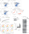
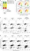
Fig. 5. Combined loss of TOPORS and RNF4 is synthetic lethal.
a, Haploid genetic fitness screen in HAP1 WT cells, presented as a fishtail plot in which genes are plotted according to ratio of sense/anti-sense orientation of gene-trap insertions (y axis) and the total number of insertions in a particular gene (x axis) (n = 4 biological replicates (independent clones)). Blue dots represent genes with significant sense bias (binomial P < 0.05, FDR corrected, across all replicates). A single representative replicate is shown. b, As in a but for HAP1 TOPORS-KO cells (n = 2 biological replicates (independent clones)). c, As in a but for HAP1 RNF4–dTAG–HA cells treated with 0.25 µM dTAG-13 for 10 d (n = 2 biological replicates (independent clones)). d. Gene rank plot for significant synthetic lethal genes for HAP1 TOPORS-KO (left) and RNF4–dTAG–HA (right) compared to HAP1 WT (Fisher’s exact test; P < 0.05; odds ratio < 0.8). The size of the dot represents the difference (Δ) in sense ratio bias. e, Normalized logarithmic proliferation quantification for U2OS cells transfected with indicated siRNAs for the indicated times, as determined by IncuCyte image-based confluence analysis (mean ± s.e.m.; n = 3 independent experiments). Imaging was started at 24 h after siRNA transfection. f, Immunoblot analysis of U2OS cells transfected with indicated siRNAs for 48 h. g, U2OS Fucci cells were transfected with indicated siRNAs for 36 h. To determine cell cycle position, CDT1 (mKO2-hCdt1+) and Geminin (mAG-hGem+) intensities were analyzed by quantitative image-based cytometry (QIBC). Quantification of cells in different cell cycle phases was done using the indicated gates. h, U2OS cells were transfected with indicated siRNAs, treated with thymidine for 18 h and released into S phase for 6 h. Cells were immunostained with γH2AX antibody and analyzed by QIBC (red bars, mean; >3,900 cells analyzed per condition). i, Immunoblot analysis of chromatin-enriched fractions of U2OS cells transfected with indicated siRNAs for 40 h. Data information: data are representative of three (f,i) and two (g,h) independent experiments with similar outcomes. Source data
Fig. 6
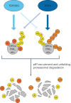
Fig. 6. Concerted action of TOPORS and RNF4 drives major STUbL-mediated processes—model.
Parallel E3 ubiquitin ligase activities of TOPORS and RNF4 provide a key driving force for major STUbL-mediated cellular processes, including SUMO-dependent DPC repair and PML body degradation. This involves a selectivity of TOPORS for targeting SUMO1-modified proteins, whereas RNF4 may have some preference for SUMO2/3-modified proteins. However, these preferences are not absolute. Accordingly, in the absence of either ligase, STUbL-driven pathways become inefficient but retain basic functionality, whereas combined loss of TOPORS and RNF4 leads to a profound defect in the ubiquitylation and proteolytic processing of SUMOylated proteins, accompanied by synthetic lethality.
Extended Data Fig. 1
Extended Data Fig. 1. TOPORS is required for SUMO-dependent DPC resolution and is stabilized by USP7.
a. Flow cytometry plots of gating strategy for FACS-based DNMT1 haploid genetic screens (Fig. 1b; Extended Data Fig. 1b). b. Mock screen for DNMT1 abundance in EdU-positive cells (n = 1). Positive and negative regulators are labeled in blue and yellow, respectively (two-sided Fisher’s exact test, FDR corrected p ≤ 0.05; non-significant genes are shown in grey). c. Immunoblot analysis of HAP1 WT and TOPORS-KO cells. d. Immunoblot analysis of U2OS cells transfected with non-targeting control (CTRL), RNF4, or TOPORS siRNAs. e. U2OS cells transfected with indicated siRNAs were released from thymidine synchronization, exposed to 5-AzadC for 30 min and collected at indicated times. Chromatin-enriched fractions were immunoblotted with indicated antibodies. f. Immunoblot analysis of chromatin-enriched fractions of siRNA-transfected U2OS cells exposed to formaldehyde for 1 h and collected at indicated times. g. Cells treated as in (f) were pre-extracted and stained with SUMO2/3 antibody. SUMO2/3 foci counts were analyzed by QIBC (red bars, mean; >4,400 cells analyzed per condition). h. Clonogenic survival of U2OS cells transfected with indicated siRNAs and exposed to formaldehyde for 30 min before replating (mean ± SEM; n = 3 independent experiments). i. Immunoblot analysis of U2OS cells treated with USP7i. Arrow indicates the band corresponding to endogenous TOPORS. j. As in (e), except USP7i was administered together with 5-AzadC where indicated. k. Immunoblot analysis of U2OS cells transfected with GFP expression plasmids and subjected to GFP IP. l. Immunoblot analysis of U2OS cells transfected with indicated GFP-TOPORS expression constructs and subjected to GFP IP under denaturing conditions. RING domain mutations (*RING) inactivating TOPORS E3 ubiquitin ligase activity (Fig. 2e) abolish its ubiquitylation, suggesting this reflects auto-ubiquitylation. m. U2OS cells transfected with GFP-TOPORS WT construct were subjected to GFP IP under denaturing conditions. Immobilized proteins were incubated with or without recombinant USP7 protein and immunoblotted with ubiquitin and GFP antibodies. Data information: Data are representative of four (d), three (f,g,i,k) and two (c,e,j,l,m) independent experiments with similar outcome. Source data
Extended Data Fig. 2
Extended Data Fig. 2. TOPORS functions as an E3 ubiquitin ligase in DPC repair.
a. Immunoblot analysis of HAP1 WT and TOPORS-KO cells transfected with control (CTRL) or TOPORS siRNAs that were released from synchronization in early S phase, pulse-labeled or not with 5-AzadC for 30 min and subjected to DNMT1 IP under denaturing conditions. b. Immunoblot analysis of U2OS cells transfected with siRNAs for 72 h, treated or not with camptothecin (CPT) and SUMO inhibitor (SUMOi) and subjected to Multi-Dsk pulldown under denaturing conditions to isolate total cellular ubiquitylated proteins. c. As in (b), except cells were treated with siRNAs for 40 h. d. U2OS cells transfected with siRNAs released from synchronization in early S phase were pulse-labeled with 5-AzadC for 30 min, collected at indicated times, pre-extracted and immunostained with DNMT1 antibody. DNMT1 foci formation was analyzed by quantitative image-based cytometry (QIBC) (red bars, mean; >2,400 cells analyzed per condition). e. siRNA-transfected U2OS cells were subjected to DNMT1 IP under denaturing conditions to isolate DNMT1 but not associated proteins. Samples were digested with trypsin and DNMT1, SUMO1, SUMO2/3, and ubiquitin peptides were identified by MS (n = 4 independent experiments). Molarity was approximated by dividing each protein’s intensity-based abundance by its molecular weight. f. Strep-HA-TOPORS proteins purified from HEK293-6E cells were analyzed by Coomassie Blue staining or immunoblotting with HA antibody. Asterisks indicate Strep-HA-TOPORS degradation products. g. Immunoblot analysis of in vitro SUMOylation reactions containing recombinant Strep-HA-TOPORS proteins, E1 (SAE1-UBA2) and E2 (UBE2I) enzymes, SUMO2, ATP and SUMO2-VS. h. Representative images of non-pre-extracted stable U2OS/GFP-TOPORS cell lines induced or not to express GFP-TOPORS proteins with Doxycycline (DOX). Scale bar, 10 μm. i. Representative images of DOX-treated U2OS/GFP-TOPORS cell lines immunostained with SUMO2/3 antibody after pre-extraction. Scale bar, 10 μm. j. U2OS cells stably expressing GFP-TOPORS *RING were released from synchronization in early S phase, pulse-treated with 5-AzadC for 30 min, pre-extracted and immunostained with DNMT1 antibody. DNMT1 foci formation in GFP-positive and -negative cells was analyzed by QIBC (red bars, mean; >479 cells analyzed per condition). Data information: Data are representative of three (b,c,f-j) and two (a,d) independent experiments with similar outcome. Source data
Extended Data Fig. 3
Extended Data Fig. 3. TOPORS is a SUMO-targeted ubiquitin ligase.
a. Immunoblot analysis of recombinant Strep-HA-TOPORS proteins incubated with free SUMO2 or poly-SUMO2 chains and subjected to Strep-Tactin pulldown. b. Representative images of DOX-treated U2OS/GFP-TOPORS cell lines immunostained with DNMT1 antibody after pre-extraction. Scale bar, 10 μm. c. Coomassie staining (top) and immunoblot analysis using SUMO2 antibody (bottom) of in vitro STUbL reactions containing recombinant RNF4 or Strep-HA-TOPORS, E1 (UBA1) and E2 enzymes, FLAG-ubiquitin, 4xSUMO2 STUbL substrate, ATP, Ub-VS and SUMO2-VS. d. U2OS cells transfected with indicated siRNAs were released from single thymidine synchronization in early S phase and pulse-treated with 5-AzadC for 30 min. Cells were collected at indicated time points, pre-extracted and immunostained with DNMT1 antibody. DNMT1 foci formation was analyzed by QIBC (red bars, mean; >6,400 cells analyzed per condition). e. Immunoblot analysis of U2OS cells transfected with non-targeting control (CTRL) or UBE2K siRNAs. Data information: Data are representative of three (a-d) and two (e) independent experiments with similar outcome. Source data
Extended Data Fig. 4
Extended Data Fig. 4. STUbL-dependent turnover of PML bodies.
a. U2OS PML-KO/YFP-PML cells transfected with indicated siRNAs were treated with arsenic and collected at indicated times. Cells were subjected to GFP IP under denaturing conditions and immunoblotted with indicated antibodies. b. Representative images of cells in Fig. 3b. Scale bar, 10 μM. c. Schematic overview of the proteomics experiment in Fig. 3e. d. U2OS cells stably expressing GFP-TOPORS WT or *RING were treated or not with Doxycycline (DOX) for 36 h. Cells were pre-extracted and immunostained with PML antibody. Intensity of PML bodies in GFP-positive and -negative cells was analyzed by QIBC and normalized to untreated GFP-negative cells (red bars, mean; n = 3; ns: not significant, one-tailed paired t-test). Data information: Data are representative of three (a,b) independent experiments with similar outcome. Source data
Extended Data Fig. 5
Extended Data Fig. 5. RNF4 and TOPORS promote p97-dependent processing of SUMOylated proteins.
a. U2OS cells released from single thymidine synchronization in early S phase and pulse-labeled with 5-AzadC for 30 min in presence or absence of p97 inhibitor (p97i) were pre-extracted and immunostained with DNMT1 antibody. DNMT1 foci formation was analyzed by QIBC (red bars, mean; >8,100 cells analyzed per condition). b. U2OS PML-KO/YFP-PML cells were exposed to arsenic in presence or absence of USP7i. Cells were collected at indicated times, pre-extracted and YFP-PML intensity was analyzed by QIBC (mean ± SEM; n = 3 independent experiments). c. Representative images of DOX-treated U2OS/GFP-TOPORS cell lines treated or not with 5-AzadC and co-immunostained with DNMT1 and p97 antibodies after pre-extraction and fixation. Scale bar, 10 μm. d. Immunoblot analysis of U2OS cells transfected with siRNAs for 40 h or treated with p97i for 10 h. e. U2OS stably expressing Ub(G76V)-GFP reporter transfected with indicated siRNAs were treated or not with p97i for 4 h. Nuclear Ub(G76V)-GFP signal intensity was analyzed by QIBC (red bars, mean; >12,000 cells analyzed per condition). f. U2OS cells transfected with indicated siRNAs were subjected to DNMT1 IP under stringent denaturing conditions. Samples were digested with trypsin, and di-glycine remnants on ubiquitin were identified by MS (n = 4 independent experiments). g. Immunoblot analysis of U2OS PML-KO/YFP-PML cells transfected with indicated siRNAs for 72 h, exposed to arsenic for 1 h and subjected to GFP IP under denaturing conditions. h. Immunoblot analysis of U2OS cells transfected with control (CTRL), RNF4, SUMO1 and/or SUMO2/3 siRNAs that were released from synchronization in early S phase, treated or not with 5-AzadC for 30 min and subjected to DNMT1 IP under denaturing conditions. i. U2OS cells transfected with control (CTRL), RNF4 and/or SUMO1 siRNAs were released from single-round thymidine synchronization in early S phase and pulse-labeled with 5-AzadC for 30 min. Cells were pre-extracted at indicated times, immunostained with DNMT1 antibody, and DNMT1 foci formation was analyzed by quantitative image-based cytometry (QIBC) (red bars, mean; >6,600 cells analyzed per condition). Data information: Data are representative of three (a,c,g-i) or two (d,e) independent experiments with similar outcome. Source data
Extended Data Fig. 6
Extended Data Fig. 6. Synthetic lethality between TOPORS and RNF4.
a. Workflow of fitness-based haploid genetic screens in Fig. 5a–d (created with Biorender). b. Immunoblot analysis of HAP1 RNF4-dTAG-HA clones treated or not with dTAG13 for 24 h. The membrane was co-stained with Ponceau S (loading control). c. Representative images of HAP1 cell lines treated with EdU in presence or absence of 10 µM 5-AzadC for 60 min. RNF4-dTAG-HA cells were pre-treated with dTAG-13 for 4 h. Cells were fixed and stained using antibodies and reagents for DNMT1, EdU and DAPI. Scale bar, 10 μM. d. Equal numbers of cells were seeded in a 6-well plate, transfected with indicated siRNAs for 72 h and stained with crystal violet. e. As in (d), except cells were transfected with siRNAs for 48 h and subsequently treated or not with USP7i for an additional 48 h prior to fixation. f. Normalized logarithmic proliferation quantification for U2OS cells transfected with control (CTRL) or RNF4 siRNAs for 48 h and then incubated or not with USP7i, as determined by Incucyte image-based confluence analysis (mean ± SD; n = 3 technical replicates). Imaging started at 72 h after siRNA transfection. g. As in (f), except cells were transfected with CTRL or TOPORS siRNAs (mean ± SD; n = 3 technical replicates). h. Immunoblot analysis of U2OS cells transfected with indicated siRNAs for 48 h and grown in presence or absence of USP7i for an additional 24 h. Data information: Data are representative of three (b,d,e) and two (c,h) independent experiments with similar outcome. Source data
Extended Data Fig. 7
Extended Data Fig. 7. Combined loss of TOPORS and RNF4 leads to defective DNA replication and cell death in S phase.
a. U2OS Fucci cells transfected with indicated siRNAs were analyzed by live-cell imaging. Expression of CDT1 (mKO2-hCdt1) and Geminin (mAG-hGem) at the point of cell death was assessed (mean ± SEM; n = 2; at least 45 cell death events were analyzed per condition per replicate). b. U2OS Fucci cells were transfected with indicated siRNAs for 24 h and grown in the absence or presence of USP7i for an additional 12 h. To determine cell cycle position, CDT1 (mKO2-hCdt1+) and Geminin (mAG-hGem+) intensities were analyzed by QIBC. Quantification of cells in different cell cycle phases was done using indicated gates. c. U2OS cells were transfected with indicated siRNAs and synchronized by treatment with thymidine for 18 h. DNA synthesis profiles of cells pulsed with EdU at indicated time points after release from thymidine arrest were determined by QIBC analysis of DAPI and EdU signal intensities (>3,900 cells analyzed per condition). d. U2OS cells were transfected with indicated siRNAs and synchronized by treatment with thymidine for 18 h. One h prior to thymidine release, USP7i was added where indicated. DNA synthesis profiles of cells pulsed with EdU at indicated time points after release from thymidine arrest were determined by QIBC analysis of DAPI and EdU signal intensities (>10,000 cells analyzed per condition). Data information: Data are representative of two (b,c,d) independent experiments with similar outcome. Source data

Similar articles

Cited by

References

    1. Vertegaal, A. C. O. Signalling mechanisms and cellular functions of SUMO. Nat. Rev. Mol. Cell Biol.23, 715–731 (2022). 10.1038/s41580-022-00500-y - DOI - PubMed
    1. Zhao, X. SUMO-mediated regulation of nuclear functions and signaling processes. Mol. Cell71, 409–418 (2018). 10.1016/j.molcel.2018.07.027 - DOI - PMC - PubMed
    1. Chang, Y. C., Oram, M. K. & Bielinsky, A. K. SUMO-targeted ubiquitin ligases and their functions in maintaining genome stability. Int. J. Mol. Sci.22, 5391 (2021). 10.3390/ijms22105391 - DOI - PMC - PubMed
    1. Lecona, E. et al. USP7 is a SUMO deubiquitinase essential for DNA replication. Nat. Struct. Mol. Biol.23, 270–277 (2016). 10.1038/nsmb.3185 - DOI - PMC - PubMed
    1. Sriramachandran, A. M. et al. Arkadia/RNF111 is a SUMO-targeted ubiquitin ligase with preference for substrates marked with SUMO1-capped SUMO2/3 chain. Nat. Commun.10, 3678 (2019). 10.1038/s41467-019-11549-3 - DOI - PMC - PubMed

LinkOut - more resources