. 2024 Oct;29(10):2997-3009.
doi: 10.1038/s41380-024-02559-9. Epub 2024 Apr 22.

Combined expansion and STED microscopy reveals altered fingerprints of postsynaptic nanostructure across brain regions in ASD-related SHANK3-deficiency

Affiliations

Combined expansion and STED microscopy reveals altered fingerprints of postsynaptic nanostructure across brain regions in ASD-related SHANK3-deficiency

Jan Philipp Delling et al. Mol Psychiatry. 2024 Oct.

Abstract

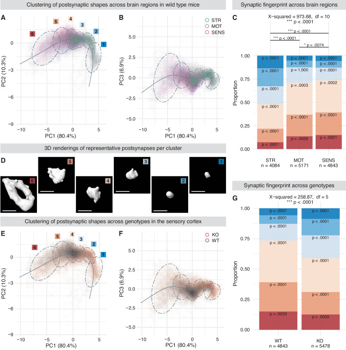
Synaptic dysfunction is a key feature of SHANK-associated disorders such as autism spectrum disorder, schizophrenia, and Phelan-McDermid syndrome. Since detailed knowledge of their effect on synaptic nanostructure remains limited, we aimed to investigate such alterations in ex11|SH3 SHANK3-KO mice combining expansion and STED microscopy. This enabled high-resolution imaging of mosaic-like arrangements formed by synaptic proteins in both human and murine brain tissue. We found distinct shape-profiles as fingerprints of the murine postsynaptic scaffold across brain regions and genotypes, as well as alterations in the spatial and molecular organization of subsynaptic domains under SHANK3-deficient conditions. These results provide insights into synaptic nanostructure in situ and advance our understanding of molecular mechanisms underlying synaptic dysfunction in neuropsychiatric disorders.


Conflict of interest statement

The authors declare no competing interests.

Figures

Fig. 1
Fig. 1. ExM and ExM-STED preserve cellular and synaptic ultrastructure.
A Murine cortical tissue stained for total protein via NHS ester (NHSE) showcasing preserved cellular ultrastructure. Organelles are magnified and shown as inlets, corresponding to the boxes within the overview. Nucleoli (NUC) show intricate structuring and surroundings of varying protein density. The pre- and postsynaptic specializations including the synaptic cleft can be readily identified in the side view projection of a neuronal synapse (SYN). Nuclear pores spanning the nuclear envelope are visible in the direct neighborhood of the endoplasmatic reticulum (NP + ER). Axons traversing the imaged plane contain mitochondria (AX + MIT). Cortical synapses in side view (B) and en face (C) projection as imaged via confocal tenfold ExM. The main image shows the NHSE total protein staining, while inlets show immunolabelings targeting HOMER1 and DLG4 of the postsynaptic density. Cortical synapses in side view (D) and en face (E) projection as imaged via ExM-STED. As compared to B and C resolution is enhanced, showing the detailed ultrastructure of neuronal synapses. F Box plots showing the analysis of AZ-PSD distances to ensure comparable local synaptic expansion factors enabling further measurements. Color-coding is according to genotype (red for SHANK3-KO and black for WT mice). One-way ANOVA (F = 0.085, p = 0.995) with pairwise Tukey post-hoc analysis does not show significant differences (p > 0.99 in all pairwise comparisons; n = 60 per animal). Detailed statistical analysis, including the code to generate the plots is provided as R script in the supplementary materials. Scale bars represent 10 µm or 3 µm in the overview or inlets of A, respectively. Scale bars in BE represent 1 µm.
Fig. 2
Fig. 2. Localization of synaptic proteins visualized via ExM-STED.
Staining for DLG4 and SHANK3 (A-C) or DLG4 and BSN (D-F) imaged via ExM-STED, showing a composite with merged channels in the main panel and inlets of the specific immunolabelings. A and D and B and E represent side view or en face projections of single murine synapses, respectively. C and F show en face projections of human synapses. DLG4 and SHANK3 are strongly associated, with SHANK3 being slightly more offset from the postsynaptic membrane (A). Together DLG4 and SHANK3 seem to be incorporated into a mosaic-like arrangement within the PSD, which can be appreciated in both en face projections of murine (B) and human (C) synapses. DLG4 and BSN are localized in the post- and presynaptic compartment separated by the synaptic cleft, respectively, with BSN showing a bell-like distribution within the presynaptic bouton (D). DLG4 and BSN are not as closely correlated in both en face projections of murine and human synapses (E, F), however seem to be organized in transsynaptic functional units possibly enabling effective neurotransmission. Scale bars represent 1 µm.
Fig. 3
Fig. 3. Morphological characteristics define brain region and genotype dependent synaptic fingerprints.
A/B, E/F Scatterplots representing shapes of postsynaptic DLG4 scaffolds (one circle per shape) projected into a three-dimensional space defined by principal components (PC) 1-3. Shapes are categorized into six groups via partitional clustering as visualized by ellipsoids representing the 95% confidence level for a multivariate t-distribution. The fitted pseudotime curve is overlayed in each 2D scatterplot. Color-coding represents brain regions striatum (STR), motor (MOT) & sensory cortex (SENS) in (A/B) and genotypes wild type (WT) and SHANK3-KO (KO) in E/F as indicated in the respective legends. D Exemplary 3D renderings of the medoids from each cluster derived from the WT sensory cortex. Synaptic DLG4 scaffolds are spherical and small in cluster 1 and gain volume as well as complexity until cluster 6, where oftentimes perforations within the scaffold can be observed. Ellipsoids in A, E are labelled with the respective cluster identity shown in D. C, G Bar plots showing the proportional distribution of clusters as visualized and color-coded in A, D and E. C Chi-square test for independence shows that the investigated brain region does influence synapse cluster identity. Pairwise Bonferroni-corrected post-hoc comparisons reveal significant differences in cluster proportions among all brain regions analyzed and that all clusters deviate significantly from the expected proportions, except for cluster 3 in the motor cortex. The striatum presents with a clear overrepresentation of small/spherical clusters 1–3, while cortical regions are dominant in voluminous/complex shaped clusters 4-6, which is also visible in the scatterplots of A/B. G Chi-square test for independence reveals that genotype (WT vs. KO) influences synapse cluster identity in the sensory cortex and that all clusters deviate significantly from the expected proportions. SHANK3-KO mice present with an overrepresentation of small/spherical clusters 1-3, while voluminous/complex shaped clusters 4–6 are less abundant compared to WT animals, as visible in scatterplots (E/F). Sample sizes as number of synapses analyzed are indicated in (C & G). *p ≤ 0.01, ***p < 0.0001. Detailed statistical analysis, including the code to generate the plots is provided as R script in the supplementary materials. Scale bars in D represent 1 µm.
Fig. 4
Fig. 4. ExM-STED enables the detection of abnormal subsynaptic organization in SHANK3-KO mice.
Representative en face images of single synapses immunolabeled for DLG4/HOMER1 (A) and DLG4/BSN (E), shown as analyzed. A was acquired in the striatal neuropil, while E was acquired in layer 2/3 of the sensory cortex of WT mice. A shows DLG4 and HOMER1 forming a postsynaptic web, while in E, DLG4 and BSN display less overlap. B/C Characterization of HOMER1 particles in the striatum of WT and KO mice, visualized by box plots. B shows that HOMER1 particles are smaller with lower protein amount, however, particle density per area is higher in KO animals compared to WT. Manders Coefficient-based correlation analysis in C reveals that both from the perspective of striatal HOMER1 and DLG4, colocalization is reduced under SHANK3-deficient conditions. (F/G) Characterization of DLG4 particles in the sensory cortex of WT and KO mice, visualized by box plots. F shows DLG4 particles are larger with higher total protein amount (however not when size-corrected), but lower particle density per area in KO animals compared to WT. Manders Coefficient-based correlation analysis in G reveals that from the perspective of cortical DLG4, colocalization with BSN is increased in SHANK3-KO. Nearest neighbor distance (NND) analysis of subsynaptic particles in the striatum (D) and sensory cortex (H) of WT and KO mice as visualized by point ranges representing the mean ± SEM per neighbor k and a fitted curve including its 95% confidence interval. Spatial pattern analysis via NND suggests a reduced association of striatal HOMER1-particles (D) or cortical DLG4 particles (H) to their respective neighborhood since distances to adjacent particles are increased. The values shown in (B, F) have been centered and scaled. In B/C and F/G, semi-parametric MANOVA with univariate Bonferroni-corrected post-hoc analysis was performed to calculate the p-values for all pairwise comparisons. Modified ANOVA-type statistics (MATS) and overall p-values are reported within the respective figure panels. Sample sizes as number of synapses analyzed in WT-/KO-mice are 54/59 in B, 54/59 in C, 129/139 in F and 69/73 in G. In D and H, repeated measures ANOVA with Benjamini-Hochberg-corrected post-hoc analysis was performed for all pairwise comparisons. The F-value and overall p-values are reported within the respective figure panels. Sample sizes as number of synapses analyzed in WT-/KO-mice are 54/59 in D and 128/138 in H. NS. p > 0.05, *p ≤ 0.05, **p < 0.01, ***p < 0.001. Detailed statistical analysis, including the code to generate the plots is provided as R script in the supplementary materials. Scale bars in (A, E) represent 1 µm.
Fig. 5
Fig. 5. Visual comparison of subsynaptic organization across genotypes and brain regions.
Representative en face images of single synapses immunolabeled for DLG4/HOMER1 (A/B) and DLG4/BSN (C/D) were acquired in the striatal neuropil and layer 2/3 of the sensory cortex, respectively. The genotype and brain region shown is indicated by row and column headings. A/B Reflecting the dominant subsynaptic changes evident from statistical analysis, representative images show that striatal HOMER1 particles are smaller with lower protein amount, but show higher density and an increased nearest neighbor distance under SHANK3-deficient conditions. Correlation of HOMER1- and DLG4-particles is also clearly reduced in SHANK3-KO animals. (C/D) Differences were not as pronounced when DLG4 and BSN were analyzed in the sensory cortex, however representative images already hint at the lower DLG4 particle density and higher nearest neighbor distance in KO animals as detected by statistical analysis. All images are shown as analyzed. Scale bars represent 1 µm in each panel.

References

    1. Hell SW, Wichmann J. Breaking the diffraction resolution limit by stimulated emission: stimulated-emission-depletion fluorescence microscopy. Opt Lett. 1994;19:780. - PubMed
    1. Klar TA, Jakobs S, Dyba M, Egner A, Hell SW. Fluorescence microscopy with diffraction resolution barrier broken by stimulated emission. Proc Natl Acad Sci. 2000;97:8206–10. - PMC - PubMed
    1. Gustafsson MGL. Surpassing the lateral resolution limit by a factor of two using structured illumination microscopy. J Microsc. 2000;198:82–87. - PubMed
    1. Heintzmann R, Cremer CG. Laterally modulated excitation microscopy: improvement of resolution by using a diffraction grating. In: Bigio IJ, Schneckenburger H, Slavik J, Svanberg K, Viallet PM, editors. Proc. SPIE, Stockholm, Sweden; 1999. p. 185-96.
    1. Bates M, Blosser TR, Zhuang X. Short-range spectroscopic ruler based on a single-molecule optical switch. Phys Rev Lett. 2005;94:108101. - PMC - PubMed

MeSH terms

Supplementary concepts