

Engineering water exchange is a safe and effective method for magnetic resonance imaging in diverse cell types
- PMID: 38649904
- PMCID: PMC11035135
- DOI: 10.1186/s13036-024-00424-5
Engineering water exchange is a safe and effective method for magnetic resonance imaging in diverse cell types
Abstract
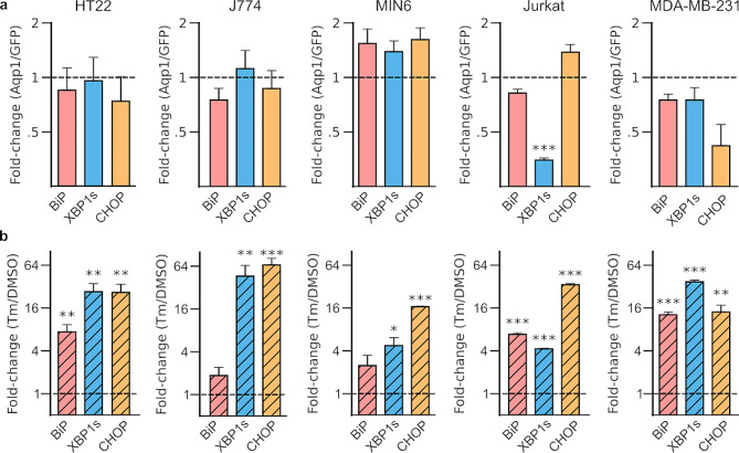
Aquaporin-1 (Aqp1), a water channel, has garnered significant interest for cell-based medicine and in vivo synthetic biology due to its ability to be genetically encoded to produce magnetic resonance signals by increasing the rate of water diffusion in cells. However, concerns regarding the effects of Aqp1 overexpression and increased membrane diffusivity on cell physiology have limited its widespread use as a deep-tissue reporter. In this study, we present evidence that Aqp1 generates strong diffusion-based magnetic resonance signals without adversely affecting cell viability or morphology in diverse cell lines derived from mice and humans. Our findings indicate that Aqp1 overexpression does not induce ER stress, which is frequently associated with heterologous expression of membrane proteins. Furthermore, we observed that Aqp1 expression had no detrimental effects on native biological activities, such as phagocytosis, immune response, insulin secretion, and tumor cell migration in the analyzed cell lines. These findings should serve to alleviate any lingering safety concerns regarding the utilization of Aqp1 as a genetic reporter and should foster its broader application as a noninvasive reporter for in vivo studies.
Keywords: Cell physiology; Diffusion; MRI; Reporter gene; Unfolded protein response; aquaporin-1.
© 2024. The Author(s).
Conflict of interest statement
The authors declare no competing interests.
Figures


Update of
-
Engineering water exchange is a safe and effective method for magnetic resonance imaging in diverse cell types.bioRxiv [Preprint]. 2023 Nov 10:2023.11.07.566095. doi: 10.1101/2023.11.07.566095. bioRxiv. 2023. Update in: J Biol Eng. 2024 Apr 22;18(1):30. doi: 10.1186/s13036-024-00424-5. PMID: 37986852 Free PMC article. Updated. Preprint.
Similar articles
-
Engineering water exchange is a safe and effective method for magnetic resonance imaging in diverse cell types.bioRxiv [Preprint]. 2023 Nov 10:2023.11.07.566095. doi: 10.1101/2023.11.07.566095. bioRxiv. 2023. Update in: J Biol Eng. 2024 Apr 22;18(1):30. doi: 10.1186/s13036-024-00424-5. PMID: 37986852 Free PMC article. Updated. Preprint.
-
Antidepressants for pain management in adults with chronic pain: a network meta-analysis.Health Technol Assess. 2024 Oct;28(62):1-155. doi: 10.3310/MKRT2948. Health Technol Assess. 2024. PMID: 39367772 Free PMC article.
-
Can a Liquid Biopsy Detect Circulating Tumor DNA With Low-passage Whole-genome Sequencing in Patients With a Sarcoma? A Pilot Evaluation.Clin Orthop Relat Res. 2025 Jan 1;483(1):39-48. doi: 10.1097/CORR.0000000000003161. Epub 2024 Jun 21. Clin Orthop Relat Res. 2025. PMID: 38905450
-
Signs and symptoms to determine if a patient presenting in primary care or hospital outpatient settings has COVID-19.Cochrane Database Syst Rev. 2022 May 20;5(5):CD013665. doi: 10.1002/14651858.CD013665.pub3. Cochrane Database Syst Rev. 2022. PMID: 35593186 Free PMC article.
-
Sexual Harassment and Prevention Training.2024 Mar 29. In: StatPearls [Internet]. Treasure Island (FL): StatPearls Publishing; 2025 Jan–. 2024 Mar 29. In: StatPearls [Internet]. Treasure Island (FL): StatPearls Publishing; 2025 Jan–. PMID: 36508513 Free Books & Documents.
Cited by
-
Noninvasive DWI tracking of hiPSCs differentiation into RTECs in AKI recovery via the KSP promoter-mediated AQP1 strategy.Theranostics. 2025 Apr 9;15(11):5106-5120. doi: 10.7150/thno.109826. eCollection 2025. Theranostics. 2025. PMID: 40303334 Free PMC article.
-
Exploring the potential of water channels for developing genetically encoded reporters and biosensors for diffusion-weighted MRI.J Magn Reson. 2024 Aug;365:107743. doi: 10.1016/j.jmr.2024.107743. Epub 2024 Jul 18. J Magn Reson. 2024. PMID: 39053029 Free PMC article.
