. 2024 Apr 22;14(1):52.
doi: 10.1186/s13578-024-01233-5.

Interruptible demyelination in avian riboflavin deficient neuropathy

Affiliations

Interruptible demyelination in avian riboflavin deficient neuropathy

Zhao Cai. Cell Biosci. .

Abstract

Background and aims: The evolution of demyelination in individual internodes remains unclear although it has been noticed the paranodal demyelination precedes internodal demyelination in neuropathies with diverse aetiologies. For therapeutic purpose, it is fundamental to know whether the demyelinating procedure in affected internodes can be interrupted. This study aimed to delineate the development of demyelination in individual internodes in avian riboflavin deficient neuropathy.

Methods: Newborn broiler meat chickens were maintained either on a routine diet containing 5.0 mg/kg riboflavin, a riboflavin deficient diet containing 1.8 mg/kg riboflavin, or initially a riboflavin deficient diet for 11 days and then routine diet plus riboflavin repletion from day 12. Evolution of demyelination in individual internodes was analyzed by teased nerve fibre studies from day 11 to 21.

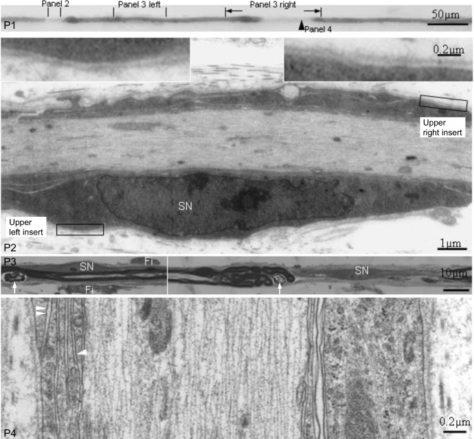
Results: In riboflavin deficient chickens, demyelination was the predominant feature: it was mainly confined to the paranodal region at day 11; extended into internodal region, but less than half of the internodal length in most affected internodes at day 16; involved more than half or whole internode at day 21. In the internode undergoing demyelination, myelin degeneration of varying degrees was noticed in the cytoplasm of the Schwann cell wrapping the internode. Two days after riboflavin repletion, co-existence of remyelination and active demyelination within individual internodes was noticed. Remyelination together with preserved short original internodes was the characteristic feature 4 and 9 days after riboflavin repletion.

Conclusion: Riboflavin repletion interrupts the progression from paranodal to internodal demyelination in riboflavin deficient chickens and promotes remyelination before complete internodal demyelination.

Keywords: Demyelination; Remyelination; Riboflavin; Schwann cell; Teased nerve fibre.


Conflict of interest statement

The author does not have competing interests.

Figures

Fig. 1
Fig. 1
Evolution of demyelination in VB2 chickens. Light micrographs. A Panel 1 (P1) and P2 show part of a TF from a VB2PH11d chicken. The nodes of Ranvier are indicated by arrows (N1 to N5). Paranodal demyelination is noted at 2 sites (N3 and N4) and normal node of Ranvier at 3 sites (N1, N2 and N5). Schwann cell nuclei (white arrowheads) are located in the middle of each internode. P3 to P5 are longitudinal sections through designated segments. P3 is through the first paranodal demyelinating area. No discernible myelin sheath is seen in the demyelinated segment. Myelin degeneration (large arrowheads in P3) is seen in the internodal region adjoining the paranodal demyelination (small arrowheads in P2). P4 is through the central part of the internode with paranodal demyelination. The myelin sheath is well preserved in this segment and a Schwann cell nucleus (SN) is present. P5 is through the second paranodal demyelinating area. No discernible myelin sheath is seen in the demyelinated segment. Myelin degeneration (large arrowheads in P5) is seen in longitudinal section through the segment (small arrowheads in P2) adjoining the paranodal demyelination. The axon is attenuated in demyelinating regions. B (adapted from Cai et al., 2007 [14] with permission from the author and publisher): P1 to P3 show a TF from a VB2PH16d chicken displaying paranodal demyelination (N1 and N4) and partial internodal demyelination (N2, and N3, N5 and N6). Paranodal swellings (small arrowheads) and surrounding pale-osmicated material representing the fibroblastic onion bulb like proliferation [14]. P4 to P6 are longitudinal sections through designated segments in the three upper panels. In the demyelinated segments, there is no discernible myelin sheath and axon (a) is attenuated. Redundant myelin foldings with varying degrees of myelin splitting and degeneration (large arrowheads) are found in longitudinal sections through paranodal swellings while the myelin sheaths in the neighbouring non-demyelinated internodal regions are intact. The fibre is surrounded by a variably thick fibroblastic proliferation consisting of fibroblast processes and collagen fibres. Numerous fibroblast nuclei (arrows), some with multiple distinct nucleoli (double arrows), are noted at both demyelinated and non-demyelinated segments. C P1 and P2 show part of a TF from a VB2PH21d chicken displaying internodal demyelination. P3 and P4 are cross sections through designated sites of the TF, showing denuded axon in the demyelinated segments (L1, 5–19), redundant myelin foldings with myelin splitting and degeneration at both ends of the myelin-maintained segment (L2 and 4). The axon size in the region with intact myelin (L3) is larger than that of demyelinated regions. Fibroblast processes and collagen ensheath the TF at all levels to some degree
Fig. 2
Fig. 2
Light micrographs, P1 and P3; Electron micrographs, P2 and P4. P1 is part of a TF from a VB2−/+PH14d chicken displaying paranodal and partial internodal demyelination according to the surface appearance. P2 is a longitudinal section through the paranodal demyelination segment. A Schwann cell nucleus (SN) is present. This Schwann cell envelops the axon under basal lamella of the nerve fibre (inserts). P3 left is a longitudinal section through the middle of the neighbouring myelin-maintained segment showing a Schwann cell nucleus (SN), 2 attached fibroblasts (Fi) and myelin debris (white arrows). P3 right is a longitudinal section through the partial internodal demyelination segment. A centrally located Schwann cell nucleus (SN) is present. Redundant myelin foldings with myelin breakdown (white arrows) is present in the paranodal region of the neighbouring myelin-maintained segment. P4 is a longitudinal section (rotate 90°) through the partial internodal demyelination region (arrowhead in panel 1) showing 4 layers of non-compacted myelin lamellae surrounding the axon under the basal lamella of the nerve fibre: an inner mesaxon (white arrowhead) connecting the axolemma and outer mesaxon (double white arrowheads) connecting Schwann cell basement membrane
Fig. 3
Fig. 3
Evolution of demyelination and remyelination in VB2−/+ chickens. Light micrographs except for electron micrograph at P6 in A. A P1 and P2 are part of a TF from a VB2−/+PH14d chicken with partial internodal demyelination. P3 to P5 are longitudinal sections through designated sites of the TF. Redundant myelin foldings are seen in externally normal paranodal regions (P3 left) and paranodal regions adjoining partial internodal demyelination (P4 and P5). A centrally located Schwann cell nucleus (SN) is noticed in the demyelinated regions (P4 and P5) and neighbouring myelin-maintained segment (P3 right). Supernumerary fibroblasts (Fi) are seen attached to the TF preparation at paranodal and internodal regions. Electron microscopy (P6) further demonstrates the fibroblast with enriched rough reticulum (white rectangle in P3 left). B P1 and P2 are part of a TF from a VB2−/+PH16d chicken, showing remyelination. P3 to P5 are longitudinal sections of designated segments. The nodes of Ranvier (N1–N13 arrows) are hardly seen from the surface appearance of the TF, but are clearly identified in longitudinal sections. The 1st (N1–N2), 4th (N4–N5), 8th (N8–N9) and 11th (N11–N12) internodes are original internodes with thick myelin sheaths. The 2nd (N2–N3), 3rd (N3–N4), 5th (N5–N6), 6th (N6–N7), 7th (N7–N8), 9th (N9–N10), 10th (N10–N11) and 12th (N12–N13) internodes are remyelinating internodes with thin myelin sheaths. Substantial variation of the internodal length is seen in both original and remyelinating internodes. The length of some remyelinating internodes, such as the 2nd (N2–N3), 3rd (N3–N4), 7th (N7–N8) and 10th (N10–N11) internodes, are similar or even longer than some original internodes, such as the 4th (N4–N5) and 8th (N8–N9) internodes. Focal myelin swellings (arrowheads) are present at the paranodal regions of original internodes. The nodal gap is not extra ordinarily large, implicating that the length of the remyelinating internode is already fixed at this stage. A Schwann cell nucleus (SN) and lipid deposition (asterisk) are present in the middle of an original internode. Myelin debris is seen in the paranodal Schwann cell cytoplasm of the original internode (double arrows). The TF is surrounded by a variably thick fibroblastic proliferation consisting of fibroblast processes and collagen fibres. Numerous fibroblast nuclei (white arrows) are noted in the longitudinal section at both original and remyelinating internodes. C P1 is part of a TF from a VB2−/+PH21d chicken, showing remyelination. The nodes of Ranvier are indicated by arrows (N1–N6). There is no considerable variation of the internodal length between the original (N2–N3 and N4–N5) and remyelinating internodes (N1–N2, N3–N4 and N5–N6). One of the remyelinating internode (N1–N2, 194 µm in length) is even longer than that of original internodes (N2–N3, 176 µm; N4–N5, 159 µm). P2 and P3 are longitudinal sections through the full length of a remyelinating and a neighbouring original internode in the first panel. Schwann cell nucleus (SN) is present in the middle of both the remyelinating and original internodes. Degenerating myelin of varying stages (double arrows) is noticed in Schwann cell cytoplasm of both original and remyelinating internodes. White arrows indicate fibroblast nuclei

Similar articles

References

    1. Dyck PJ, Dyck PJB, Engelstad J, et al. Pathologic alterations of nerve. In: Dyck PJ, Thomas PK, Griffin J, et al., editors. Peripheral neuropathy. 4. Philadelphia, PA: Elsevier; 2005. pp. 733–829.
    1. Koike H, Kadoya M, Kaida KI, et al. Paranodal dissection in chronic inflammatory demyelinating polyneuropathy with anti-neurofascin-155 and anti-contactin-1 antibodies. J Neurol Neurosurg Psychiatry. 2017;88:465–473. doi: 10.1136/jnnp-2016-314895. - DOI - PubMed
    1. Koike H, Katsuno M. Macrophages and autoantibodies in demyelinating diseases. Cells. 2021;10:844. doi: 10.3390/cells10040844. - DOI - PMC - PubMed
    1. Allt G, Cavanagh JB. Ultrastructural changes in the region of the node of Ranvier in the rat caused by diphtheria toxin. Brain. 1969;92:459–468. doi: 10.1093/brain/92.2.459. - DOI - PubMed
    1. Uncini A, England JD, Rhee EK, et al. Tellurium-induced demyelination: an electrophysiological and morphological study. Muscle Nerve. 1988;11:871–879. doi: 10.1002/mus.880110812. - DOI - PubMed

LinkOut - more resources