. 2024 Apr 22;19(1):257.
doi: 10.1186/s13018-024-04727-7.

CircPRKD3/miR-6783-3p responds to mechanical force to facilitate the osteogenesis of stretched periodontal ligament stem cells

Affiliations

CircPRKD3/miR-6783-3p responds to mechanical force to facilitate the osteogenesis of stretched periodontal ligament stem cells

Jiani Liu et al. J Orthop Surg Res. .

Abstract

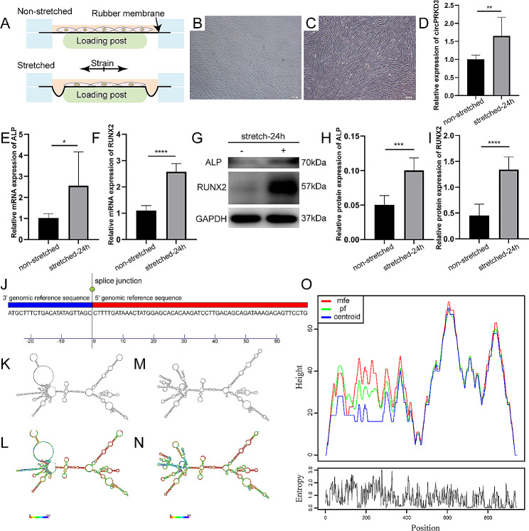
Background: The mechanotransduction mechanisms by which cells regulate tissue remodeling are not fully deciphered. Circular RNAs (circRNAs) are crucial to various physiological processes, including cell cycle, differentiation, and polarization. However, the effects of mechanical force on circRNAs and the role of circRNAs in the mechanobiology of differentiation and remodeling in stretched periodontal ligament stem cells (PDLSCs) remain unclear. This article aims to explore the osteogenic function of mechanically sensitive circular RNA protein kinase D3 (circPRKD3) and elucidate its underlying mechanotransduction mechanism.

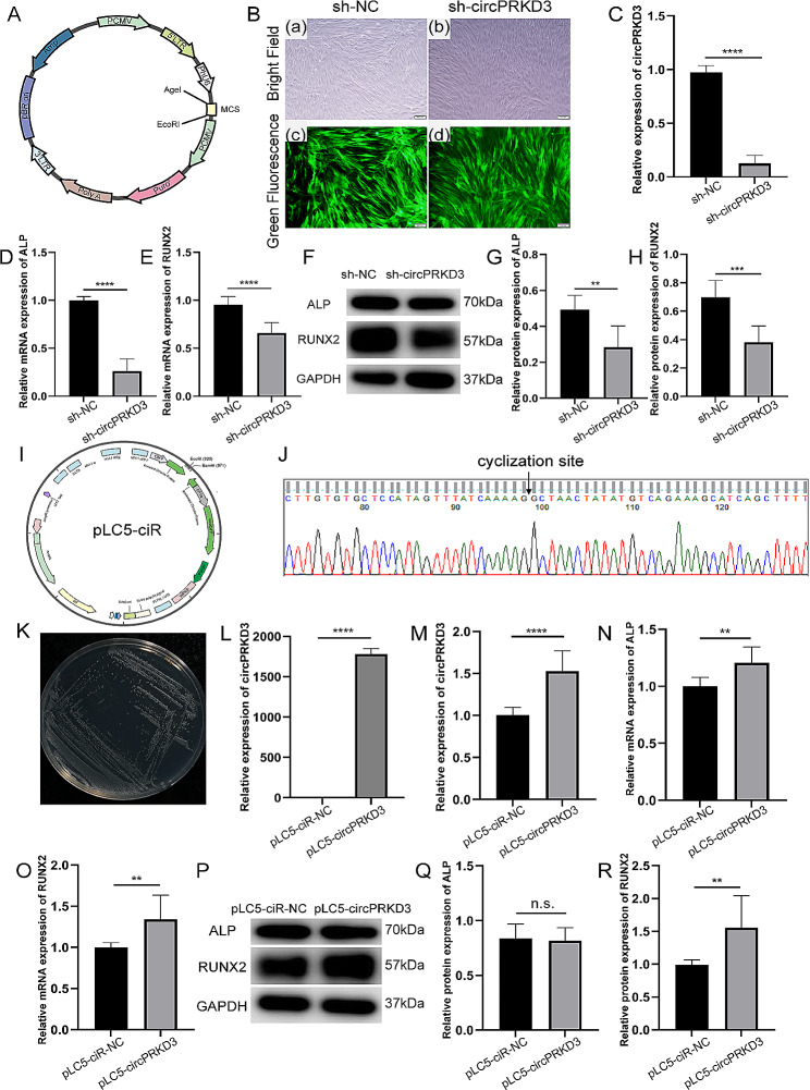
Materials and methods: PDLSCs were elongated with 8% stretch at 0.5 Hz for 24 h using the Flexcell® FX-6000™ Tension System. CircPRKD3 was knockdown or overexpressed with lentiviral constructs or plasmids. The downstream molecules of circPRKD3 were predicted by bioinformatics analysis. The osteogenic effect of related molecules was evaluated by quantitative real-time PCR (qRT-PCR) and western blot.

Results: Mechanical force enhanced the osteogenesis of PDLSCs and increased the expression of circPRKD3. Knockdown of circPRKD3 hindered PDLSCs from osteogenesis under mechanical force, while overexpression of circPRKD3 promoted the early osteogenesis process of PDLSCs. With bioinformatics analysis and multiple software predictions, we identified hsa-miR-6783-3p could act as the sponge of circPRKD3 to indirectly regulate osteogenic differentiation of mechanically stimulated PDLSCs.

Conclusions: Our results first suggested that both circPRKD3 and hsa-miR-6783-3p could enhance osteogenesis of stretched PDLSCs. Furthermore, hsa-miR-6783-3p could sponge circPRKD3 to indirectly regulate RUNX2 during the periodontal tissue remodeling process in orthodontic treatment.

Keywords: CircPRKD3; Mechanical force; PDLSCs; ceRNAs; miR-6783-3p.


Conflict of interest statement

The authors declare no competing interests.

Figures

Fig. 1
Fig. 1
Identification and biological characteristics of PDLSCs. (A) Morphology of PDLSCs (P0) from periodontal tissue blocks (scale bar 200 μm). (B) Identification of MSCs surface markers in PDLSCs by flow cytometry. ALP staining of PDLSCs cultured for 7 days without (C) or with (D) osteogenic induction (scale bar 200 μm). Alizarin red staining of PDLSCs cultured for 28 days without (E) or with (F) osteogenic induction (scale bar 200 μm). Oil red O staining of PDLSCs cultured for 21 days without (G) or with (H) adipogenic induction (scale bar 50 μm)
Fig. 2
Fig. 2
CircPRKD3 was upregulated during osteogenic development of mechanically stimulated PDLSCs. (A) Schematic diagram of mechanically stimulated PDLSCs in vitro. Morphology of PDLSCs without (B) or with (C) stretch inducement for 24 h (scale bar 200 μm). (D) Relative gene expression level of circPRKD3 in non-stretched and stretched-24 h groups. (E-F) The mRNA expression level of ALP and RUNX2 in non-stretched and stretched-24 h groups. (G-I) The protein level of ALP and RUNX2 and the quantitative data analyzed by image J in non-stretched and stretched-24 h groups. Full-length blots were presented in Supplementary Fig. 1 of additional file 1. (J) The genetic sequence of circPRKD3 on circBase. (K) Centroid plain structure drawing of circPRKD3. (L) Centroid structure drawing encoding positional entropy of circPRKD3. (M) MFE plain structure drawing of circPRKD3. (N) MFE structure drawing encoding positional entropy of circPRKD3. (O) Peak diagram of circPRKD3 with minimum free energy. Data are presented as mean ± SD of three independent experiments. (*p < 0.05; **p < 0.01; ***p < 0.001; ****p < 0.0001)
Fig. 3
Fig. 3
Regulation of circPRKD3 affected osteogenesis of mechanically stimulated PDLSCs. (A) Schematic diagram of virus construction. Immunofluorescence staining (B) and qRT-PCR verification (C) of transfection efficiency in sh-NC and sh-circPRKD3 groups. (D-E) The mRNA expression level of ALP and RUNX2 in mechanically stimulated sh-NC and sh-circPRKD3 groups after transfection for 24 h. (F-H) The protein level of ALP and RUNX2 and the quantitative data analyzed by image J in mechanically stimulated sh-NC groups and sh-circPRKD3 groups after transfection for 24 h. Full-length blots were presented in Supplementary Fig. 2 of additional file 1. (I) Schematic diagram of plasmids construction. (J) The head-to‐tail splicing of circPRKD3 as a qRT‐PCR product was verified by Sanger sequencing. (K) Pure E. coli containing circPRKD3-overexpressed plasmids were filtered out by ampicillin. Verification of transfection efficiency in 293T (L) and PDLSCs (M) was assayed by qRT‐PCR. (N-O) The mRNA expression level of ALP and RUNX2 in mechanically stimulated pLC5‐ciR-NC groups and pLC5‐circPRKD3 groups after transfection for 24 h. (P-R) The protein level of ALP and RUNX2 and the quantitative data analyzed by image J in mechanically stimulated pLC5‐ciR-NC groups and pLC5‐circPRKD3 groups after transfection for 24 h. Full-length blots were presented in Supplementary Fig. 3 of additional file 1. Data are presented as mean ± SD of three independent experiments. (n.s., no significance; **p < 0.01; ****p < 0.0001)
Fig. 4
Fig. 4
Screening and validation of circPRKD3 downstream targets. Immunofluorescence staining (A) and quantitative analysis (B) of circPRKD3 distribution in nucleus and cytoplasm. (C) Prediction of miRNAs combined with circPRKD3 through different algorithms. (D) Force-sensitive miRNAs were sought among the 10 predicted miRNAs above by qRT-PCR analysis. (E) CircPRKD3 overexpression-associated miRNAs among force-sensitive miRNAs were evaluated by qRT‐PCR in mechanically stimulated pLC5‐ciR-NC groups and pLC5‐circPRKD3 groups after transfection for 24 h. (F) CircPRKD3 knockdown-associated miRNAs among force-sensitive miRNAs were assayed by qRT‐PCR in mechanically stimulated sh-NC and sh-circPRKD3 groups after transfection for 24 h. (G) Schematic diagram of luciferase assay and related sequence information. (H) Relative luciferase activity was measured in circPRKD3-WT groups and circPRKD3-mut groups with or without miR-6783-3p mimics. Data are presented as mean ± SD of three independent experiments. (n.s., no significance; *p < 0.05; **p < 0.01; ***p < 0.001; ****p < 0.0001)
Fig. 5
Fig. 5
Interaction between miRNA-6783-3p and circPRKD3 regulated osteogenesis of mechanically stimulated PDLSCs. (A) Validation of miR-6783-3p overexpression efficiency by qRT-PCR analysis in PDLSCs. (B-C) The mRNA expression level of ALP and RUNX2 in mechanically stimulated NC-mimics groups and mimics-miR-6783-3p groups after transfection for 24 h. (D-F) The protein level of ALP and RUNX2 and the quantitative data analyzed by image J in mechanically stimulated NC-mimics groups and mimics-miR-6783-3p groups after transfection for 24 h. Full-length blots were presented in Supplementary Fig. 4 of additional file 1. (G-I) The protein level of ALP and RUNX2 and the quantitative data analyzed by image J after co-transfection of pLC5‐circPRKD3 plasmids and mimics-miR-6783-3p for 24 h under mechanical force. Full-length blots were presented in Supplementary Fig. 5 of additional file 1. (J) Predicted sites where circPRKD3 binds to different RUNX2 protein isoforms sequences on the catRAPID website. (a) Binding sites prediction on RUNX2 isoform a. (b) Binding sites prediction on RUNX2 isoform b. (c) Binding sites prediction on RUNX2 isoform d. (d) Binding sites prediction on RUNX2 isoform e. (K) The working model that miR-6783-3p acts as a sponge of circPRKD3 to indirectly regulate the osteogenesis of PDLSCs under mechanical force. Data are presented as mean ± SD of three independent experiments. (n.s., no significance; *p < 0.05; **p < 0.01; ***p < 0.001; ****p < 0.0001)

Similar articles

Cited by

References

    1. Li M, Zhang C, Yang Y. Effects of mechanical forces on osteogenesis and osteoclastogenesis in human periodontal ligament fibroblasts: a systematic review of in vitro studies. Bone Joint Res. 2019;8(1):19–31. doi: 10.1302/2046-3758.81.BJR-2018-0060.R1. - DOI - PMC - PubMed
    1. Jin SS, He DQ, Wang Y, Zhang T, Yu HJ, Li ZX, et al. Mechanical force modulates periodontal ligament stem cell characteristics during bone remodelling via TRPV4. Cell Prolif. 2020;53(10):e12912. doi: 10.1111/cpr.12912. - DOI - PMC - PubMed
    1. Liu M, Zhang H, Li Y, Wang S. Noncoding RNAs interplay in Ovarian Cancer Therapy and Drug Resistance. Cancer biotherapy & radiopharmaceuticals; 2022. - PubMed
    1. Amorim M, Salta S, Henrique R, Jerónimo C. Decoding the usefulness of non-coding RNAs as breast cancer markers. J Translational Med. 2016;14:265. doi: 10.1186/s12967-016-1025-3. - DOI - PMC - PubMed
    1. Ali T, Grote P. Beyond the RNA-dependent function of LncRNA genes. eLife. 2020;9. - PMC - PubMed