. 2024 Apr 22;12(1):65.
doi: 10.1186/s40478-024-01777-0.

Extracellular vesicle encapsulated nicotinamide delivered via a trans-scleral route provides retinal ganglion cell neuroprotection

Affiliations

Extracellular vesicle encapsulated nicotinamide delivered via a trans-scleral route provides retinal ganglion cell neuroprotection

Myungjin Kim et al. Acta Neuropathol Commun. .

Abstract

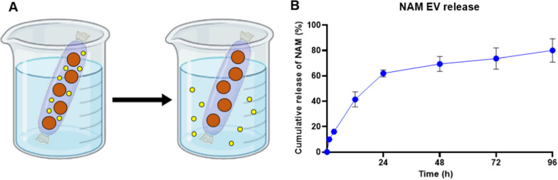
The progressive and irreversible degeneration of retinal ganglion cells (RGCs) and their axons is the major characteristic of glaucoma, a leading cause of irreversible blindness worldwide. Nicotinamide adenine dinucleotide (NAD) is a cofactor and metabolite of redox reaction critical for neuronal survival. Supplementation with nicotinamide (NAM), a precursor of NAD, can confer neuroprotective effects against glaucomatous damage caused by an age-related decline of NAD or mitochondrial dysfunction, reflecting the high metabolic activity of RGCs. However, oral supplementation of drug is relatively less efficient in terms of transmissibility to RGCs compared to direct delivery methods such as intraocular injection or delivery using subconjunctival depots. Neither method is ideal, given the risks of infection and subconjunctival scarring without novel techniques. By contrast, extracellular vesicles (EVs) have advantages as a drug delivery system with low immunogeneity and tissue interactions. We have evaluated the EV delivery of NAM as an RGC protective agent using a quantitative assessment of dendritic integrity using DiOlistics, which is confirmed to be a more sensitive measure of neuronal health in our mouse glaucoma model than the evaluation of somatic loss via the immunostaining method. NAM or NAM-loaded EVs showed a significant neuroprotective effect in the mouse retinal explant model. Furthermore, NAM-loaded EVs can penetrate the sclera once deployed in the subconjunctival space. These results confirm the feasibility of using subconjunctival injection of EVs to deliver NAM to intraocular targets.

Keywords: Extracellular vesicle; Glaucoma; Neuroprotection; Nicotinamide; Retinal ganglion cell; Trans-scleral route.


Conflict of interest statement

The authors declare that they have no competing interests.

Figures

Fig. 1
Fig. 1
Comparison of response to IOP elevation with RGC soma counts and dendritic arbourisations in a mouse glaucoma model. A IOP elevation was induced using intracameral injection of polyurethane microbeads in mouse eyes (26 retinas from 13 mice). B IOP curves of four groups which were divided by the IOP of 17 mmHg at week 1. C The Cumulative IOP graph shows that the IOP elevation was statistically significant in both the RBPMS high group and the DiOlistics high group compared to the RBPMS low group and the DiOlistics low group, respectively. D In the RBPMS low OHT group, the count of RBPMS+ RGCs was similar to the control group. However, the count of RBPMS+ RGCs was lower, with marginal significance in the RBPMS high OHT group compared to the control group. Each value of the box plot is an average of the RGC counts in one retina. Scale bar = 20 μm. E Whereas the statistically significant change of dendritic arbour was apparent in both the DiOlistics low group as well as the DiOlistics high group compared to the control group. Scale bar = 50 μm. NT normal tension (control group), OHT ocular hypertension (treated group). ap = 0.05, *p < 0.05, all comparison was performed between each treated group and NT
Fig. 2
Fig. 2
Engineering and characterisation of NAM-loaded EV. A Schematic illustration of the fabrication method for NAM-loaded ASC EV (NAM-EV). B The size distribution of both ASC EVs and NAM-EVs falls within the typical size range for EVs. C The pH of the ASC EV and NAM-EV were found to be neutral. D Confirmation by western blot of surface marker (CD63), internal marker (TSG101), and negative marker (ApoA1) of ASC EVs and NAM-EVs proved their identity as EVs. E The cup-shaped structural morphology of the TEM image confirmed the presence of EVs. Scale bars = 100 nm. F The loading efficiency of nicotinamide into EVs. ASC adipose-derived stem cell
Fig. 3
Fig. 3
Mouse retinal explant model in four groups (control, NAM, EV, and NAM-EV). A Schematic illustration of mouse retinal explant and culture method. B Division into four groups based on the presence or absence of additives in the media: control (3DEV), NAM, EV, and NAM-EV, respectively (16 retinas from 8 mice). C Representative images of dendritic arbourisation in each group. Scale bar = 50 μm. D Sholl analysis graphs for each group show significant preservation of dendritic arbour in the NAM and NAM-EV groups compared to the control group (3DEV). DEV days ex vivo. *p < 0.05, All comparison was performed between each treated group and control group (3DEV)
Fig. 4
Fig. 4
Scleral penetration of DiO-labelled EV via subconjunctival injection. A Schematic illustration of the purification of DiO-labelled EVs. B The DiO intensity of all size exclusion chromatography factions. C By confirming the DiO intensity of the concentrated DiO-labelled EV and PBS solutions, we proved that the concentrated DiO-labelled EV solution contained DiO-EV. D The size distribution of DiO-labelled EVs demonstrates they fit in the size range of EVs. E Detection of DiO-labelled EV penetration via sclera after subconjunctival injection in NZW rabbit. Note that the depth of EV penetration increases as time goes by (2 h > 1 h > 0 h). Scale bars = 200 μm. F High magnification images at 2 h after injection. The DiO-labelled EVs were detected in the retinal tissue. Scale bars = 10 μm
Fig. 5
Fig. 5
The releasing profile of NAM from EV. A Schematic illustration of the method for determining the nicotinamide release profile from NAM-EV. B The release profile of nicotinamide released from NAM-EVs showed an initial burst up to 24 h, followed by a sustained release pattern

Similar articles

Cited by

References

    1. Afarid M, Mahmoodi S, Baghban R. Recent achievements in nano-based technologies for ocular disease diagnosis and treatment, review and update. J Nanobiotechnol. 2022;20:1–36. doi: 10.1186/s12951-022-01567-7. - DOI - PMC - PubMed
    1. Ahmed I, Gokhale RD, Shah MV, Patton TF. Physicochemical determinants of drug diffusion across the conjunctiva, sclera, and cornea. J Pharm Sci. 1987;76:583–586. doi: 10.1002/jps.2600760802. - DOI - PubMed
    1. Allen TM, Hansen CB, de Menezes DEL. Pharmacokinetics of long-circulating liposomes. Adv Drug Deliv Rev. 1995;16:267–284. doi: 10.1016/0169-409X(95)00029-7. - DOI
    1. Alvarez-Erviti L, Seow Y, Yin H, Betts C, Lakhal S, Wood MJ. Delivery of siRNA to the mouse brain by systemic injection of targeted exosomes. Nat Biotechnol. 2011;29:341–345. doi: 10.1038/nbt.1807. - DOI - PubMed
    1. Ambati J, Adamis AP. Transscleral drug delivery to the retina and choroid. Prog Retin Eye Res. 2002;21:145–151. doi: 10.1016/s1350-9462(01)00018-0. - DOI - PubMed

Publication types

MeSH terms