

Comparison of vestibular aqueduct visualization on computed tomography and magnetic resonance imaging in patients with Ménière's disease
- PMID: 38649991
- PMCID: PMC11034041
- DOI: 10.1186/s12880-024-01275-8
Comparison of vestibular aqueduct visualization on computed tomography and magnetic resonance imaging in patients with Ménière's disease
Abstract
Background: The vestibular aqueduct (VA) serves an essential role in homeostasis of the inner ear and pathogenesis of Ménière's disease (MD). The bony VA can be clearly depicted by high-resolution computed tomography (HRCT), whereas the optimal sequences and parameters for magnetic resonance imaging (MRI) are not yet established. We investigated VA characteristics and potential factors influencing MRI-VA visibility in unilateral MD patients.
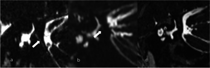
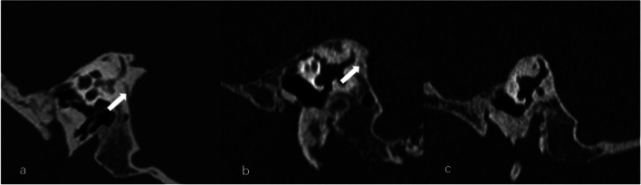
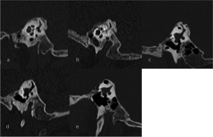
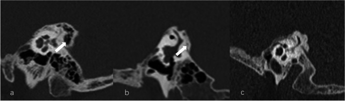
Methods: One hundred patients with unilateral MD underwent MRI with three-dimensional sampling perfection with application optimized contrasts using different flip angle evolutions (3D-SPACE) sequence and HRCT evaluation. The imaging variables included MRI-VA and CT-VA visibility, CT-VA morphology and CT-peri-VA pneumatization.
Results: The most frequent type of MRI-VA and CT-VA visualization was invisible VA and continuous VA, respectively. The MRI-VA visibility was significantly lower than CT-VA visibility. MRI-VA visibility had a weak positive correlation with ipsilateral CT-VA visualization. For the affected side, the MRI-VA visualization was negatively correlated with the incidence of obliterated-shaped CT-VA and positively with that of tubular-shaped CT-VA. MRI-VA visualization was not affected by CT-peri-VA pneumatization.
Conclusion: In patients with MD, the VA visualization on 3D-SPACE MRI is poorer than that observed on CT and may be affected by its osseous configuration. These findings may provide a basis for further characterization of VA demonstrated by MRI and its clinical significance.
Keywords: Computed tomography; Magnetic resonance imaging; Ménière’s disease; Vestibular aqueduct.
© 2024. The Author(s).
Conflict of interest statement
The authors declare no competing interests.
Figures
References
Publication types
MeSH terms
Grants and funding
LinkOut - more resources
Full Text Sources
Medical
