Review
. 2024 Apr 22;23(1):117.
doi: 10.1186/s12944-024-02108-x.

Updated mechanisms of MASLD pathogenesis

Affiliations
Review

Updated mechanisms of MASLD pathogenesis

Yuxuan Li et al. Lipids Health Dis. .

Abstract

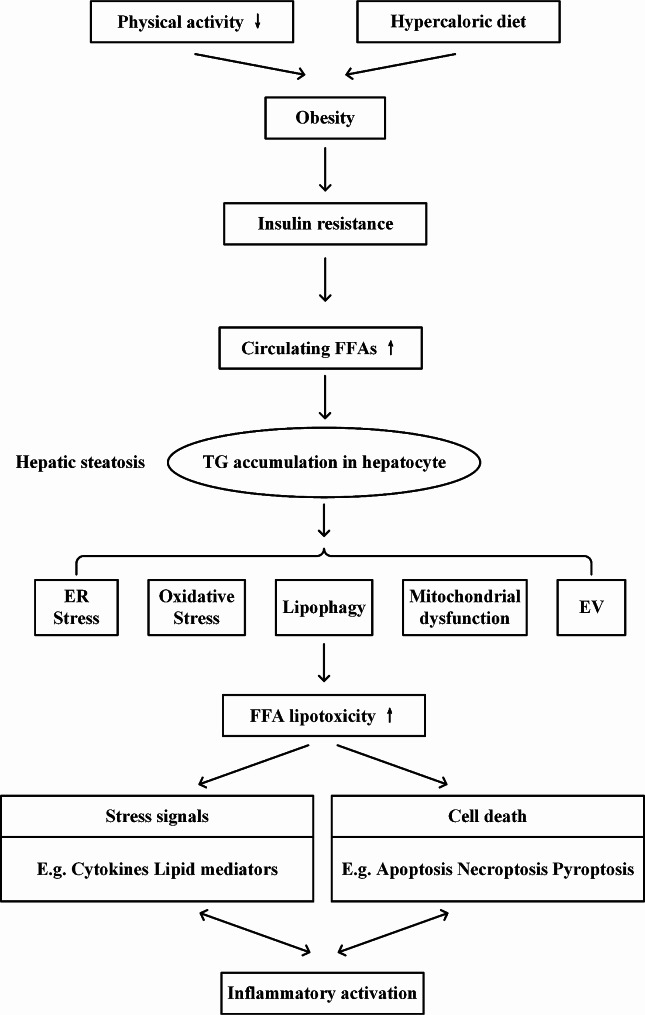
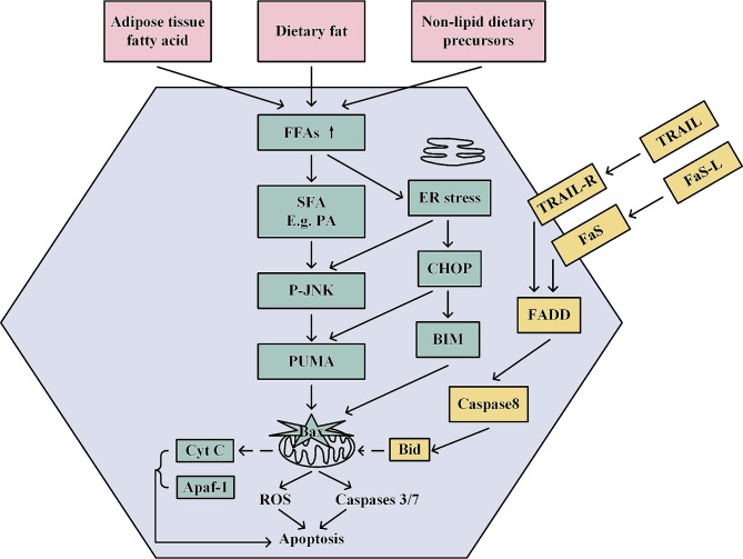
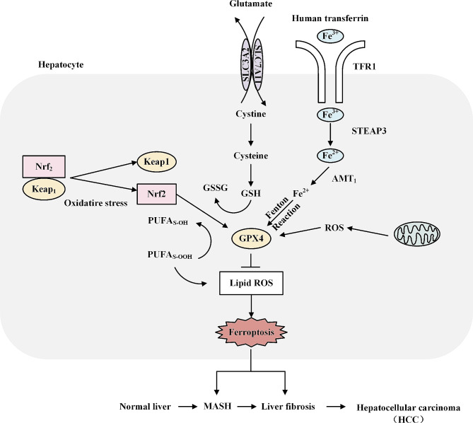
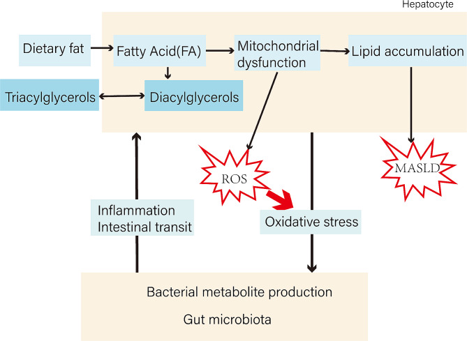
Metabolic dysfunction-associated steatotic liver disease (MASLD) has garnered considerable attention globally. Changing lifestyles, over-nutrition, and physical inactivity have promoted its development. MASLD is typically accompanied by obesity and is strongly linked to metabolic syndromes. Given that MASLD prevalence is on the rise, there is an urgent need to elucidate its pathogenesis. Hepatic lipid accumulation generally triggers lipotoxicity and induces MASLD or progress to metabolic dysfunction-associated steatohepatitis (MASH) by mediating endoplasmic reticulum stress, oxidative stress, organelle dysfunction, and ferroptosis. Recently, significant attention has been directed towards exploring the role of gut microbial dysbiosis in the development of MASLD, offering a novel therapeutic target for MASLD. Considering that there are no recognized pharmacological therapies due to the diversity of mechanisms involved in MASLD and the difficulty associated with undertaking clinical trials, potential targets in MASLD remain elusive. Thus, this article aimed to summarize and evaluate the prominent roles of lipotoxicity, ferroptosis, and gut microbes in the development of MASLD and the mechanisms underlying their effects. Furthermore, existing advances and challenges in the treatment of MASLD were outlined.

Keywords: Lipid metabolism; Lipotoxicity; MASLD; Therapeutics.


Conflict of interest statement

The authors declare no competing interests.

Figures

Fig. 1
Fig. 1
Potential sources and mechanisms of hepatic fat accumulation. Genetic risk, lifestyle, and metabolic factors all contribute to hepatic steatosis. Lipid accumulation in hepatocytes leads to lipotoxicity, which activates oxidative stress-related molecules and signals and transmits them between cells in the form of extracellular vesicles or diffusion, thereby triggering the cell death program and pushing hepatic steatosis toward inflammation and fibrosis. FFA, free fatty acid; TG, triglyceride; ER, endoplasmic reticulum; EV, extracellular vesicles
Fig. 2
Fig. 2
Molecular mechanisms associated with hepatocyte lipotoxicity and apoptosis. There are 3 major sources of fatty acids in the liver, including dietary intake, self-synthesis from scratch, and catabolism by peripheral adipose tissue. FFA is transported into the hepatocytes to synthesize triglycerides, which leads to hepatic steatosis. Lipotoxicity induces death receptor signaling pathways that recruit caspase 8 to cleave Bid and regulate apoptosis. Excess SFA accumulates in the ER and induces ER stress, which in turn induces the transcription factor CHOP and mediates the onset of JNK. CHOP not only interacts with activated c-jun to upregulate the transcription of the pro-apoptotic BH3 protein, PUMA but also increases the expression of another BH3-only protein, Bim, which synergistically activates the pro-apoptotic protein, Bax. Bim and PUMA synergistically activate the pro-apoptotic protein Bax, which causes mitochondrial dysfunction and induces apoptosis through the release of cyt C and the activation of caspases proteases. Mitochondrial dysfunction, on the other hand, also leads to the overproduction of reactive ROS, which causes oxidative stress and further induces cell death. FFA, free fatty acid; SFA, saturated fatty acid; ER, endoplasmic reticulum; oxygen species
Fig. 3
Fig. 3
The regulatory mechanisms of ferroptosis in MASLD and its effects on the progression of MASLD. Cystine enters the cell via SLC7A11 and SLC3A2 embedded in the surface of the cell membrane and is then oxidized to cysteine, which is catalytically synthesized into GSH.GPX4 utilizes the ability of GSH to convert lipid peroxidation of L-OOH to L-OH, losing its peroxidative activity and thus protecting against the induction of ferroptosis. Nrf2 inhibits ferroptosis by regulating GPX4 and iron metabolism. PUFA binds to phosphatidylethanolamine(PE) to form polyunsaturated fatty acids phospholipids, the latter of which are susceptible to lipoxygenase(LOX)-mediated free radical-induced oxidation that induces ferroptosis. Fe3 + is uptaken by TFR1, reduced to Fe2 + by STEAP3, and later transported into the cytoplasmic unstable iron pool (LIP), a regulator of iron metabolism, and two proteins in the family of zinc-iron-modulated proteins (ZIP8/14), however, in the presence of excess Fe2+, can induce cellular ferroptosis by increasing reactive oxygen species generation and promoting lipid peroxidation formation via the fenton pathway. GSH, glutathione; GPX4, glutathione peroxidase 4; Nrf2, nuclear erythroid-related factor 2; PUMA, P53-up-regulated modulator of apoptosis; TFR1, transferrin receptor 1 P53-up-regulated modulator of apoptosis; TFR1, transferrin receptor 1
Fig. 4
Fig. 4
Schematic representation illustrating part of the pathogenesis of MASLD. Excessive dietary fat intake triggers multiple hepatic mechanisms, including mitochondrial dysfunction, which induces hepatic lipid accumulation, leading to MASLD. on the other hand, gut microbial dysbiosis is an essential component in the study of MASLD development, and the “liver-gut axis” has been proposed as a new target to drive the complex progression of MASLD.

Similar articles

Cited by

References

    1. Scorletti E, Carr RM. A new perspective on NAFLD: focusing on lipid droplets. J Hepatol. 2022;76:934–45. doi: 10.1016/j.jhep.2021.11.009. - DOI - PubMed
    1. Lazarus JV, Mark HE, Anstee QM, Arab JP, Batterham RL, Castera L, et al. Advancing the global public health agenda for NAFLD: a consensus statement. Nat Rev Gastroenterol Hepatol. 2022;19:60–78. doi: 10.1038/s41575-021-00523-4. - DOI - PubMed
    1. Liu C, Liu T, Zhang Q, Jia P, Song M, Zhang Q, et al. New-onset age of nonalcoholic fatty liver Disease and Cancer Risk. JAMA Netw Open. 2023;6:e2335511. doi: 10.1001/jamanetworkopen.2023.35511. - DOI - PMC - PubMed
    1. Tilg H, Moschen AR. Evolution of inflammation in nonalcoholic fatty liver disease: the multiple parallel hits hypothesis. Hepatology (Baltimore MD) 2010;52:1836–46. doi: 10.1002/hep.24001. - DOI - PubMed
    1. Loomba R, Friedman SL, Shulman GI. Mechanisms and disease consequences of nonalcoholic fatty liver disease. Cell. 2021;184:2537–64. doi: 10.1016/j.cell.2021.04.015. - DOI - PubMed

LinkOut - more resources