. 2024 Apr 22;22(1):236.
doi: 10.1186/s12964-024-01604-y.

Interleukin-4 protects retinal ganglion cells and promotes axon regeneration

Affiliations

Interleukin-4 protects retinal ganglion cells and promotes axon regeneration

Zhaoyang Zuo et al. Cell Commun Signal. .

Abstract

Background: The preservation of retinal ganglion cells (RGCs) and the facilitation of axon regeneration are crucial considerations in the management of various vision-threatening disorders. Therefore, we investigate the efficacy of interleukin-4 (IL-4), a potential therapeutic agent, in promoting neuroprotection and axon regeneration of retinal ganglion cells (RGCs) as identified through whole transcriptome sequencing in an in vitro axon growth model.

Methods: A low concentration of staurosporine (STS) was employed to induce in vitro axon growth. Whole transcriptome sequencing was utilized to identify key target factors involved in the molecular mechanism underlying axon growth. The efficacy of recombinant IL-4 protein on promoting RGC axon growth was validated through in vitro experiments. The protective effect of recombinant IL-4 protein on somas of RGCs was assessed using RBPMS-specific immunofluorescent staining in mouse models with optic nerve crush (ONC) and N-methyl-D-aspartic acid (NMDA) injury. The protective effect on RGC axons was evaluated by anterograde labeling of cholera toxin subunit B (CTB), while the promotion of RGC axon regeneration was assessed through both anterograde labeling of CTB and immunofluorescent staining for growth associated protein-43 (GAP43).

Results: Whole-transcriptome sequencing of staurosporine-treated 661 W cells revealed a significant upregulation in intracellular IL-4 transcription levels during the process of axon regeneration. In vitro experiments demonstrated that recombinant IL-4 protein effectively stimulated axon outgrowth. Subsequent immunostaining with RBPMS revealed a significantly higher survival rate of RGCs in the rIL-4 group compared to the vehicle group in both NMDA and ONC injury models. Axonal tracing with CTB confirmed that recombinant IL-4 protein preserved long-distance projection of RGC axons, and there was a notably higher number of surviving axons in the rIL-4 group compared to the vehicle group following NMDA-induced injury. Moreover, intravitreal delivery of recombinant IL-4 protein substantially facilitated RGC axon regeneration after ONC injury.

Conclusion: The recombinant IL-4 protein exhibits the potential to enhance the survival rate of RGCs, protect RGC axons against NMDA-induced injury, and facilitate axon regeneration following ONC. This study provides an experimental foundation for further investigation and development of therapeutic agents aimed at protecting the optic nerve and promoting axon regeneration.

Keywords: Axon regeneration; Nerve excitotoxicity; Optic nerve crush; Retinal ganglion cells.


Conflict of interest statement

The authors declare no competing interests.

Figures

Fig. 1
Fig. 1
Induction of axon outgrowth in 661 W cells by STS. A: Light microscopy showed that 661 W cells treated with 50 nM STS for 6 h extended long axon-like structures (indicated by red arrows). B: The extended axon-like structures of 661 W cells treated with 50 nM STS showed positive immunofluorescence staining for βIII-tubulin. C: Neurite tracing showed that the extended neurites of 661 W cells treated with 50 nM STS were longer than those treated with vehicle. D, E: Higher fluorescence intensity of NeuN (red) and βIII-tubulin (green) was observed in 661 W cells treated with STS. F: Western blot results showed that the protein levels of NeuN and βIII-tubulin were higher in 661 W cells treated with STS. n = 3 per group, ** indicates p < 0.01, *** indicates p < 0.005, **** indicates p < 0.001
Fig. 2
Fig. 2
Enrichment analysis of upregulated mRNAs in STS-treated 661 W cells. A: Differentially expressed mRNAs in 661 W cells treated with STS compared with 661 W cells treated with vehicle were ranked according to the changes in transcriptional levels. The top twenty upregulated or downregulated mRNAs are listed. IL-4 was located at the 6th position of all upregulated mRNAs. B: Enrichment analysis of biological process, cellular component and molecular function of upregulated mRNAs. C: KEGG pathway enrichment analysis results. D: Protein‒protein interaction analysis of the top 200 upregulated mRNAs. IL-4 was the central node
Fig. 3
Fig. 3
Induction of axon outgrowth by recombinant IL-4 protein in 661 W cells. A: Light microscopy image of 661 W cells treated with vehicle and 1 µg/mL rIL-4 for 24 h. Axon-like structures (indicated by red arrows) extended from 661 W cells in the rIL-4 group. B: Immunofluorescence staining of βIII-tubulin showed that the axon-like structure extended from 661 W cells in the rIL-4 group was βIII-tubulin positive. C: Neurite tracing showed that the neurite extension length of the rIL-4 group was significantly longer than that of the vehicle group at the same treatment time. D, E, F: The fluorescence intensity of NeuN (red) and βIII-tubulin (green) in the rIL-4 group was higher than that in the vehicle group. Western blot results showed that the protein levels of βIII-tubulin and NeuN in the rIL-4 group were higher than those in the vehicle group. n = 3 per group, * indicates p < 0.05, *** indicates p < 0.005
Fig. 4
Fig. 4
IL-4 receptors expressed on mouse RGCs. RGCs were labeled with RBPMS (green), and IL-4 receptors were labeled with an IL-4R antibody (red). First row: control group without IL-4 receptor antibody incubation; Row 2: IL-4 receptor was expressed in the RGC layer. The red fluorescence labeled IL-4 receptor coincided with the green fluorescence labeled RGC, suggesting that IL-4 receptors were expressed on RGCs
Fig. 5
Fig. 5
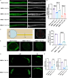
Recombinant IL-4 protein reduced RGC cell death caused by NMDA injury. A: Immunofluorescence staining of retinal ganglion cells in the negative control group, vehicle group and rIL-4 group on day 7 and day 14 showed that the density of RGCs in the rIL-4 group was higher than that in the vehicle group at the same treatment time. B: Schematic diagram of the RGC counting method. A rectangle (red) indicates a high-power field of 0.38 mm×0.285 mm. The number of RGCs in each high-power field was counted, and the mean RGC number in a high-power field was calculated. The proportion of the mean RGC number of the vehicle group or rIL-4 group relative to the mean RGC number of the negative control group was calculated as the relative RGC survival rate. C: The relative RGC survival rate of each group. The relative RGC survival rate of the IL-4 group was greater than that of the vehicle group with the same treatment time. n = 9 eyes per group, *** indicates p < 0.005, **** indicates p < 0.001
Fig. 6
Fig. 6
Recombinant IL-4 protein attenuated NMDA-induced axon degeneration and maintained long-range axon projections in RGCs. A, B: Immunofluorescence images of retinal ganglion cell axons traced by CTB-Alexa Fluor 488. The average relative fluorescence intensity in the rIL-4 group was higher than that in the vehicle group on days 7 and 14. C, D: The cross-sectional area where axons were quantified was 1 cm distal to the injury site. The number of axons labeled with CTB was counted in five areas on a cross section of the optic nerve. Each area was 0.02 × 0.02 (mm2). The mean number of CTB-labeled axons in an area was calculated. E, F: On day 7, the mean axon density labeled by CTB in the rIL-4 group was greater than that in the vehicle group. G, H: CTB labeling in the lateral geniculate body and superior colliculus of mice. On day 7, the relative fluorescence intensity of CTB in the LGN and SC in the contralateral side of rIL-4 treated eyes was significantly higher than that in the contralateral side of vehicle treated eyes. n = 4 nerves or brains, *** indicates p < 0.005, **** indicates p < 0.001
Fig. 7
Fig. 7
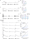
rIL-4 treatment improves ERG performance in mice with NMDA-induced retina injury. A: Representative PhNR waveforms from eyes in blank control group, vehicle group and rIL-4 group, after 7-day NMDA damage. The PhNR was elicited with flash stimulus at an intensity of 10.0 cd·s/m2 and measured from the baseline to the bottom of the trough following the b wave. B: Average amplitudes of PhNR in each group. C: Representative pSTR waveforms in each group. The pSTR was elicited with a flash stimulus at an intensity of 0.001 cd·s/m2 and measured from the baseline to the following positive peak of the waveform. D: Average amplitudes of pSTR in each group. E: Representative OP waveforms in each group. The OPs were elicited with the white flashes of 3.0 cd·s/m2 and recorded via bandpass filtering between 50 and 170 Hz. F: Average amplitudes of OPs (OP1, OP2, and OP3) in each group. G: Representative scotopic ERG responses in response to the increasing flash intensity in each group. The flash intensities used to elicit the responses are presented to the left. H: Average amplitudes of a- and b- waves in scotopic ERG responses, respectively. n = 4 eyes per group, * indicates p < 0.05, ** indicates p < 0.01, *** indicates p < 0.005
Fig. 8
Fig. 8
Recombinant IL-4 protein reduced RGC cell death caused by ONC injury. A: Immunofluorescence staining of retinal ganglion cells in the negative control group, vehicle group and IL-4 group on day 7 and day 14 showed that the density of RGCs in the rIL-4 group was higher than that in the vehicle group at the same treatment time. B: Schematic diagram of the RGC counting method. The calculation method of the relative RGC survival rate was similar to the method of the NMDA model. C: Relative RGC survival rate of each group. The relative RGC survival rate of the rIL-4 group was greater than that of the vehicle group with the same treatment time. n = 9 eyes per group, ** indicates p < 0.01, **** indicates p < 0.001
Fig. 9
Fig. 9
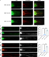
RGC axon regeneration after ONC injury induced by recombinant IL-4 protein. A: CTB fluorescence images and GAP43 immunofluorescence staining images of axons distal to the injury site. In the rIL-4 group, there were axons labeled with both CTB (green) and GAP43 (red) distal to the injury site on days 7 and 14. The length of double-labeled axons distal to the injury site was longer in the rIL-4 treatment group on day 14 than on day 7. B, C: The number of CTB-labeled axons beyond the injury site. The number of regenerated axons with lengths greater than 500 μm beyond the injury site in the rIL-4 group was significantly higher than that on day 7 in the rIL-4 group and that on day 14 in the vehicle group. D, E: The number of axons beyond the injury site traced by GAP43 immunofluorescence staining. The number of regenerated axons with lengths greater than 500 μm beyond the injury site in the rIL-4 group was significantly higher than that on day 7 in the rIL-4 group and that on day 14 in the vehicle group. n = 5 nerves per group, * indicates p < 0.05, ** indicates p < 0.01, **** indicates p < 0.001

Similar articles

Cited by

References

    1. Koh JY, Wie MB, Gwag BJ, Sensi SL, Canzoniero LM, Demaro J, et al. Staurosporine-induced neuronal apoptosis. Exp Neurol. 1995;135(2):153–9. doi: 10.1006/exnr.1995.1074. - DOI - PubMed
    1. Kim YH, Gum SI, Lee TY, Shin JY, Ma JY, Kim I, et al. The neuroprotective effect of staurosporine on mouse retinal ganglion cells after optic nerve injury. Int J Clin Exp Pathol. 2017;10(7):7920–8. - PMC - PubMed
    1. Min JY, Park MH, Park MK, Park KW, Lee NW, Kim T, et al. Staurosporin induces neurite outgrowth through ROS generation in HN33 hippocampal cell lines. J Neural Transm (Vienna) 2006;113(11):1821–6. doi: 10.1007/s00702-006-0500-z. - DOI - PubMed
    1. Wakita S, Izumi Y, Nakai T, Adachi K, Takada-Takatori Y, Kume T, et al. Staurosporine induces dopaminergic neurite outgrowth through AMP-activated protein kinase/mammalian target of rapamycin signaling pathway. Neuropharmacology. 2014;77:39–48. doi: 10.1016/j.neuropharm.2013.09.012. - DOI - PubMed
    1. Kim YH, Chang Y, Jung JC. Staurosporine induces ganglion cell differentiation in part by stimulating urokinase-type plasminogen activator expression and activation in the developing chick retina. Biochem Biophys Res Commun. 2012;423(1):67–72. doi: 10.1016/j.bbrc.2012.05.084. - DOI - PubMed

Publication types

MeSH terms

LinkOut - more resources