Paediatric cutaneous lymphomas including rare subtypes: A 40-year experience at a tertiary referral centre
- PMID: 38650545
- PMCID: PMC11664474
- DOI: 10.1111/jdv.20028
Paediatric cutaneous lymphomas including rare subtypes: A 40-year experience at a tertiary referral centre
Abstract
Background: Primary cutaneous lymphomas are neoplasms of the immune system with a distinct tropism for the skin and an absence of extracutaneous manifestations at the time of diagnosis. Studies focusing on cutaneous lymphomas in children and adolescents remain scarce and often do not encompass the rare subtypes.
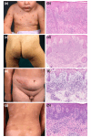
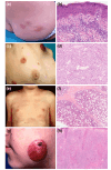
Objectives: To address this knowledge gap by describing the clinical, histological and molecular characteristics of a large group of paediatric patients affected by primary cutaneous lymphoma. We also provided the Paediatric Primary Cutaneous Lymphoma Atlas that illustrates the clinicopathological spectrum of observed presentations, in the hope of supporting other physicians in the diagnostic process.
Methods: Retrospective chart review of paediatric patients diagnosed with primary cutaneous lymphomas between 1980 and 2022 at the Paediatric Dermatology Unit of Fondazione IRCCS Ca' Granda Ospedale Maggiore Policlinico, Milan.
Results: A total of 101 patients (58 males, 43 females) met the inclusion criteria. The most common subtypes were lymphomatoid papulosis (n = 48) and mycosis fungoides (n = 31). These were followed by primary cutaneous CD4+ small/medium T-cell lymphoproliferative disorders (n = 7), primary cutaneous anaplastic large-cell lymphomas (n = 5), primary cutaneous marginal zone B-cell lymphomas (n = 3), primary cutaneous follicle centre lymphomas (n = 2), subcutaneous panniculitis-like T-cell lymphomas (n = 2), primary cutaneous peripheral T-cell lymphoma not otherwise specified (n = 1), primary cutaneous precursor B-lymphoblastic lymphoma (n = 1) and Sézary syndrome (n = 1). Clinical follow-up data covering a median of 70.8 months (range 1-324) were available for 74 patients, of whom three died due to cutaneous lymphoma.
Conclusions: Our findings shed light on the peculiar aspects and long-term outcomes of paediatric cutaneous lymphomas, particularly emphasizing their distinctive features in comparison to their adult counterparts and exploring the less common subtypes. Further larger-scale studies are warranted to better characterize these entities and to achieve a more rapid and accurate diagnosis.
© 2024 The Author(s). Journal of the European Academy of Dermatology and Venereology published by John Wiley & Sons Ltd on behalf of European Academy of Dermatology and Venereology.
Conflict of interest statement
The authors declare no conflicts of interest.
Figures


References
-
- Kempf W, Kazakov DV, Belousova IE, Mitteldorf C, Kerl K. Paediatric cutaneous lymphomas: a review and comparison with adult counterparts. J Eur Acad Dermatol Venereol. 2015;29(9):1696–1709. - PubMed
-
- Dobos G, de Masson A, Ram‐Wolff C, Beylot‐Barry M, Pham‐Ledard A, Ortonne N, et al. Epidemiological changes in cutaneous lymphomas: an analysis of 8593 patients from the French Cutaneous Lymphoma Registry. Br J Dermatol. 2021;184(6):1059–1067. - PubMed
-
- Boccara O, Blanche S, de Prost Y, Brousse N, Bodemer C, Fraitag S. Cutaneous hematologic disorders in children. Pediatr Blood Cancer. 2012;58:226–232. - PubMed
MeSH terms
Supplementary concepts
LinkOut - more resources
Full Text Sources
Medical
Research Materials
Miscellaneous

