. 2024 Apr 8:17:1268013.
doi: 10.3389/fnmol.2024.1268013. eCollection 2024.

Allelic heterogeneity and abnormal vesicle recycling in PLAA-related neurodevelopmental disorders

Affiliations

Allelic heterogeneity and abnormal vesicle recycling in PLAA-related neurodevelopmental disorders

Michele Iacomino et al. Front Mol Neurosci. .

Abstract

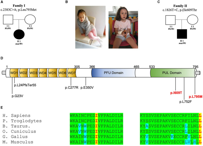
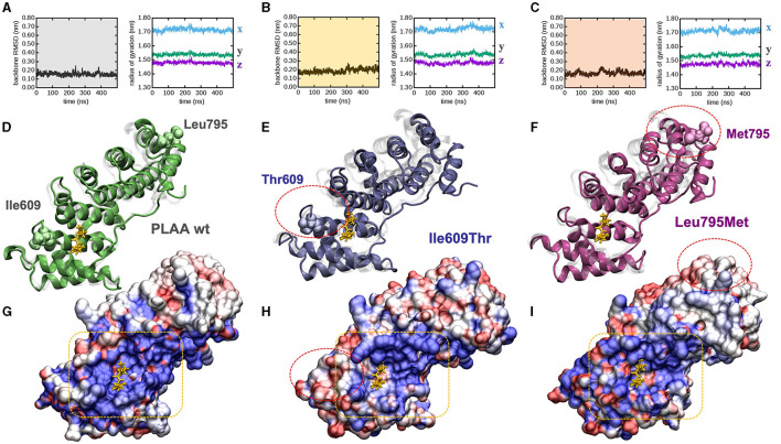
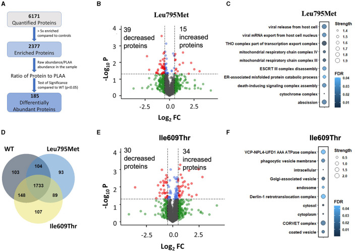
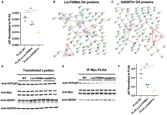
The human PLAA gene encodes Phospholipase-A2-Activating-Protein (PLAA) involved in trafficking of membrane proteins. Through its PUL domain (PLAP, Ufd3p, and Lub1p), PLAA interacts with p97/VCP modulating synaptic vesicles recycling. Although few families carrying biallelic PLAA variants were reported with progressive neurodegeneration, consequences of monoallelic PLAA variants have not been elucidated. Using exome or genome sequencing we identified PLAA de-novo missense variants, affecting conserved residues within the PUL domain, in children affected with neurodevelopmental disorders (NDDs), including psychomotor regression, intellectual disability (ID) and autism spectrum disorders (ASDs). Computational and in-vitro studies of the identified variants revealed abnormal chain arrangements at C-terminal and reduced PLAA-p97/VCP interaction, respectively. These findings expand both allelic and phenotypic heterogeneity associated to PLAA-related neurological disorders, highlighting perturbed vesicle recycling as a potential disease mechanism in NDDs due to genetic defects of PLAA.

Keywords: PLAA gene; SNAREopathies; de novo variants; developmental regression; neurodevelopmental disorders; synaptic transmission.


Conflict of interest statement

The authors declare that the research was conducted in the absence of any commercial or financial relationships that could be construed as a potential conflict of interest.

Figures

Figure 1
Figure 1
PLAA intragenic de novo variants identified in this study. (A) Family 1 pedigree: squares and circles represent males and females, respectively. Closed symbols denote affected individuals. Open symbols denote unaffected individuals. Electropherogram showing heterozygous mutation c.C2383A [p.L795M] and wild-type (wt) sequences. (B) Photos of the patients. (C) Family 2 pedigree: squares and circles represent males and females, respectively. Closed symbols denote affected individuals. Open symbols denote unaffected individuals. Electropherogram showing heterozygous mutation c.T1826C [p.I609T] and wild-type (wt) sequences. (D) Representation of the wild-type (Leu795) PLAP vs. the mutant (Met795) and representation of the wild-type (Ile609) PLAP vs. the mutant (Thr609). (E) Multiple alignment showing PLAA protein complete conservation across species alignment for p.Leu795 and Ile609 aminoacids in red squares.
Figure 2
Figure 2
PLAA molecular modeling and dynamic simulations. (A–C) Molecular dynamics trajectories analysis: backbone root mean squared deviation (left) and x, y, x, components of the backbone radius of gyration (right) for (A) wt, (B) Ile609Thr, and (C) Leu795Met. (D, E) End-simulation configuration for (D) wt, (E) Ile609Thr, and (F) Leu795Met, the configurations are superposed to the crystallographic structure 3EBB, chain A, where PLAA (shaded in gray) is bound to a the p97 fragment (highlighted in yellow). (G–I) Solvent accessible surface area of (G) wt, (H) Ile609Thr, and (I) Leu795Met, also in this case the configurations are superposed to the crystallographic structure 3EBB and the p97 fragment is highlighted (yellow), the surfaces are colored according to the calculated electrostatic potential at each surface point: positive (blue), negative (red), and neutral/hydrophobic (white). For ease of comparison among panels red circles highlight the mutations in (E–I), and a yellow box highlights the PLAA positively charged groove (G–I). All configurations are snapshots taken at 500 ns. Water molecules have been omitted for ease of interpretation.
Figure 3
Figure 3
Mass Spectrometry of wild type, p. Ile609Thr and p. Leu795Met Immunoprecipitation. (A) A flow-through of the mass spectrometry data analysis steps. Briefly, proteins were first filtered based on enrichment relative to controls (>5x), then for each protein, a two-tailed student's t-test was used to compare each of the mutants to the wild type. (B–E) Volcano plot showing the differentially pulled down proteins in the Leu795Met and Ile609Thr samples compared to wild type. Cutoff lines are set to p = 0.05 and |Log2FC|>0.5. (D) Venn diagram of the remaining proteins after filtering, i.e., proteins which are >5x enriched in each of the IP conditions compared to untransfected controls. (C–F) Gene ontology enrichment analysis. Analysis was performed through STRING website. All terms (including BP, CC, and MF) were combined and ranked in descending order based on Strength. The top 10 terms were kept and displayed.
Figure 4
Figure 4
Effect of the two PLAA mutations on the PLAA/p97 interaction. (A) Plot of the p97 abundance (as a ratio to PLAA) from the IP Mass Spectrometry data (*p < 0.05, **p < 0.01). (B, C) Map of the interaction network of the differentially pulled down proteins in the Leu795Met and Ile609Thr samples compared to wild type. The red arrow indicated VCP/p97. The red triangle indicates a network of SNARE associated proteins (D) Western blots of the whole cell lysates from which have undergone immunoprecipitation (E) Western blots from the IP elution samples. (F) Quantification of p97 from the IP Myc-PLAA elution samples. p97 was divided by the amount of PLAA in the corresponding sample (top Myc-PLAA band) before plotting (*p < 0.05, **p < 0.01).

References

    1. American Psychiatric Association (2013). Diagnostic and Statistical Manual of Mental Disorders: DSM-5, 5th Edn. Arlington, VA: American Psychiatric Association.
    1. Azzariti D. R., Hamosh A. (2020). Genomic data sharing for novel mendelian disease gene discovery: the matchmaker exchange. Annu. Rev. Genom. Hum. Genet. 21, 305–326. 10.1146/annurev-genom-083118-014915 - DOI - PubMed
    1. Baldassari S., Musante I., Iacomino M., Zara F., Salpietro V., Scudieri P., et al. . (2020). Brain organoids as model systems for genetic neurodevelopmental disorders. Front. Cell Dev. Biol. 8:590119. 10.3389/fcell.2020.590119 - DOI - PMC - PubMed
    1. Bell S., Rousseau J., Peng H., Aouabed Z., Priam P., Theroux J., et al. . (2019). Mutations in ACTL6B cause neurodevelopmental deficits and epilepsy and lead to loss of dendrites in human neurons. Am. J. Hum. Genet. 104, 815–834. 10.1016/j.ajhg.2019.03.022 - DOI - PMC - PubMed
    1. Coleman J., Jouannot O., Ramakrishnan S. K., Zanetti M. N., Wang J., Salpietro V., et al. . (2018). PRRT2 regulates synaptic fusion by directly modulating SNARE complex assembly. Cell Rep. 22, 820–831. 10.1016/j.celrep.2017.12.056 - DOI - PMC - PubMed

LinkOut - more resources