

Novel Pseudomonas Species Prevent the Growth of the Phytopathogenic Fungus Aspergillus flavus
- PMID: 38651488
- PMCID: PMC11036216
- DOI: 10.3390/biotech13020008
Novel Pseudomonas Species Prevent the Growth of the Phytopathogenic Fungus Aspergillus flavus
Abstract
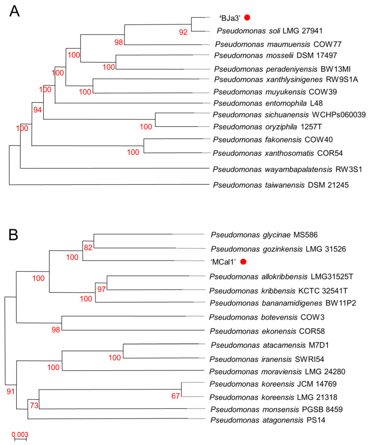
In response to the escalating demand for sustainable agricultural methodologies, the utilization of microbial volatile organic compounds (VOCs) as antagonists against phytopathogens has emerged as a viable eco-friendly alternative. Microbial volatiles exhibit rapid diffusion rates, facilitating prompt chemical interactions. Moreover, microorganisms possess the capacity to emit volatiles constitutively, as well as in response to biological interactions and environmental stimuli. In addition to volatile compounds, these bacteria demonstrate the ability to produce soluble metabolites with antifungal properties, such as APE Vf, pyoverdin, and fragin. In this study, we identified two Pseudomonas strains (BJa3 and MCal1) capable of inhibiting the in vitro mycelial growth of the phytopathogenic fungus Aspergillus flavus, which serves as the causal agent of diseases in sugarcane and maize. Utilizing GC/MS analysis, we detected 47 distinct VOCs which were produced by these bacterial strains. Notably, certain volatile compounds, including 1-heptoxydecane and tridecan-2-one, emerged as primary candidates for inhibiting fungal growth. These compounds belong to essential chemical classes previously documented for their antifungal activity, while others represent novel molecules. Furthermore, examination via confocal microscopy unveiled significant morphological alterations, particularly in the cell wall, of mycelia exposed to VOCs emitted by both Pseudomonas species. These findings underscore the potential of the identified BJa3 and MCal1 Pseudomonas strains as promising agents for fungal biocontrol in agricultural crops.
Keywords: Pseudomonas; bacteria volatile compounds; biological control; microbial antagonistic activity; phytopathogenic fungus.
Conflict of interest statement
Authors are involved in the development of a patent related to the research topic investigated in this paper. RS-R is employed by ByMyCell Inova Simples.
Figures


Similar articles
-
Bacterial volatile organic compounds induce adverse ultrastructural changes and DNA damage to the sugarcane pathogenic fungus Thielaviopsis ethacetica.Environ Microbiol. 2022 Mar;24(3):1430-1453. doi: 10.1111/1462-2920.15876. Epub 2022 Jan 7. Environ Microbiol. 2022. PMID: 34995419
-
Identification of Volatile Organic Compounds Produced by Xenorhabdus indica Strain AB and Investigation of Their Antifungal Activities.Appl Environ Microbiol. 2022 Jul 12;88(13):e0015522. doi: 10.1128/aem.00155-22. Epub 2022 Jun 21. Appl Environ Microbiol. 2022. PMID: 35727028 Free PMC article.
-
Biocontrol Activity of Volatile-Producing Bacillus megaterium and Pseudomonas protegens Against Aspergillus and Penicillium spp. Predominant in Stored Rice Grains: Study II.Mycobiology. 2018 Mar 27;46(1):52-63. doi: 10.1080/12298093.2018.1454015. eCollection 2018. Mycobiology. 2018. PMID: 29998033 Free PMC article.
-
The power of the smallest: The inhibitory activity of microbial volatile organic compounds against phytopathogens.Front Microbiol. 2023 Jan 4;13:951130. doi: 10.3389/fmicb.2022.951130. eCollection 2022. Front Microbiol. 2023. PMID: 36687575 Free PMC article. Review.
-
Impact of Volatile Organic Compounds on the Growth of Aspergillus flavus and Related Aflatoxin B1 Production: A Review.Int J Mol Sci. 2022 Dec 8;23(24):15557. doi: 10.3390/ijms232415557. Int J Mol Sci. 2022. PMID: 36555197 Free PMC article. Review.
Cited by
-
An environmental isolate of Pseudomonas, 20EI1, reduces Aspergillus flavus growth in an iron-dependent manner and alters secondary metabolism.Front Microbiol. 2025 Jan 20;15:1514950. doi: 10.3389/fmicb.2024.1514950. eCollection 2024. Front Microbiol. 2025. PMID: 39902287 Free PMC article.
References
-
- Coomes T.J., Sanders J.C. The Detection and Estimation of Aflatoxin in Groundnuts and Groundnut Materials. Part I. Paper-Chromatographic Procedure. Analyst. 1963;88:209. doi: 10.1039/an9638800209. - DOI
-
- Silva J.J., Puel O., Lorber S., Ferranti L.S., Ortiz L.F., Taniwaki M.H., Iamanaka B.T., Fungaro M.H.P. Occurrence and Diversity of Aspergillus in Commercial Yerba Mate Elaborated for the Brazilian Beverage ‘Chimarrão’. Food Res. Int. 2019;121:940–946. doi: 10.1016/j.foodres.2019.01.023. - DOI - PubMed
Grants and funding
LinkOut - more resources
Full Text Sources
Research Materials
Miscellaneous