Reducing False-Positives Due to Urinary Stagnation in the Prostatic Urethra on 18 F-DCFPyL PSMA PET/CT With MRI
- PMID: 38651785
- PMCID: PMC11150104
- DOI: 10.1097/RLU.0000000000005220
Reducing False-Positives Due to Urinary Stagnation in the Prostatic Urethra on 18 F-DCFPyL PSMA PET/CT With MRI
Abstract
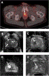
Purpose: Prostate-specific membrane antigen (PSMA)-targeting PET radiotracers reveal physiologic uptake in the urinary system, potentially misrepresenting activity in the prostatic urethra as an intraprostatic lesion. This study examined the correlation between midline 18 F-DCFPyL activity in the prostate and hyperintensity on T2-weighted (T2W) MRI as an indication of retained urine in the prostatic urethra.
Patients and methods: Eighty-five patients who underwent both 18 F-DCFPyL PSMA PET/CT and prostate MRI between July 2017 and September 2023 were retrospectively analyzed for midline radiotracer activity and retained urine on postvoid T2W MRIs. Fisher's exact tests and unpaired t tests were used to compare residual urine presence and prostatic urethra measurements between patients with and without midline radiotracer activity. The influence of anatomical factors including prostate volume and urethral curvature on urinary stagnation was also explored.
Results: Midline activity on PSMA PET imaging was seen in 14 patients included in the case group, whereas the remaining 71 with no midline activity constituted the control group. A total of 71.4% (10/14) and 29.6% (21/71) of patients in the case and control groups had urethral hyperintensity on T2W MRI, respectively ( P < 0.01). Patients in the case group had significantly larger mean urethral dimensions, larger prostate volumes, and higher incidence of severe urethral curvature compared with the controls.
Conclusions: Stagnated urine within the prostatic urethra is a potential confounding factor on PSMA PET scans. Integrating PET imaging with T2W MRI can mitigate false-positive calls, especially as PSMA PET/CT continues to gain traction in diagnosing localized prostate cancer.
Copyright © 2024 Wolters Kluwer Health, Inc. All rights reserved.
Conflict of interest statement
Conflicts of interest and sources of funding: none declared.
Figures


References
-
- Siegel RL, Miller KD, Fuchs HE, Jemal A. Cancer statistics, 2022. CA Cancer J Clin. 2022;72(1):7–33. - PubMed
-
- Wright GL Jr., Haley C, Beckett ML, Schellhammer PF. Expression of prostate-specific membrane antigen in normal, benign, and malignant prostate tissues. Urol Oncol. 1995;1(1):18–28. - PubMed
-
- Wang Y, Galante JR, Haroon A, Wan S, Afaq A, Payne H, et al. The future of PSMA PET and WB MRI as next-generation imaging tools in prostate cancer. Nat Rev Urol. 2022;19(8):475–93. - PubMed
-
- Hofman MS, Lawrentschuk N, Francis RJ, Tang C, Vela I, Thomas P, et al. Prostate-specific membrane antigen PET-CT in patients with high-risk prostate cancer before curative-intent surgery or radiotherapy (proPSMA): a prospective, randomised, multicentre study. Lancet. 2020;395(10231):1208–16. - PubMed
MeSH terms
Substances
Grants and funding
LinkOut - more resources
Full Text Sources
Medical
Miscellaneous

