Review
. 2024 Apr;311(1):e231934.
doi: 10.1148/radiol.231934.

MRI in the Evaluation of Cryptogenic Stroke and Embolic Stroke of Undetermined Source

Affiliations
Review

MRI in the Evaluation of Cryptogenic Stroke and Embolic Stroke of Undetermined Source

Jiayu Xiao et al. Radiology. 2024 Apr.

Abstract

Cryptogenic stroke refers to a stroke of undetermined etiology. It accounts for approximately one-fifth of ischemic strokes and has a higher prevalence in younger patients. Embolic stroke of undetermined source (ESUS) refers to a subgroup of patients with nonlacunar cryptogenic strokes in whom embolism is the suspected stroke mechanism. Under the classifications of cryptogenic stroke or ESUS, there is wide heterogeneity in possible stroke mechanisms. In the absence of a confirmed stroke etiology, there is no established treatment for secondary prevention of stroke in patients experiencing cryptogenic stroke or ESUS, despite several clinical trials, leaving physicians with a clinical dilemma. Both conventional and advanced MRI techniques are available in clinical practice to identify differentiating features and stroke patterns and to determine or infer the underlying etiologic cause, such as atherosclerotic plaques and cardiogenic or paradoxical embolism due to occult pelvic venous thrombi. The aim of this review is to highlight the diagnostic utility of various MRI techniques in patients with cryptogenic stroke or ESUS. Future trends in technological advancement for promoting the adoption of MRI in such a special clinical application are also discussed.


Conflict of interest statement

Disclosures of conflicts of interest: J.X. No relevant relationships. R.A.P. Grant from the Southern California Clinical and Translational Science Institute (UL1TR001855). A.L. No relevant relationships. P.L.N. No relevant relationships. J.W.S. Recipient of the American Heart Association Career Development Award (938082); associate editor for Radiology Advances. N.S. No relevant relationships. A.G.W. Consulting fees from Canon Medical Images. S.S.S. Grants or contracts from the National Institutes of Health–National Institute of Neurological Disorders and Stroke (StrokeNet grant U19NS115388) and Genentech (protocol no. ML40787); consulting fees from Kandu Health; honorarium for Stroke CME lecture; expert witness provided for stroke clinic patient who was in litigation for workers compensation; patents planned, issued, or pending with Cedars-Sinai Medical Center. P.D.L. Consultant for Apex Innovations. J.L.S. No relevant relationships. B.A.W. No relevant relationships. Z.F. No relevant relationships.

Figures

None
Graphical abstract
Illustration shows MRI applications in different areas of the body for
determining the etiology of stroke. ASL = arterial spin labeling, CMR = cardiac
MRI, DSC = dynamic susceptibility contrast, DWI = diffusion-weighted imaging,
FLAIR = fluid-attenuated inversion recovery, MRA = MR angiography, MRV = MR
venography, SWI = susceptibility-weighted imaging, T2WI = T2-weighted imaging,
T2*WI = T2*-weighted imaging, VWI = vessel wall imaging.
Illustration was created using BioRender.
Figure 1:
Illustration shows MRI applications in different areas of the body for determining the etiology of stroke. ASL = arterial spin labeling, CMR = cardiac MRI, DSC = dynamic susceptibility contrast, DWI = diffusion-weighted imaging, FLAIR = fluid-attenuated inversion recovery, MRA = MR angiography, MRV = MR venography, SWI = susceptibility-weighted imaging, T2WI = T2-weighted imaging, T2*WI = T2*-weighted imaging, VWI = vessel wall imaging. Illustration was created using BioRender.
Brain MRI scans show example findings at different stages of ischemic
stroke, with arrows indicating ischemic lesions on various contrast-weighted
images. ADC = apparent diffusion coefficient, DWI = diffusion-weighted imaging,
FLAIR = fluid-attenuated inversion recovery, T1WI = T1-weighted imaging, T2WI =
T2-weighted imaging.
Figure 2:
Brain MRI scans show example findings at different stages of ischemic stroke, with arrows indicating ischemic lesions on various contrast-weighted images. ADC = apparent diffusion coefficient, DWI = diffusion-weighted imaging, FLAIR = fluid-attenuated inversion recovery, T1WI = T1-weighted imaging, T2WI = T2-weighted imaging.
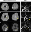
Example diffusion-weighted imaging (DWI) and MR angiography (MRA) in
patients with stroke. (A) Images in a 54-year-old male patient diagnosed with
cryptogenic stroke show multiple bilateral cortical-subcortical and
periventricular infarcts on the DWI scan (arrows, left), while MRA (right) and
transesophageal echocardiography were unrevealing. (B) Images in a 44-year-old
male patient with stroke attributed to large artery atherosclerosis show
multiple unilateral small subcortical infarcts on the DWI scan (arrows, left)
and severe focal stenosis of the ipsilateral middle cerebral artery on the MR
angiogram (arrowhead, right). (C) Images in an 82-year-old male patient with
stroke attributed to a cardioembolic source show large territorial infarcts on
the DWI scan (arrows, left) and occlusion of the left posterior cerebral artery
on the MR angiogram (arrowhead, right).
Figure 3:
Example diffusion-weighted imaging (DWI) and MR angiography (MRA) in patients with stroke. (A) Images in a 54-year-old male patient diagnosed with cryptogenic stroke show multiple bilateral cortical-subcortical and periventricular infarcts on the DWI scan (arrows, left), while MRA (right) and transesophageal echocardiography were unrevealing. (B) Images in a 44-year-old male patient with stroke attributed to large artery atherosclerosis show multiple unilateral small subcortical infarcts on the DWI scan (arrows, left) and severe focal stenosis of the ipsilateral middle cerebral artery on the MR angiogram (arrowhead, right). (C) Images in an 82-year-old male patient with stroke attributed to a cardioembolic source show large territorial infarcts on the DWI scan (arrows, left) and occlusion of the left posterior cerebral artery on the MR angiogram (arrowhead, right).
MRI in a 58-year-old female patient initially diagnosed with
cryptogenic stroke. (A) Diffusion-weighted image shows multiple unilateral
infarcts in the distribution of the left middle cerebral artery. (B) MR
angiogram shows a failure to identify stenosis. (C) Precontrast vessel wall
images (parallel and perpendicular views) show a nonstenotic plaque
(arrowhead) in the left middle cerebral artery, with focal eccentric wall
thickening and positive remodeling (corresponding to the arrow in B). (D)
Postcontrast vessel wall images show the plaque with strong enhancement
(arrowhead). The etiology was subsequently reclassified as intracranial
large artery atherosclerosis.
Figure 4:
MRI in a 58-year-old female patient initially diagnosed with cryptogenic stroke. (A) Diffusion-weighted image shows multiple unilateral infarcts in the distribution of the left middle cerebral artery. (B) MR angiogram shows a failure to identify stenosis. (C) Precontrast vessel wall images (parallel and perpendicular views) show a nonstenotic plaque (arrowhead) in the left middle cerebral artery, with focal eccentric wall thickening and positive remodeling (corresponding to the arrow in B). (D) Postcontrast vessel wall images show the plaque with strong enhancement (arrowhead). The etiology was subsequently reclassified as intracranial large artery atherosclerosis.
Brain MRI in a 71-year-old male patient diagnosed with cryptogenic
stroke. (A) T2*-weighted image shows the susceptibility vessel sign
(arrow) in the M2 segment of the right middle cerebral artery. (B)
Diffusion-weighted image shows the corresponding ischemic area.
Figure 5:
Brain MRI in a 71-year-old male patient diagnosed with cryptogenic stroke. (A) T2*-weighted image shows the susceptibility vessel sign (arrow) in the M2 segment of the right middle cerebral artery. (B) Diffusion-weighted image shows the corresponding ischemic area.
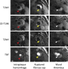
MR images of carotid plaque show the complicated characteristics of
American Heart Association (AHA) type VI plaques. The presence of at least one
of the following criteria defines plaques as type VI: intraplaque hemorrhage
(red arrowheads, left), ruptured fibrous cap (yellow arrowheads, middle), or
mural thrombus indicating juxtaluminal hemorrhage (white arrowheads, right). CE
= contrast-enhanced, TOF = time of flight, T1WI = T1-weighted imaging, T2WI =
T2-weighted imaging. (Adapted, with permission, from reference 48.)
Figure 6:
MR images of carotid plaque show the complicated characteristics of American Heart Association (AHA) type VI plaques. The presence of at least one of the following criteria defines plaques as type VI: intraplaque hemorrhage (red arrowheads, left), ruptured fibrous cap (yellow arrowheads, middle), or mural thrombus indicating juxtaluminal hemorrhage (white arrowheads, right). CE = contrast-enhanced, TOF = time of flight, T1WI = T1-weighted imaging, T2WI = T2-weighted imaging. (Adapted, with permission, from reference .)
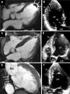
Example contrast-enhanced cardiac MRI (CMR) and conventional
transthoracic echocardiography for detecting left ventricular (LV) thrombi
in patients diagnosed with embolic stroke of undetermined source. (A, B)
Images in an 85-year-old male patient show a large apical thrombus (arrows)
on both the CMR scan (A) and the transthoracic echocardiogram (B). (C, D)
Images in an 82-year-old male patient show a small apical thrombus on the
CMR scan (arrow in C) but not on the transthoracic echocardiogram (D)
because the apical view image was diminished by an artifact. (E, F) Images
in a 74-year-old male patient show a mural thrombus of the posterior wall on
the CMR scan (arrows in E) but not on the transthoracic echocardiogram (F).
Ao = ascending aorta, LA = left atrium. (Reprinted, with permission, from
reference 56.)
Figure 7:
Example contrast-enhanced cardiac MRI (CMR) and conventional transthoracic echocardiography for detecting left ventricular (LV) thrombi in patients diagnosed with embolic stroke of undetermined source. (A, B) Images in an 85-year-old male patient show a large apical thrombus (arrows) on both the CMR scan (A) and the transthoracic echocardiogram (B). (C, D) Images in an 82-year-old male patient show a small apical thrombus on the CMR scan (arrow in C) but not on the transthoracic echocardiogram (D) because the apical view image was diminished by an artifact. (E, F) Images in a 74-year-old male patient show a mural thrombus of the posterior wall on the CMR scan (arrows in E) but not on the transthoracic echocardiogram (F). Ao = ascending aorta, LA = left atrium. (Reprinted, with permission, from reference .)
(A) Three-dimensional aortic wall image in an 86-year-old male patient
with cryptogenic stroke shows the region of atherosclerotic plaque (arrow)
in the proximal descending aorta (DAo). (B) Images show four-dimensional
flow MRI for assessing aortic flow reversal. The direction of forward and
reverse flow is determined for each voxel. Then forward flow, reverse flow,
and the flow reversal fraction (FRF) are determined for each voxel during
the cardiac cycle and quantified as mean parametric maps. Retrograde flow
provides an explanation for how plaque in the descending aorta causes
ischemic stroke. Color bars in the forward flow and reverse flow panels
indicate the mean flow velocity in each voxel (milliliters per cardiac
cycle). The color bar in the flow reversal fraction panel indicates the
level of the flow reversal fraction. AAo = ascending aorta. (Adapted, with
permission, from reference 69.)
Figure 8:
(A) Three-dimensional aortic wall image in an 86-year-old male patient with cryptogenic stroke shows the region of atherosclerotic plaque (arrow) in the proximal descending aorta (DAo). (B) Images show four-dimensional flow MRI for assessing aortic flow reversal. The direction of forward and reverse flow is determined for each voxel. Then forward flow, reverse flow, and the flow reversal fraction (FRF) are determined for each voxel during the cardiac cycle and quantified as mean parametric maps. Retrograde flow provides an explanation for how plaque in the descending aorta causes ischemic stroke. Color bars in the forward flow and reverse flow panels indicate the mean flow velocity in each voxel (milliliters per cardiac cycle). The color bar in the flow reversal fraction panel indicates the level of the flow reversal fraction. AAo = ascending aorta. (Adapted, with permission, from reference .)
(A) Illustration of May-Thurner syndrome, which is clinically associated
with cryptogenic stroke, shows the left iliac vein compressed by the right iliac
artery. (B) Two-dimensional time-of-flight MR angiographic images in a
30-year-old female patient diagnosed with cryptogenic stroke show narrowing of
the left iliac vein caused by the overlying right iliac artery, consistent with
May-Thurner syndrome.
Figure 9:
(A) Illustration of May-Thurner syndrome, which is clinically associated with cryptogenic stroke, shows the left iliac vein compressed by the right iliac artery. (B) Two-dimensional time-of-flight MR angiographic images in a 30-year-old female patient diagnosed with cryptogenic stroke show narrowing of the left iliac vein caused by the overlying right iliac artery, consistent with May-Thurner syndrome.

References

    1. Adams HP Jr , Bendixen BH , Kappelle LJ , et al. . Classification of subtype of acute ischemic stroke. Definitions for use in a multicenter clinical trial. TOAST. Trial of Org 10172 in Acute Stroke Treatment . Stroke 1993. ; 24 ( 1 ): 35 – 41 . - PubMed
    1. Elgendy AY , Saver JL , Amin Z , et al. . Proposal for Updated Nomenclature and Classification of Potential Causative Mechanism in Patent Foramen Ovale-Associated Stroke . JAMA Neurol 2020. ; 77 ( 7 ): 878 – 886 . - PubMed
    1. Hart RG , Diener HC , Coutts SB , et al. . Embolic strokes of undetermined source: the case for a new clinical construct . Lancet Neurol 2014. ; 13 ( 4 ): 429 – 438 . - PubMed
    1. Ntaios G . Embolic Stroke of Undetermined Source: JACC Review Topic of the Week . J Am Coll Cardiol 2020. ; 75 ( 3 ): 333 – 340 . - PubMed
    1. Hart RG , Sharma M , Mundl H , et al. . Rivaroxaban for Stroke Prevention after Embolic Stroke of Undetermined Source . N Engl J Med 2018. ; 378 ( 23 ): 2191 – 2201 . - PubMed

MeSH terms

LinkOut - more resources