. 2024 Apr 23;9(10):e178372.
doi: 10.1172/jci.insight.178372.

A tryptophan-derived uremic metabolite/Ahr/Pdk4 axis governs skeletal muscle mitochondrial energetics in chronic kidney disease

Affiliations

A tryptophan-derived uremic metabolite/Ahr/Pdk4 axis governs skeletal muscle mitochondrial energetics in chronic kidney disease

Trace Thome et al. JCI Insight. .

Abstract

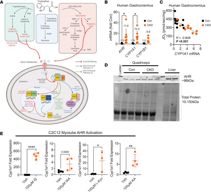
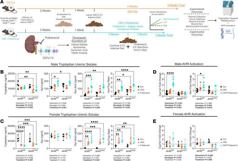
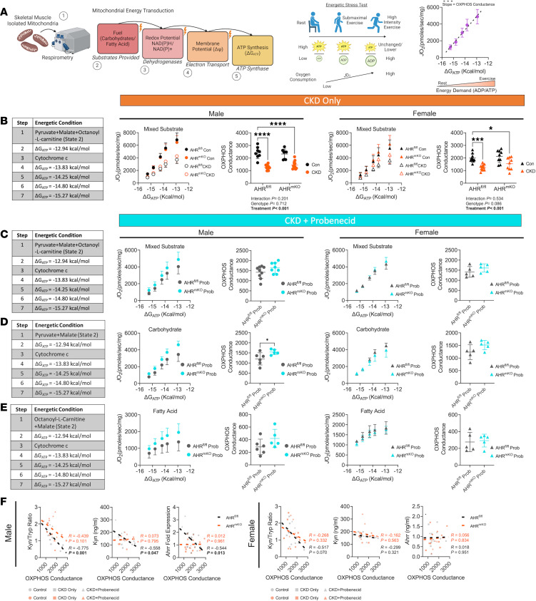
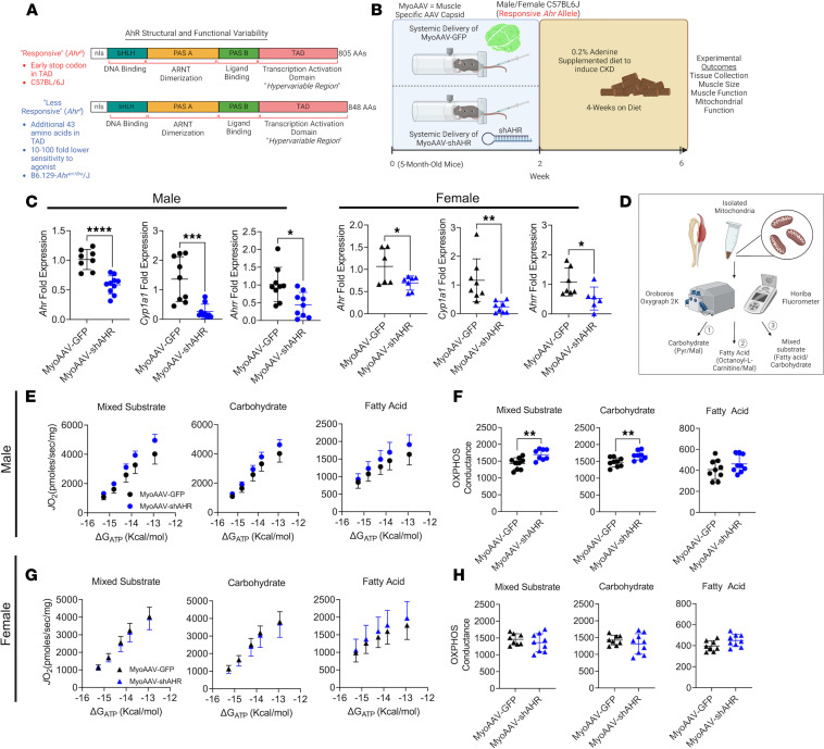
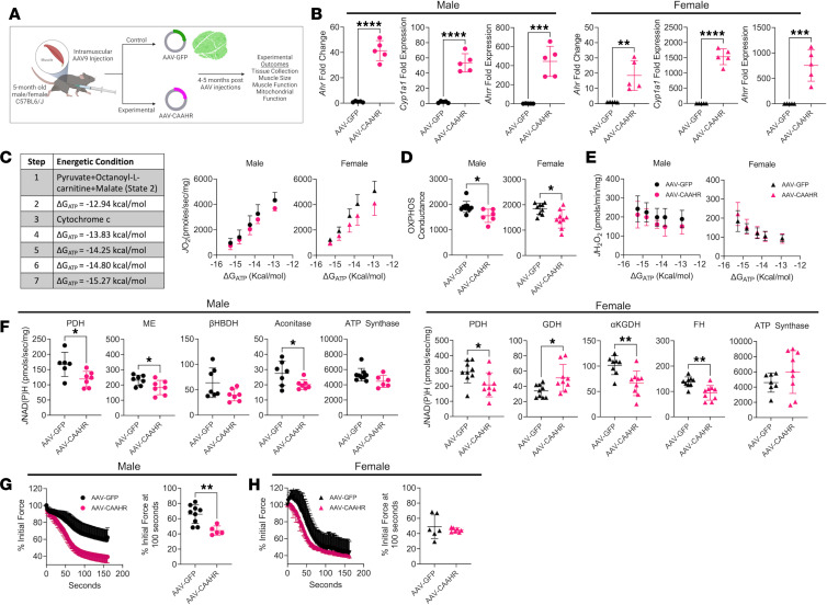
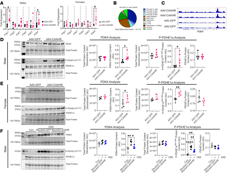
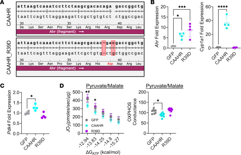
Chronic kidney disease (CKD) causes accumulation of uremic metabolites that negatively affect skeletal muscle. Tryptophan-derived uremic metabolites are agonists of the aryl hydrocarbon receptor (AHR), which has been shown to be activated in CKD. This study investigated the role of the AHR in skeletal muscle pathology of CKD. Compared with controls with normal kidney function, AHR-dependent gene expression (CYP1A1 and CYP1B1) was significantly upregulated in skeletal muscle of patients with CKD, and the magnitude of AHR activation was inversely correlated with mitochondrial respiration. In mice with CKD, muscle mitochondrial oxidative phosphorylation (OXPHOS) was markedly impaired and strongly correlated with the serum level of tryptophan-derived uremic metabolites and AHR activation. Muscle-specific deletion of the AHR substantially improved mitochondrial OXPHOS in male mice with the greatest uremic toxicity (CKD + probenecid) and abolished the relationship between uremic metabolites and OXPHOS. The uremic metabolite/AHR/mitochondrial axis in skeletal muscle was verified using muscle-specific AHR knockdown in C57BL/6J mice harboring a high-affinity AHR allele, as well as ectopic viral expression of constitutively active mutant AHR in mice with normal renal function. Notably, OXPHOS changes in AHRmKO mice were present only when mitochondria were fueled by carbohydrates. Further analyses revealed that AHR activation in mice led to significantly increased pyruvate dehydrogenase kinase 4 (Pdk4) expression and phosphorylation of pyruvate dehydrogenase enzyme. These findings establish a uremic metabolite/AHR/Pdk4 axis in skeletal muscle that governs mitochondrial deficits in carbohydrate oxidation during CKD.

Keywords: Chronic kidney disease; Mitochondria; Muscle biology; Nephrology; Skeletal muscle.


Conflict of interest statement

Conflict of interest: The authors have declared that no conflicts of interest exist.

Figures

Figure 1
Figure 1. AHR activation is present in CKD skeletal muscle and associates with mitochondrial respiratory function.
(A) Graphical depiction of tryptophan metabolism and the AHR signaling pathway. (B) qPCR quantification of AHR, CYP1A1, and CYP1B1 mRNA signaling in gastrocnemius muscle biopsies from patients without (n = 5) and with CKD (n = 8–10). (C) Relationship between muscle mitochondrial oxygen consumption (JO2) and CYP1A1 in patients with and without CKD. (D) Immunoblotting of the AHR protein in skeletal muscle of mice. (E) qPCR quantification of Cyp1a1 mRNA levels in C2C12 myotubes treated with tryptophan-derived uremic metabolites indoxyl sulfate (IS), indole-3-acetic acid (IAA), L-kynurenine (L-Kyn), and kynurenic acid (KA) (n = 3–4 biological replicates/group). Statistical analyses performed using 2-tailed Student’s t test. Data are shown as mean ± SD. *P < 0.05, **P < 0.01, and ****P < 0.0001.
Figure 2
Figure 2. Uremic metabolite accumulation drives AHR activation in CKD muscle, which is abolished by muscle-specific AHR deletion.
(A) Experimental treatment timeline. (B) Concentrations of tryptophan-derived uremic metabolites in plasma from male AHRfl/fl and AHRmKO mice without CKD, with CKD, and with CKD plus daily probenecid treatment (n = 4–5/group/genotype). (C) Concentrations of tryptophan-derived uremic metabolites in plasma from female AHRfl/fl and AHRmKO mice without CKD, with CKD, and with CKD plus daily probenecid treatment (n = 4–5/group/genotype). (D) qPCR quantification of Cyp1a1 and Ahrr levels in skeletal muscle of male AHRfl/fl and AHRmKO mice without CKD, with CKD, and with CKD plus daily probenecid treatment (n = 5–7/group/genotype). (E) qPCR quantification of Cyp1a1 and Ahrr levels in skeletal muscle of female AHRfl/fl and AHRmKO mice without CKD, with CKD, and with CKD plus daily probenecid treatment (n = 5–6/group/genotype). Statistical analyses performed using 2-way ANOVA with Dunnett’s post hoc testing for multiple comparisons. Data are shown as mean ± SD. *P < 0.05, **P < 0.01, ***P < 0.001, and ****P < 0.0001.
Figure 3
Figure 3. Muscle-specific AHR deletion improves mitochondrial OXPHOS with high tryptophan-derived uremic metabolite levels.
(A) Graphical depiction of mitochondrial OXPHOS system and the use of a creatine kinase clamp to measure oxygen consumption (JO2) across physiologically relevant energetic demands (ΔGATP). (B) Experimental conditions quantification JO2 at each level of ΔGATP, as well as the OXPHOS conductance in male and female AHRfl/fl and AHRmKO mice with or without CKD (n = 8–12/group/genotype). (CE) Experimental conditions and quantification JO2 at each level of ΔGATP, as well as the OXPHOS conductance in male and female AHRfl/fl and AHRmKO mice with CKD plus daily probenecid treatment (n = 5–9/group/genotype) for mixed substrates (C), pyruvate/malate (D), and octanoylcarnitine/malate (E). (F) Pearson correlational analyses of quantified OXPHOS conductance (mixed substrates) and kynurenine to tryptophan ratio, kynurenine concentrations, and Ahrr mRNA in male and female AHRfl/fl and AHRmKO mice across control, CKD, and CKD plus probenecid daily. Data were analyzed by 2-way ANOVA with Dunnett’s post hoc testing for multiple comparisons in B. Two-tailed Student’s t test was performed in CE. Data are shown as mean ± SD. *P < 0.05, ***P < 0.001, and ****P < 0.0001.
Figure 4
Figure 4. Muscle-specific AHR knockdown improves mitochondrial OXPHOS in mice harboring the high-affinity AHR allele.
(A) Graphical depiction of polymorphisms in the AHR that confer differences in ligand affinity. (B) Experimental timeline of delivery of MyoAAV-GFP or MyoAAV-shAHR in high-affinity C57BL/6J mice with CKD. (C) qPCR validation of Ahr knockdown and subsequent reduction in Cyp1a1 and Ahrr mRNA induction in MyoAAV-shAHR mice (n = 6–10/group). (D) Graphical depiction of analytical approach for mitochondrial OXPHOS assessments. (E) Relationship between JO2 and ΔGATP in isolated mitochondria from the gastrocnemius muscle in different substrate conditions in male mice with CKD (n = 8–9/group). (F) Quantification of OXPHOS conductance in male mice (n = 8–9/group). (G) Relationship between JO2 and ΔGATP in isolated mitochondria from the gastrocnemius muscle in different substrate conditions in female mice with CKD (n = 8–9/group). (H) Quantification of OXPHOS conductance in female mice (n = 8–9/group). Statistical analyses were performed using 2-tailed Student’s t test. Data are shown as mean ± SD. *P < 0.05, **P < 0.01, ***P < 0.001, and ****P < 0.0001.
Figure 5
Figure 5. Ectopic expression of a constitutively active AHR impairs muscle mitochondrial OXPHOS in mice with normal kidney function.
(A) Experimental design for muscle-specific delivery of mutant constitutively active AHR (CAAHR). (B) qPCR of Ahr, Cyp1a1, and Ahrr in male and female mice treated with AAV-GFP and AAV-CAAHR (n = 5/group). (C) Substrate conditions and quantification of the relationship between JO2 and ΔGATP in male and female mice treated with AAV-GFP or AAV-CAAHR (n = 6–10/group). (D) OXPHOS conductance in male and female mice (n = 6–10/group). (E) Mitochondrial JH2O2 and ΔGATP in male and female mice (n = 6–10/group). (F) Quantification of mitochondrial matrix dehydrogenase enzyme activity in male and female mice (n = 6–9/group). (G and H) Analysis of extensor digitorum longus muscle fatigue in male and female mice (n = 5–9/group). Data analyzed using 2-tailed Student’s t test. Data are shown as mean ± SD. *P < 0.05, **P < 0.01, ***P < 0.001, and ****P < 0.0001.
Figure 6
Figure 6. AHR activation increased PDK4 expression and PDH phosphorylation.
(A) qPCR of Pdp1, Pdp2, Pdk1, Pdk2, Pdk3, and Pdk4 in male and female mice treated with AAV-GFP and AAV-CAAHR (n = 5–6/group). (B) Peak annotation pie charts for ATAC-Seq peaks in AAV-GFP versus AAV-CAAHR muscles (n = 3/group). (C) IGV snapshots of the Pdk4 gene showing chromatin accessibility, with the red-dashed box highlighting the promoter region. (D) Western blotting of PDK4, phosphorylated PDHE1αSer300, and total PDHE1α protein expression in male AAV-GFP or AAV-CAAHR gastrocnemius muscle (n = 4–5/group). (E) Western blotting of PDK4, phosphorylated PDHE1αSer300, and total PDHE1α protein expression in female AAV-GFP or AAV-CAAHR gastrocnemius muscle (n = 5/group). (F) Western blotting of PDK4, phosphorylated PDHE1αSer300, and total PDHE1α protein expression in male control, CKD MyoAAV-GFP, and CKD MyoAAV-shAHR gastrocnemius muscle (n = 4/group). Data in A, D, and E were analyzed using 2-tailed Student’s t test. Data in F were analyzed using 1-way ANOVA with Tukey’s post hoc test. *P < 0.05, **P < 0.01, and ****P < 0.0001.
Figure 7
Figure 7. Expression of a transcriptionally inept CAAHR abolishes Pdk4 expression and pyruvate-supported OXPHOS impairment in C2C12 muscle cells.
(A) Sequencing results demonstrating the introduction of point mutation that converted arginine-39 to aspartate (R39D). (B) qPCR validation of the overexpression of Ahr and lack of transcriptional activity (Cyp1a1) in the R39D mutant. A GFP control plasmid was also tested (n = 4/group). (C) Pdk4 mRNA expression (fold GFP) (n = 4/group). (D) Pyruvate supported respiration in muscle cells and quantified OXPHOS conductance (n = 8/group). Data are shown as mean ± SD. Data were analyzed using 1-way ANOVA with Tukey’s post hoc test. *P < 0.05, ***P < 0.001, and ****P < 0.0001. #P < 0.05 indicates CAAHR versus R39D.

References

    1. Global Burden of Disease Study 2013 Collaborators Global, regional, and national incidence, prevalence, and years lived with disability for 301 acute and chronic diseases and injuries in 188 countries, 1990-2013: a systematic analysis for the Global Burden of Disease Study 2013. Lancet. 2015;386(9995):743–800. doi: 10.1016/S0140-6736(15)60692-4. - DOI - PMC - PubMed
    1. Gamboa JL, et al. Skeletal muscle mitochondrial dysfunction is present in patients with CKD before initiation of maintenance hemodialysis. Clin J Am Soc Nephrol. 2020;15(7):926–936. doi: 10.2215/CJN.10320819. - DOI - PMC - PubMed
    1. Gregg LP, et al. Fatigue in CKD: epidemiology, pathophysiology, and treatment. Clin J Am Soc Nephrol. 2021;16(9):1445–1455. doi: 10.2215/CJN.19891220. - DOI - PMC - PubMed
    1. Kestenbaum B, et al. Impaired skeletal muscle mitochondrial bioenergetics and physical performance in chronic kidney disease. JCI Insight. 2020;5(5):e133289. doi: 10.1172/jci.insight.133289. - DOI - PMC - PubMed
    1. Mori K. Maintenance of skeletal muscle to counteract sarcopenia in patients with advanced chronic kidney disease and especially those undergoing hemodialysis. Nutrients. 2021;13(5):1538. doi: 10.3390/nu13051538. - DOI - PMC - PubMed

MeSH terms

Substances