. 2024 May 28;43(5):114122.
doi: 10.1016/j.celrep.2024.114122. Epub 2024 Apr 22.

HSV-1 employs UL56 to antagonize expression and function of cGAMP channels

Affiliations

HSV-1 employs UL56 to antagonize expression and function of cGAMP channels

Henry T W Blest et al. Cell Rep. .

Abstract

DNA sensing is important for antiviral immunity. The DNA sensor cGAS synthesizes 2'3'-cyclic GMP-AMP (cGAMP), a second messenger that activates STING, which induces innate immunity. cGAMP not only activates STING in the cell where it is produced but cGAMP also transfers to other cells. Transporters, channels, and pores (including SLC19A1, SLC46A2, P2X7, ABCC1, and volume-regulated anion channels (VRACs)) release cGAMP into the extracellular space and/or import cGAMP. We report that infection with multiple human viruses depletes some of these cGAMP conduits. This includes herpes simplex virus 1 (HSV-1) that targets SLC46A2, P2X7, and the VRAC subunits LRRC8A and LRRC8C for degradation. The HSV-1 protein UL56 is necessary and sufficient for these effects that are mediated at least partially by proteasomal turnover. UL56 thereby inhibits cGAMP uptake via VRAC, SLC46A2, and P2X7. Taken together, HSV-1 antagonizes intercellular cGAMP transfer. We propose that this limits innate immunity by reducing cell-to-cell communication via the immunotransmitter cGAMP.

Keywords: CP: Immunology; CP: Microbiology; DNA sensing; LRRC8A; P2X7; SLC46A2; STING; UL56; VRAC; cGAMP; cGAMP transport; herpes simplex virus.


Conflict of interest statement

Declaration of interests The authors declare no competing interests.

Figures

Figure 1
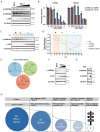
Figure 1. Viral infection affects protein levels of cGAMP channels, transporters and pores.
(A) HEK293 cells were infected with HSV-1 (MOI – 0.02, 0.006, 0.002), VACV (MOI – 0.2, 0.066, 0.022), AdV (MOI – 1500, 150, 15), HIV-1 (1:10, 1:50, 1:250), HIV-2 (1:10, 1:50, 1:250), ZIKV (MOI – 1, 0.25, 0.06) and IAV (MOI – 10, 5, 2.5). 48 hours later, cells were lysed, and abundance of the indicated proteins was assessed by western blot. β-Actin served as a loading control. (B) Summary of the protein levels of cGAMP conduits following infection with different viruses. Data in (A) are representative of two (AdV) and three biological repeats (all other viruses) and the numbers provided in (B) indicate the number of biological repeats for each protein tested for the indicated infections. See also Figures S1-S3.
Figure 2
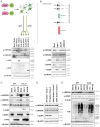
Figure 2. HSV-1 targets VRAC.
(A) HEK293 cells were infected with HSV-1 (MOI = 0.06, 0.12, 0.25, 0.5 and 1). After 24 hours, cells were lysed, and the indicated proteins were detected by western blot. β-Actin served as a loading control. The arrow indicates a faster migrating band detected by the α-LRRC8C antibody. (B) Total RNA was extracted from cells infected in (A). LRRC8A and LRRC8C mRNA levels were determined by RT-qPCR and normalised to GAPDH mRNA. The western blot in (A) was quantified by densitometry. Data were set to 1 in mock infected cells (M). (C, D) HEK293 cells were infected with HSV-1 (MOI = 1) and lysed at the indicated time points. Samples were analysed as in (A) by western blot (C), which was quantified by densitometry (D). Data for LRRC8C are from Figure S4A. (E) Schematic showing phylogeny of human herpes viruses. (F) HEK293T cells were infected with HSV-2 (MOI = 1, 3, 5 and 10). After 24 hours, cells were lysed, and the indicated proteins were detected by western blot. (G) VZV-infected MeWo cells were co-cultured with uninfected MeWo cells at a ratio of 1:5 (infected : uninfected) for one hour. Infected inoculum cells were then washed off and cells were incubated for 24 hours. The indicated proteins were detected by western blot in cell lysates. (H) Overview showing how three candidate genes were identified amongst the 84 genes encoded by HSV-1. Data in (A), (B) and (G) are representative of three biological repeats. In (B), data points show technical triplicates of the RT-qPCR. Data in (C), (D) and (F) are representative of two biological repeats. See also Figures S4 and S5 and Tables S1 and S2.
Figure 3
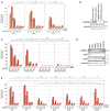
Figure 3. UL56 is necessary and sufficient for LRRC8A and LRRC8C degradation.
(A) HEK293T cells were transiently transfected with expression plasmids encoding UL56-GFP or UL6-GFP. 24 hours later, GFP- and GFP+ cells were enriched by FACS. The gating strategy is shown in Figure S6. Untreated cells (Mock), lipofectamine treated cells (Lipo) and HSV-1 infected cells (MOI = 3, 24 hours) were included as controls without sorting. The experimental strategy is shown (top) and samples were analysed by western blot using the indicated antibodies (bottom). β-Actin served as a loading control. (B) Motifs and domains found in UL56 are shown (top). RR, arginine-rich region; TM, transmembrane domain. HEK293 cells were infected with HSV-1 of the indicated genotypes (MOI = 3, 24 hours). Cell lysates were analysed by western blot. (C) HFFs or HaCaT cells were infected with HSV-1 of the indicated genotypes (MOI = 1, 48 hours). Cell lysates were analysed by western blot. (D) Gene targeting in HEK293 cells was performed using CRISPR-Cas9 with non-targeting (NT) or GOPC-targeting sgRNAs. Clonal cell lines were obtained and analysed by immunoblot. (E) HEK293T cells stably transduced with GFP or UL56 were treated with either DMSO, lactacystin (20 μM) or MG132 (5 μM) for 24 hours. Cell lysates were then analysed by western blot. Data are representative of three biological repeats. See also Figure S6.
Figure 4
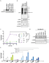
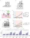
Figure 4. UL56 inhibits VRAC-mediated cGAMP uptake.
(A) HEK293T cells were transiently transfected with three plasmids: p125-FLuc (IFNβ promoter reporter), pRL-TK (constitutively expresses Renilla luciferase) and pcDNA3.2-STING. After 24 hours, cells were incubated in low salt (NaCl) buffers containing cGAMP at the indicated concentrations. Where indicated, cells were also treated with vehicle control (DMSO) or DCPIB (20 μM). After one hour, these buffers were removed and replaced with medium. After an additional 24 hours, FLuc activity was measured and normalised to Renilla luciferase activity. The averages of all 150 mM NaCl / 0 μM cGAMP conditions was then set to 1. (B) HEK293 LRRC8A knockout clonal cell lines were generated using CRISPR-Cas9 and lysates were analysed by western blot using the indicated antibodies. (C) The cell lines shown in (B) were treated and analysed as in (A). (D) HEK293T stably transduced with GFP, UL56 or the indicated UL56 mutants were lysed and analysed by western blot using the indicated antibodies. (E) The cells shown in (D) were treated and analysed as in (A). Data are representative of three independent biological repeats. In (A), (C) and (E), averages of three technical replicates from one experiment are shown, and statistical analysis was done using grouped two-way ANOVA – Tukey. *=p<0.05, **=p<0.01, ***=p<0.001, ****=p<0.0001, ns=not significant. See also Figure S7.
Figure 5
Figure 5. UL56 inhibits SLC46A2 mediated cGAMP uptake.
(A) HEK293T cells stably transduced with SLC46A2-V5 were treated and analysed as described in Figure 3A. (B) HEK293T-SLC46A2-V5 were infected and analysed as described in Figure 3B. (C) HEK293T-SLC46A2-V5 cells were transfected as described in Figure 4A. After 24 hours, cGAMP was added to the medium at the indicated concentrations. Cells were also treated with vehicle control (DMSO) or SSZ (1 mM). After an additional 24 hours, IFNβ promoter reporter induction was assessed as in Figure 4A, setting the averages of all mock conditions to 1. (D) HEK293T-SLC46A2-V5 cells were additionally transduced with GFP, UL56 or the indicated UL56 mutants. Cell lysates were analysed by western blot using the indicated antibodies. (E) The cells shown in (D) were treated and analysed as in (C). Data in (A) and (D) are representative of two independent biological repeats. Data in (B), (C) and (E) are representative of three independent biological repeats. In (C) and (E), averages of three technical replicates from one experiment are shown with SD, and statistical analysis was done using grouped two-way ANOVA – Tukey. *=p<0.05, **=p<0.01, ***=p<0.001, ****=p<0.0001, ns=not significant. See also Figures S6 and S7.
Figure 6
Figure 6. UL56 inhibits P2X7’s activity.
(A) HEK293T cells stably transduced with P2X7-V5 were treated and analysed as described in Figure 3A. (B) HEK293T cells stably transduced with P2X7-V5 were infected and analysed as described in Figure 3B. (C) Schematic showing YO-PRO-1 uptake through P2X7. (D) HEK293T cells stably expressing GFP or P2X7-V5 were incubated YO-PRO-1 and green fluorescence was measured at 45 second intervals. After 10 minutes (vertical grey line), BzATP (80 µM) was added or not. P2X7-V5 cells were also treated with A74003 (100 mM) or DMSO as indicated. Fluorescence signals were normalised to the average fluorescence before BzATP was added (F/F0). (E) HEK293T-P2X7-V5 cells were additionally transduced with GFP, UL56 or the indicated UL56 mutants. Cell lysates were analysed by western blot using the indicated antibodies. (F) The cells shown in (E) were treated and analysed as in (D). (G) HEK293T-P2X7-V5 cells were transfected as described in Figure 4A. After 24 hours, BzATP and cGAMP were added to the medium at the indicated concentrations. After an additional 24 hours, IFNβ promoter reporter induction was assessed as in Figure 4A. The averages of all 0 μM BzATP / 0 μM cGAMP conditions was set to 1. Data are representative of three independent biological repeats. In (D) and (F) data points show averages of 3 technical replicates with SD, and statistical analysis was done using grouped two-way ANOVA – Tukey. Statistical analysis in (D), (F) and (G) was done using grouped two-way ANOVA – Tukey. *=p<0.05, **=p<0.01, ***=p<0.001, ****=p<0.0001, ns=not significant. See also Figures S6 and S7.

References

    1. Medzhitov R. Approaching the asymptote: 20 years later. Immunity. 2009;30:766–775. - PubMed
    1. Sun L, Wu J, Du F, Chen X, Chen ZJ. Science (New York, NY) American Association for the Advancement of Science; 2013. Cyclic GMP-AMP Synthase Is a Cytosolic DNA Sensor That Activates the Type I Interferon Pathway. - DOI - PMC - PubMed
    1. Mankan AK, Schmidt T, Chauhan D, Goldeck M, Honing K, Gaidt M, Kubarenko AV, Andreeva L, Hopfner KP, Hornung V. Cytosolic RNA:DNA hybrids activate the cGAS-STING axis. The EMBO journal. 2014;33:2937–2946. doi: 10.15252/embj.201488726. - DOI - PMC - PubMed
    1. Ablasser A, Chen ZJ. cGAS in action: Expanding roles in immunity and inflammation. Science. 2019;363:eaat8657. - PubMed
    1. Guey B, Ablasser A. Emerging dimensions of cellular cGAS-STING signaling. Curr Opin Immunol. 2022;74:164–171. - PubMed

Publication types

Substances

LinkOut - more resources