. 2024 Apr 30;121(18):e2317760121.
doi: 10.1073/pnas.2317760121. Epub 2024 Apr 23.

Decorin suppresses tumor lymphangiogenesis: A mechanism to curtail cancer progression

Affiliations

Decorin suppresses tumor lymphangiogenesis: A mechanism to curtail cancer progression

Dipon K Mondal et al. Proc Natl Acad Sci U S A. .

Abstract

The complex interplay between malignant cells and the cellular and molecular components of the tumor stroma is a key aspect of cancer growth and development. These tumor-host interactions are often affected by soluble bioactive molecules such as proteoglycans. Decorin, an archetypical small leucine-rich proteoglycan primarily expressed by stromal cells, affects cancer growth in its soluble form by interacting with several receptor tyrosine kinases (RTK). Overall, decorin leads to a context-dependent and protracted cessation of oncogenic RTK activity by attenuating their ability to drive a prosurvival program and to sustain a proangiogenic network. Through an unbiased transcriptomic analysis using deep RNAseq, we identified that decorin down-regulated a cluster of tumor-associated genes involved in lymphatic vessel (LV) development when systemically delivered to mice harboring breast carcinoma allografts. We found that Lyve1 and Podoplanin, two established markers of LVs, were markedly suppressed at both the mRNA and protein levels, and this suppression correlated with a significant reduction in tumor LVs. We further identified that soluble decorin, but not its homologous proteoglycan biglycan, inhibited LV sprouting in an ex vivo 3D model of lymphangiogenesis. Mechanistically, we found that decorin interacted with vascular endothelial growth factor receptor 3 (VEGFR3), the main lymphatic RTK, and its activity was required for the decorin-mediated block of lymphangiogenesis. Finally, we identified that Lyve1 was in part degraded via decorin-evoked autophagy in a nutrient- and energy-independent manner. These findings implicate decorin as a biological factor with antilymphangiogenic activity and provide a potential therapeutic agent for curtailing breast cancer growth and metastasis.

Keywords: VEGFR3; breast cancer; ex vivo lymphatic ring assay; lymphatic vessels; small leucine-rich proteoglycan.


Conflict of interest statement

Competing interests statement:The authors declare no competing interest.

Figures

Fig. 1.
Fig. 1.
Role of decorin in breast cancer. (A) TCGA RNAseq of DCN expression in normal (n = 806) and malignant (n = 1,075) breast carcinoma tissues. (B) Single-cell RNAseq of a cohort of triple-negative breast cancer patients based on an analysis of ~2.8 × 10⁴ genes. (C) Representative immunohistochemical images of stromal DCN expression in ductal and lobular carcinomas. The images were selected from among 27 and 12 different samples of ductal and lobular carcinomas, respectively. (D) DCN expression in 3 different breast cancer datasets stratified by grade; below: samples/grade; **P < 0.01 and ***P < 0.001. (E) Relapse-free survival in breast cancers (n= 2,765) vis-à-vis DCN expression. The numbers below are color-coded for high and low DCN expression.
Fig. 2.
Fig. 2.
Effects of decorin on E0771 breast carcinoma allografts. (A) SDS-PAGE of endotoxin-free decorin and BSA stained with Colloidal Coomassie Blue. (B) Tumor growth following i.p. injections of PBS or 5 mg/kg DecorinHis6. Five injections were given at 2-d intervals, starting from day 10 following tumor injection. n = 7 each; ***P < 0.001. (C) Immunofluorescence images of frozen sections from vehicle- or decorin-treated allografts reacted with anti-His antibody. Bar ~ 100 µm. (D) Western blot of β-catenin and CD31 from mammary carcinoma allografts treated with PBS or 5 mg/kg Decorin. N = 3 independent experiments from unique tumors. (E and F) Immunofluorescence images of vehicle- and decorin-treated tumors immunostained for β-catenin or CD31. Representative images are shown here from at least 4 independent experiments of different tumor sections. Bar ~ 100 µm. (G) Quantification of relative fluorescence; n = 5, 10 fields/case. (H) Relative quantification of β catenin and CD31 protein levels from the immunoblots, n = 4, unpaired t test. ***P < 0.001, **P < 0.01, and *P < 0.05. (I) Volcano plot with five highlighted genes down-regulated >threefold, P < 0.001 adjusted for multiple comparisons; n = 7 each for vehicle and decorin. (J) STRING analysis of a subset of RNA-encoded proteins as the interaction network with a minimum score of 0.4 (medium confidence). The connecting lines between protein nodes indicate evidence of text mining (green), coexpression (black), and known interactions from databases (blue) or experiments (pink). Nodes are colored according to indicated GO terms. (K) Validation by qPCR from 500 ng of RNA of frozen tumors ±decorin encompassing the 5 down-regulated genes highlighted in red. Actin was used as a housekeeping control.
Fig. 3.
Fig. 3.
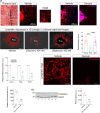
Detection of lymphatic markers in the frozen sections of breast carcinoma allografts. (A) Mammary allografts (n = 5 each) ± systemic delivery recombinant decorin or biglycan, 5 mg/kg, five injections were given at 2-d intervals, starting from day 10 following tumor implantation. One-way analysis ANOVA *P < 0.05. (B) Distribution of Lyve1 (green) and CD31 (red) in normal mouse organs and DAPI (blue); bar ~ 50 µm. (C) Distribution of Lyve1 (red) immunostaining in the intra- and peritumor areas with and without decorin treatment. DAPI (blue). Images were taken at 20× magnification via a Leica fluorescence microscope. Bar ~ 10 µm. (D) Quantification of relative fluorescence intensity from the images captured at constant exposure time, gain, and intensity; n = 3 to 6 independent replicas, 10 fields/case; unpaired two-tailed t test *P < 0.05. (E) Immunostaining of Podoplanin (green) in intra- and peritumor LVs with and without decorin treatment at 20×, DAPI (blue). Bar ~ 10 µm. (F) Quantification of relative Podoplanin fluorescence, 10 fields/case, captured at identical exposure time and intensity; n = 5 independent biological replicates; unpaired t test, **P < 0.01. (G and H) Western blot and quantification of tumor lysates immunoreacted for Has2 and Gapdh; n = 5 to 6 independent biological replicates, unpaired t test, **P < 0.01.
Fig. 4.
Fig. 4.
Effects of decorin in an ex vivo model of lymphangiogenesis in the 3D collagen matrix. (A) Inner view of the thorax after removal of the heart and lungs. The thoracic duct has absorbed EB and can be observed to the left of the thoracic aorta. (B) Confocal image of LR sprouts immunostained for Lyve1 (red), confirming its lymphatic origin, DAPI (blue). (C and D) Confocal images of Lyve1-labeled lymphatic sprouts ± decorin. (EG) Colorized bright field images of LRs after day 6 of explant in 3D Collagen I. Recombinant decorin or biglycan (400 nM each) was added for the last 3 d in culture. Bar ~ 400 µm. (H) Quantification of radial distance from five independent experiments using 10 mice, 40 to 45 rings/condition; one-way ANOVA **P < 0.01. (I) qPCR analysis of 500 ng from 3 pooled LR sprouts ± decorin. Actin was used as a housekeeping control; mean ± SD; unpaired two-tailed t test, n = 3 to 5 biological replicates for each gene tested **P < 0.01 and ***P < 0.001. (J and K) Confocal images of podoplanin immunostaining (red) in LRs ± decorin; DAPI (blue). Bar ~ 150 µm. (L) Quantification of mean relative fluorescence intensity captured at constant exposure time, gain, and intensity; n = 3 to 5 independent biological replicas; unpaired t test ***P < 0.001. (M and N) Western blot and quantification of podoplanin expression from 3 pooled LR lysates ± decorin. GAPDH was used as a housekeeping control. n = 3 independent biological samples; unpaired t test, **P < 0.01,
Fig. 5.
Fig. 5.
Decorin evokes autophagic degradation of Lyve1 in ex vivo LR assays and in primary cultures of LECs. (A) qPCR analysis from 3 pooled LR sprouts (500 ng of total RNA) ±3 d of constant decorin. Actin was used as a housekeeping control; mean ± SD; n = 3 independent biological replicas. Unpaired t test **P < 0.01 and *P < 0.05. Western blot analysis in three pooled LR lysates, with or without 400 nM decorin treatment for 3 d, was conducted for Beclin-1 and relative quanitfication using Gapdh as a housekeeping control is shown (B and C). The same is done for LC3 (D and E); n = 3 biological replicates. Unpaired t test **P < 0.01 and *P < 0.05. (F and G) Respective immunoblots for Beclin-1 and Gapdh protein expression for mouse LECs treated with or without 400 nM decorin for 6 along with respective quantification; n = 3 independent replicas; unpaired two-tailed t test ***P < 0.001. (H–J) Immunodetection of LC3 ±decorin (400 nM) for 6 or 18 h in mouse LECs, as indicated; bar ~ 10 µm. (K-M) Immunodetection of Lyve1 and LC3 ±decorin (400 nM) for 6 or 18 h in mouse LECs, as indicated; bar ~ 10 μm. (N–P) Immunodetection of Beclin-1 and LC3 ± decorin (400 nM) for 6 or 18 h in mouse LECs, as indicated; bar ~ 10 µm. (Q and R) Immunodetection of LC3 and Lyve1 in LECs treated with mTOR inhibitors Torin-1 or INK128 (20 nM each) for 6 h. Arrows point to LC3+ or LC3+/Lyve1+ puncta. Bars ~ 10 µm. (S and T) Immunoblotting of primary mouse LECs for Lyve1 and Gapdh incubated with indicated time point of decorin and respective quantification; n = 3 independent replicas; unpaired t test ***P < 0.001. (U and V) Autophagic flux experiments maintaining constant decorin (400 nM) for 4 h in mouse LECs and 500 nM Bafilomycin A1 was added at the indicated times; representative blots for Lyve1 and Gapdh are shown here from 3 independent experiments; *P < 0.05.
Fig. 6.
Fig. 6.
Decorin protein core binds and down-regulates VEGFR3 and activates Akt in a VEGFR3-dependent manner in LECs. (A and B) Ligand-binding assays using sVEGFR3 or decorin protein core (100 ng each) as immobilized substrates or as soluble ligands as indicated. (C) Displacement of decorin protein core (constant 50 nM) bound to immobilized sVEGFR3 by increasing concentrations of VEGFC; n = 3 independent experiments run in triplicates. (D) Dose–response immunoblotting of VEGFR3 and Gapdh with increasing decorin concentrations as indicated for 6 h. (E) Immunoblotting of P-Akt induction in LECs via VEGFC at the indicated time points. (F) Immunoblotting of P-Akt blockage evoked by the TKi, SAR. Primary cultures of human LECs were pretreated for 30 min with 5 mM SAR after which decorin and VEGFC were added, 400 nM and 500 ng/mL respectively, for 20 min. (G) Quantification of Akt phosphorylation at Ser473; n = 4 to 7 biological replicates; unpaired t test **P < 0.01; ***P < 0.001. (H) Immunoblotting of P-Akt blockage evoked by the TKi, Anlotinib, and respective quantification. Cells were treated identically as in panel F. (I) Quantification of Akt phosphorylation at Ser473 in the presence of decorin, VEGFC, or Anlotinib as indicated. n = 4 to 7 biological replicates; unpaired t test; **P < 0.01;***P < 0.001. (J) Quantification of the relative P-VEGFR3 level at Tyr residues by sandwich ELISAs in LECs treated for 20 min with either VEGFC or decorin and blocked with SAR as indicated; n = 5 biological replicates; unpaired t test; **P < 0.01 and ***P < 0.001.

Update of

References

    1. Escobedo N., Oliver G., Lymphangiogenesis: Origin, specification, and cell fate determination. Annu. Rev. Cell Dev. Biol 32, 677–691 (2016). - PubMed
    1. Jalkanen S., Salmi M., Lymphatic endothelial cells of the lymph node. Nat. Rev. Immunol. 20, 566–578 (2020). - PubMed
    1. Royston D., Jackson D. G., Mechanisms of lymphatic metastasis in human colorectal adenocarcinoma. J. Pathol. 217, 608–619 (2009). - PubMed
    1. Jones D., Pereira E. R., Padera T. P., Growth and immune evasion of lymph node metastasis. Front. Oncol. 8, 36 (2018). - PMC - PubMed
    1. Jana S., Muscarella R. A. Jr., Jones D., The multifaceted effects of breast cancer on tumor-draining lymph nodes. Am. J. Pathol. 191, 1353–1363 (2021). - PMC - PubMed

Publication types

MeSH terms

LinkOut - more resources