. 2024 May 9;187(10):2536-2556.e30.
doi: 10.1016/j.cell.2024.03.027. Epub 2024 Apr 22.

DrugMap: A quantitative pan-cancer analysis of cysteine ligandability

Mariko Takahashi  1 Harrison B Chong  2 Siwen Zhang  2 Tzu-Yi Yang  2 Matthew J Lazarov  2 Stefan Harry  3 Michelle Maynard  4 Brendan Hilbert  4 Ryan D White  4 Heather E Murrey  4 Chih-Chiang Tsou  4 Kira Vordermark  2 Jonathan Assaad  2 Magdy Gohar  2 Benedikt R Dürr  2 Marianne Richter  2 Himani Patel  2 Gregory Kryukov  4 Natasja Brooijmans  4 Aliyu Sidi Omar Alghali  5 Karla Rubio  5 Antonio Villanueva  5 Junbing Zhang  2 Maolin Ge  2 Farah Makram  2 Hanna Griesshaber  2 Drew Harrison  2 Ann-Sophie Koglin  2 Samuel Ojeda  2 Barbara Karakyriakou  2 Alexander Healy  2 George Popoola  2 Inbal Rachmin  6 Neha Khandelwal  2 Jason R Neil  4 Pei-Chieh Tien  2 Nicholas Chen  7 Tobias Hosp  2 Sanne van den Ouweland  2 Toshiro Hara  5 Lillian Bussema  5 Rui Dong  5 Lei Shi  2 Martin Q Rasmussen  2 Ana Carolina Domingues  2 Aleigha Lawless  8 Jacy Fang  9 Satoshi Yoda  2 Linh Phuong Nguyen  2 Sarah Marie Reeves  2 Farrah Nicole Wakefield  2 Adam Acker  2 Sarah Elizabeth Clark  2 Taronish Dubash  2 John Kastanos  2 Eugene Oh  10 David E Fisher  6 Shyamala Maheswaran  10 Daniel A Haber  11 Genevieve M Boland  12 Moshe Sade-Feldman  10 Russell W Jenkins  13 Aaron N Hata  10 Nabeel M Bardeesy  10 Mario L Suvà  14 Brent R Martin  4 Brian B Liau  15 Christopher J Ott  10 Miguel N Rivera  14 Michael S Lawrence  16 Liron Bar-Peled  17
Affiliations

DrugMap: A quantitative pan-cancer analysis of cysteine ligandability

Mariko Takahashi et al. Cell. .

Abstract

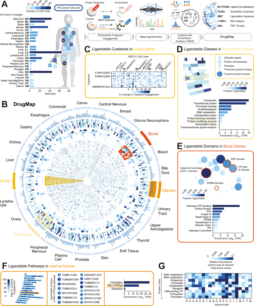
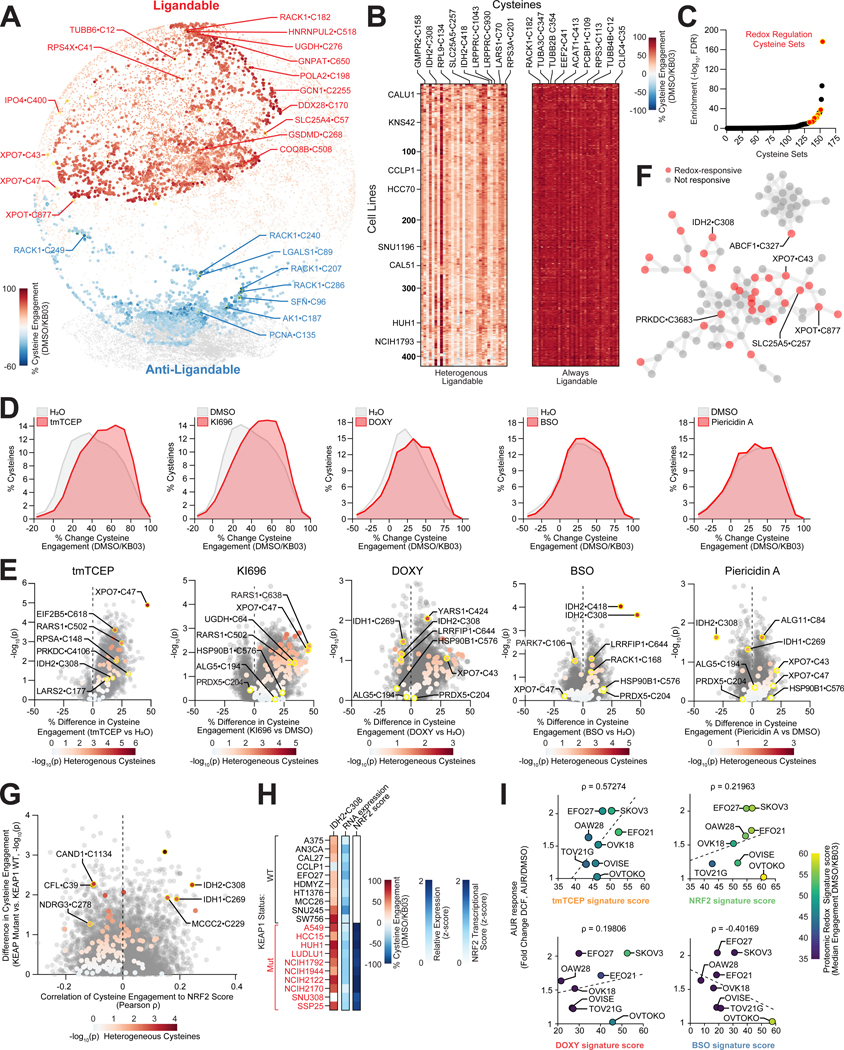
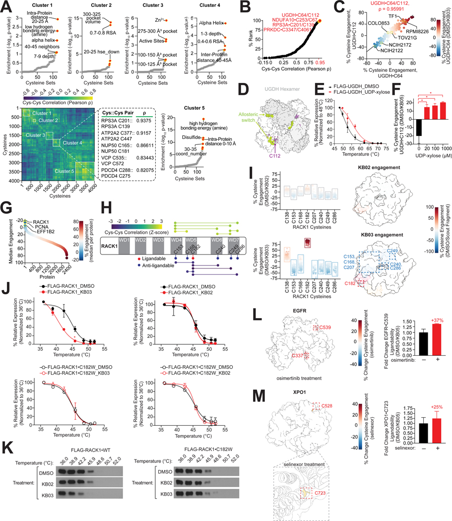
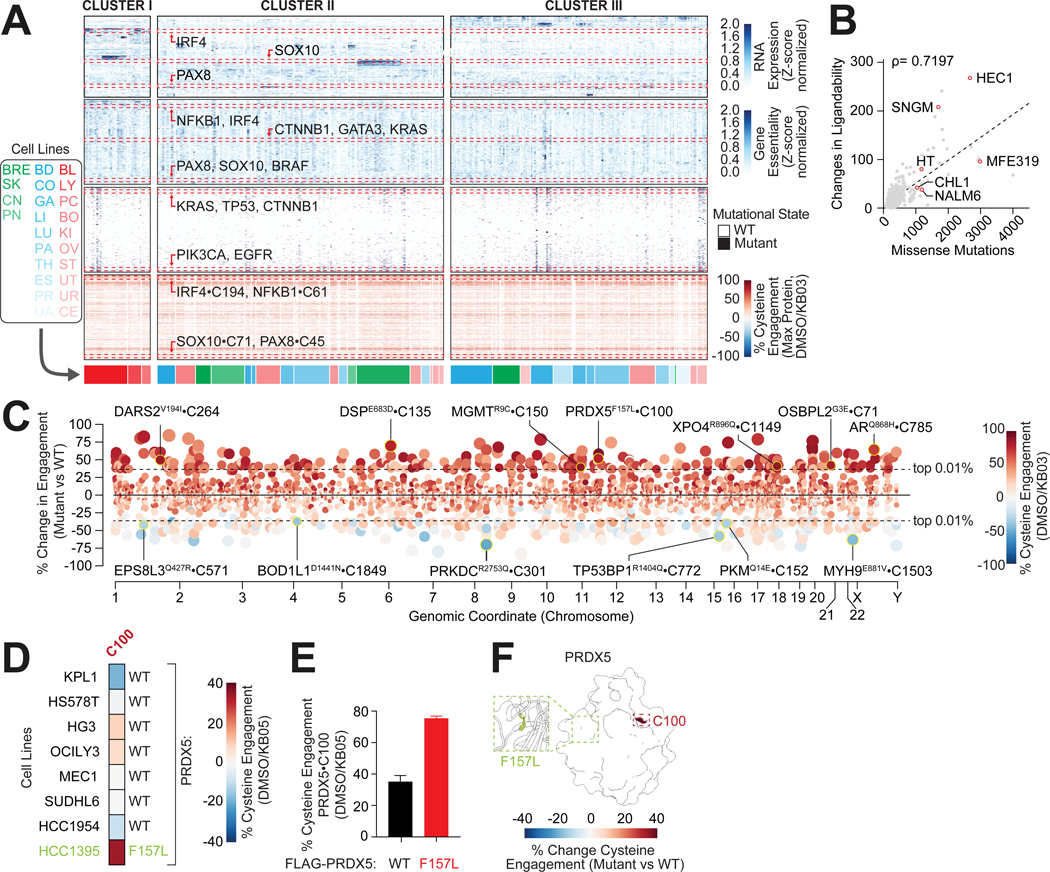
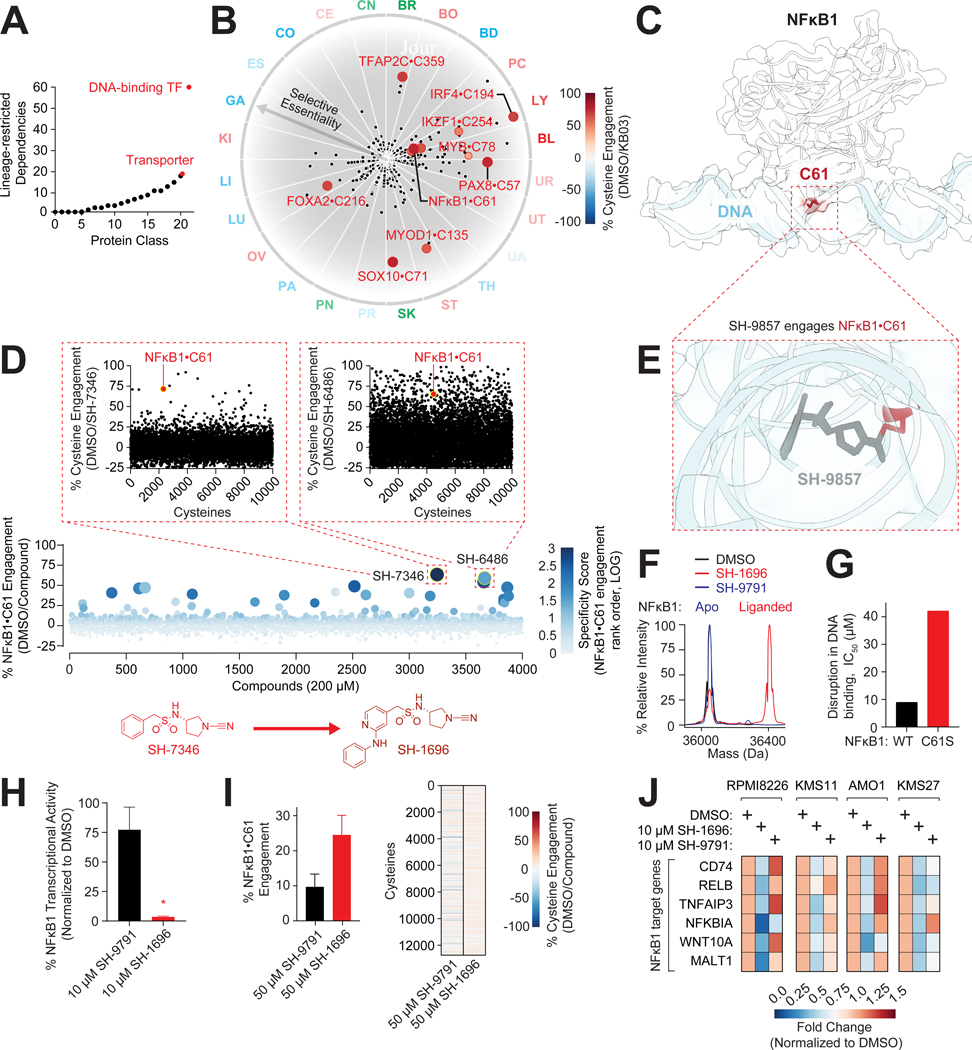
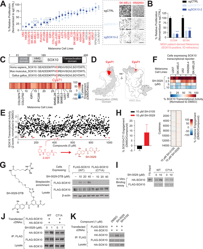
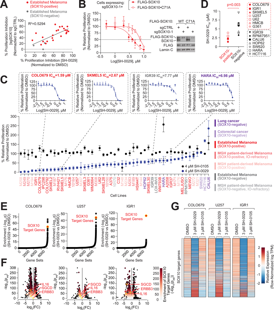
Cysteine-focused chemical proteomic platforms have accelerated the clinical development of covalent inhibitors for a wide range of targets in cancer. However, how different oncogenic contexts influence cysteine targeting remains unknown. To address this question, we have developed "DrugMap," an atlas of cysteine ligandability compiled across 416 cancer cell lines. We unexpectedly find that cysteine ligandability varies across cancer cell lines, and we attribute this to differences in cellular redox states, protein conformational changes, and genetic mutations. Leveraging these findings, we identify actionable cysteines in NF-κB1 and SOX10 and develop corresponding covalent ligands that block the activity of these transcription factors. We demonstrate that the NF-κB1 probe blocks DNA binding, whereas the SOX10 ligand increases SOX10-SOX10 interactions and disrupts melanoma transcriptional signaling. Our findings reveal heterogeneity in cysteine ligandability across cancers, pinpoint cell-intrinsic features driving cysteine targeting, and illustrate the use of covalent probes to disrupt oncogenic transcription-factor activity.

Keywords: chemical proteomics; covalent inhibitors; cysteine ligandability; transcription factors.


Conflict of interest statement

Declaration of interests D.E.F. has a financial interest in Soltego. S.M. and D.A.H. are cofounders of TellBio. G.M.B. has sponsored research agreements with Olink Proteomics, Teiko Bio, InterVenn Biosciences, and Palleon Pharmaceuticals; served on advisory boards for Iovance, Merck, Nektar Therapeutics, Novartis, and Ankyra Therapeutics; consults for Merck, InterVenn Biosciences, Iovance, and Ankyra Therapeutics; and holds equity in Ankyra Therapeutics. M.S.-F. received funding from Calico Life Sciences, Bristol-Myers Squibb, Istari Oncology, and consultants for Galvanize Therapeutics. R.W.J. is a member of the advisory board for/has a financial interest in Xsphera Biosciences Inc. R.W.J. has received honoraria from Incyte (invited speaker), G1 Therapeutics (advisory board), and Bioxcel Therapeutics (invited speaker). R.W.J. has ownership interest in U.S. patents US20200399573A9 and US20210363595A1. A.N.H. has received grants/research support from Amgen, Blueprint Medicines, BridgeBio, Bristol-Myers Squibb, C4 Therapeutics, Eli Lilly, Novartis, Nuvalent, Pfizer, Roche/Genentech, and Scorpion Therapeutics. A.N.H. has served as a compensated consultant for Amgen, Engine Biosciences, Nuvalent, Oncovalent, Pfizer, TigaTx, and Tolremo Therapeutics. M.L.S. is an equity holder and scientific co-founder/advisory board member of Immunitas Therapeutics. C.J.O. received funding from Scorpion Therapeutics, Gilead Sciences, and eFFECTOR Therapeutics. L.B.-P. is a founder/consultant/holds privately held equity in Scorpion Therapeutics. All relationships for investigators have been reviewed/managed by their respective institutions in accordance with their conflict-of-interest policies. M.M., B.H., R.D.W., H.E.M., C.-C.T., G.K., N.B., J.R.N., and B.R.M. are employees of Scorpion Therapeutics and some hold equity.

Figures

Figure 1.
Figure 1.. Defining a cysteine ligandability map across cancer.
(A) Cancer cell lines analyzed in this study and schematic of DrugMap development (see also Table S1). (B) Circular heatmap depicting differences among cysteine, domain, class and pathway ligandability (from inner- to outermost layers, respectively) across 416 cell lines (see also Tables S2, STAR Methods). (C) Examples of differences in cysteine ligandability among 64 lung cancer cell lines. (D) Cysteine set enrichment analysis (CSEA) of protein classes identified among twelve pancreatic cancer cell lines. (see Figure S2A, text, and STAR Methods). (E) CSEA of protein domains identified among nine bone cancer cell lines. (F) CSEA of biological pathways identified among nine uterine cancer cell lines. (G) Heatmap depicting relative abundance of amino acid residues within a 6 Å radius of ligandable cysteines for each protein class (see also Figures S3F–G and STAR Methods).
Figure 2.
Figure 2.. Heterogeneity in cysteine ligandability is driven by cellular redox state.
(A) Spherical embedding of ~18,000 cysteines displaying ligandable and anti-ligandable cysteines (see also Figure 3N, Table S2 and STAR Methods). (B) Examples of ligandable or heterogeneous cysteine ligandability (see also Table S2). (C) Redox-regulated cysteines are enriched among heterogeneous cysteines as detected by CSEA. (D-E) Redox perturbations alter cysteine liganability. Ligandability (D) and volcano (E) plots comparing changes in KB03 engagement in K562 cells pre-treated with tmTCEP, KI696,, doxycycline (DOXY), L-Buthionine-sulfoximine (BSO), and Piericidin A. Heterogenous cysteines are colored (E) (see also Figure S4B–E, Table S3). (F) Network analysis of heterogeneous cysteine ligandability. (G) Heterogenous cysteine ligandability is altered between KEAP1-mutant and KEAP1-WT cell lines. (H) IDH2•C308 is differentially ligandable in KEAP1-mutant cell lines. (I) DCF fluorescence changes induced by auranofin correlate with proteomic redox signatures.
Figure 3.
Figure 3.. Heterogenous cysteine ligandability is driven by protein conformational changes.
(A) Heatmap depicting correlations of 4,000+ cys-cys pairs. Surrounding plots display enrichment of structural elements in each cluster. Inset, cys-cys correlations identified in Cluster 4. (B-C) UGDH•C64/C112 ligandability is highly correlated (B) across hundreds of cell lines (C). (D) UGDH•C112 is proximal to the UGDH allosteric switch and oligomerization interface (PDB:5VR8). (E) UDP-Xylose increases UGDH thermal stability in HEK-293T lysates overexpressing FLAG-UGDH (see also Figure S4H). (F) UDP-Xylose increases UGDH•C112 cysteine ligandability in K562 lysates (see also Table S3). (G) RACK1 is anti-ligandable. (H) Schematic depicting RACK1 cysteine engagement and cys-cys correlations. (I) RACK1•C182 liganding by KB03 induces cysteine anti-ligandability. Left, RACK1 cysteine reactivity changes following treatment with KB02 or KB03 across > 200 cell lines. Right, RACK1 structure displaying cysteine ligandability (PDB:4AOW). (J-K) RACK1•C182 engagement alters protein stability. Thermal shift plots (J) for FLAG-RACK1 and FLAG-RACK1•C182W following treatment with 100 μM KB03 or KB02 determined by immunoblot (K) (see also STAR Methods). (L-M) Changes in EGFR or XPO1 cysteine ligandability in PC9 lysates pre-treated with 1 μM of Osimertinib (L, PDB:3QWQ) or in K562 lysates pre-treated with 1 μM of selinexor (M, PDB:3GB8) (see also Figures S4M–N). Data are represented as mean ± SD. *p < 0.05. Student’s t-test (two-tailed, unpaired) were used to determine statistical significance.
Figure 4.
Figure 4.. Genetic determinants of cysteine ligandability.
(A) Multi-omic clustering of mRNA expression, gene essentiality, mutational status and cysteine ligandability across 300 cell lines. (B) Increased mutational burden correlates with changes in cysteine ligandability. (C) Identification of mutations that associate with changes in cysteine ligandability in the same protein (see STAR Methods). (D-F) Mutation of Phe157–>Leu correlates with increased PRDX5•C100 ligandability. Heatmap depicting differential engagement of PRDX5•C100 in cell lines harboring WT or PRDX5•F157L (D). FLAG-PRDX5•F157L in HEK-293T has increased PRDX5•C100 engagement (E). Structure of PRDX5 (F) highlighting the locations of F157 and C100 (PDB:1HD2).
Figure 5.
Figure 5.. Development of a NFκB1 probe that disrupts DNA interaction.
(A) Transcription factors (TFs) display lineage-restricted essentiality (see STAR Methods). (B) Plot comparing differential transcription factor essentiality, expression and cysteine ligandability. (C) NFκB1 DNA binding domain (white, PDB:2O61) bound to DNA (blue) with adjacent Cys61 (red). (D) Top, iso-TMT screen of 4,000+ cysteine reactive compounds identified SH-7346 as a covalent ligand for NFκB1•C61. Insets, iso-TMT engagement profile for SH-7346 and SH-6486. Bottom, development of SH-7346 to SH-1696 (see also Figure S5I–J, STAR Methods). (E) Structural solution of NFκB1•C61 engagement by SH-9857 (SH-7346 analog) predicts clashing with DNA binding (See also Figure S5K, Table S7). (F) Intact mass spectrometry analysis of SH-1696 and SH-9791 (negative control) binding to recombinant NFκB1. (G) SH-1696 disrupts NFκB1 DNA binding in a C61S-dependent manner (see STAR Methods). (H) SH-1696 disrupts NFκB1 transcriptional activity in HEK-293 cells (see also Figure S5L). (I) NFκB1•C61 engagement determined by iso-TMT in MM1S lysates treated with SH-1696 or SH-9791 (see also Table S3). (J) SH-1696 disrupts the expression of NFκB1 target genes in different hematopoietic cell lines as determined by qPCR (see also Figure S5M). Data are represented as mean ± SD. *p< 0.05. Student’s t-test (two-tailed, unpaired) were used to determine statistical significance.
Figure 6.
Figure 6.. Development of a chemical probe that disrupts SOX10 activity in melanomas.
(A-B) Relative proliferation of established melanoma cell lines (A) and immunotherapy-resistant, patient-derived lines (B) expressing the indicated sgRNAs targeting SOX10. Inset, micrographs of SKMEL5 and WM266–4 expressing the indicated sgRNAs. Scale bar=200 μm. (C) Top, SOX10•C71 is localized to the SOXE dimerization domain. Bottom, SOX10•C71 engagement with KB03 across 33 melanoma cell lines. (D) Predicted SOX10 structure (AF-P56693-F1) encompassing amino acids 62–172. (E) Developing a SOX10 probe. Top, SOX10 transcriptional reporter screen with a library of 1,000 cysteine-reactive compounds in U257 cells. Bottom, development of hit compound 2-A01 to SH-0029 (see also Figures S6D–F, STAR Methods). (F) SOX10 transcriptional activity in melanoma cell lines treated with SH-0029. (G) SH-0029 ligands SOX10•C71. Immunoblot analysis of HEK-293T cells expressing FLAG-SOX10 or FLAG-SOX10•C71A following enrichment with biotinylated SH-0029 analog (SH-0029-DTB). (H) SOX10•C71 engagement determined by iso-TMT in SKMEL5 lysates treated with SH-0029 or SH-0105. Right, heatmap displaying cysteinome-wide reactivity changes (see also Figure S6H, Table S3). (I) SH-0029 increases SOX10-SOX10 interactions in vitro in a Cys71 dependent manner (see also Figures S6I–K, STAR Methods). (J-K) SH-0029 increases SOX10-SOX10 interactions in cells. Immunoblot analysis of SOX10 interactions following treatment with SH-0029 (J) or SH-105 (K) (see also Figures S6L). Data are represented as mean ± SD. ***p< 0.0001. Student’s t-test (two-tailed, unpaired) were used to determine statistical significance.
Figure 7.
Figure 7.. SOX10 covalent inhibitor blocks SOX10 activity in melanomas.
(A) SH-0029 proliferation effects correlate with depletion of SOX10 in melanomas. (B) SOX10•C71A mutation partially rescues SH-0029 proliferation. Left, relative proliferation following SH-0029 treatment in U257 cells expressing sgSOX10_1 and PAM-resistant FLAG-SOX10 or FLAG-SOX10•C71A. Right, immunoblot analysis of SOX10 levels (see STAR Methods). (C) Proliferation assay in 51 cancer cell lines treated with SH-0029 or SH-0105. Inset, SH-0029 IC50 values for the indicated cell lines. (D) SH-0029 IC50 values correlate with SOX10 expression. (E-F) SH-0029 disrupts SOX10 transcriptional signaling in melanomas. GSEA analysis in melanoma cell lines following treatment with SH-0029 (E) and corresponding volcano plots highlighting SOX10-regulated genes (F). (G) SOX10-regulated genes are differentially impacted in melanoma cell lines following SH-0029 treatment (see also Figure S7F, STAR Methods). Data are represented as mean ± SD. *p< 0.05. Student’s t-test (two-tailed, unpaired) was used to determine statistical significance.

Update of

  • DrugMap: A quantitative pan-cancer analysis of cysteine ligandability.
    Takahashi M, Chong HB, Zhang S, Lazarov MJ, Harry S, Maynard M, White R, Murrey HE, Hilbert B, Neil JR, Gohar M, Ge M, Zhang J, Durr BR, Kryukov G, Tsou CC, Brooijmans N, Alghali ASO, Rubio K, Vilanueva A, Harrison D, Koglin AS, Ojeda S, Karakyriakou B, Healy A, Assaad J, Makram F, Rachman I, Khandelwal N, Tien PC, Popoola G, Chen N, Vordermark K, Richter M, Patel H, Yang TY, Griesshaber H, Hosp T, van den Ouweland S, Hara T, Bussema L, Dong R, Shi L, Rasmussen MQ, Domingues AC, Lawless A, Fang J, Yoda S, Nguyen LP, Reeves SM, Wakefield FN, Acker A, Clark SE, Dubash T, Fisher DE, Maheswaran S, Haber DA, Boland G, Sade-Feldman M, Jenkins R, Hata A, Bardeesy N, Suva ML, Martin B, Liau B, Ott C, Rivera MN, Lawrence MS, Bar-Peled L. Takahashi M, et al. bioRxiv [Preprint]. 2023 Oct 23:2023.10.20.563287. doi: 10.1101/2023.10.20.563287. bioRxiv. 2023. Update in: Cell. 2024 May 9;187(10):2536-2556.e30. doi: 10.1016/j.cell.2024.03.027. PMID: 37961514 Free PMC article. Updated. Preprint.

Comment in

  • Mapping cysteine ligandability.
    Crunkhorn S. Crunkhorn S. Nat Rev Drug Discov. 2024 Jun;23(6):420. doi: 10.1038/d41573-024-00074-8. Nat Rev Drug Discov. 2024. PMID: 38710826 No abstract available.

Similar articles

Cited by

References

    1. Hanahan D. (2022). Hallmarks of Cancer: New Dimensions. Cancer Discov 12, 31–46. 10.1158/2159-8290.Cd-21-1059. - DOI - PubMed
    1. Hahn WC, Bader JS, Braun TP, Califano A, Clemons PA, Druker BJ, Ewald AJ, Fu H, Jagu S, Kemp CJ, et al. (2021). An expanded universe of cancer targets. Cell 184, 1142–1155. 10.1016/j.cell.2021.02.020. - DOI - PMC - PubMed
    1. Quintanal-Villalonga Á, Chan JM, Yu HA, Pe’er D, Sawyers CL, Sen T, and Rudin CM (2020). Lineage plasticity in cancer: a shared pathway of therapeutic resistance. Nat Rev Clin Oncol 17, 360–371. 10.1038/s41571-020-0340-z. - DOI - PMC - PubMed
    1. Garraway LA, and Lander ES (2013). Lessons from the cancer genome. Cell 153, 17–37. 10.1016/j.cell.2013.03.002. - DOI - PubMed
    1. Chang L, Ruiz P, Ito T, and Sellers WR (2021). Targeting pan-essential genes in cancer: Challenges and opportunities. Cancer Cell 39, 466–479. 10.1016/j.ccell.2020.12.008. - DOI - PMC - PubMed

Publication types