. 2024 May 2;111(5):896-912.
doi: 10.1016/j.ajhg.2024.03.017. Epub 2024 Apr 22.

Gene-specific somatic epigenetic mosaicism of FDFT1 underlies a non-hereditary localized form of porokeratosis

Affiliations

Gene-specific somatic epigenetic mosaicism of FDFT1 underlies a non-hereditary localized form of porokeratosis

Sonoko Saito et al. Am J Hum Genet. .

Abstract

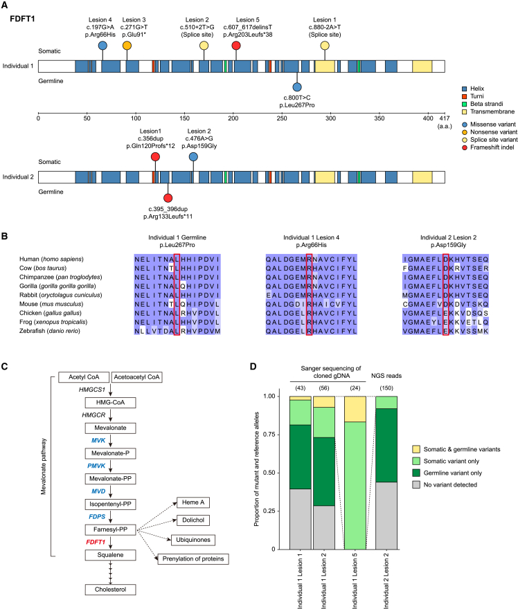
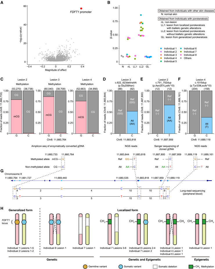
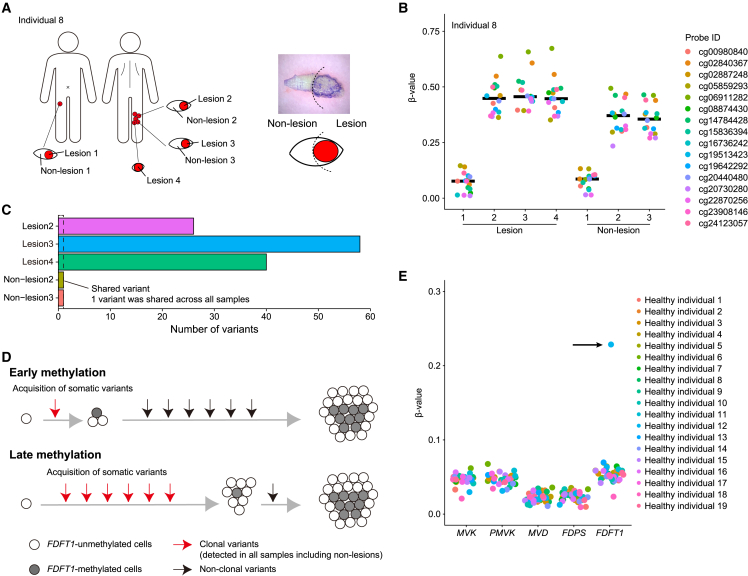
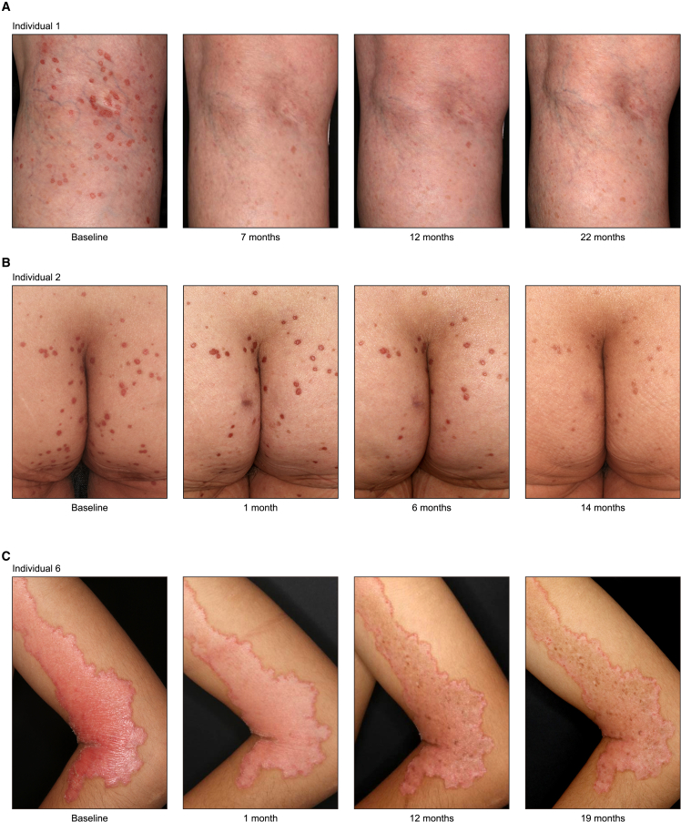
Porokeratosis is a clonal keratinization disorder characterized by solitary, linearly arranged, or generally distributed multiple skin lesions. Previous studies showed that genetic alterations in MVK, PMVK, MVD, or FDPS-genes in the mevalonate pathway-cause hereditary porokeratosis, with skin lesions harboring germline and lesion-specific somatic variants on opposite alleles. Here, we identified non-hereditary porokeratosis associated with epigenetic silencing of FDFT1, another gene in the mevalonate pathway. Skin lesions of the generalized form had germline and lesion-specific somatic variants on opposite alleles in FDFT1, representing FDFT1-associated hereditary porokeratosis identified in this study. Conversely, lesions of the solitary or linearly arranged localized form had somatic bi-allelic promoter hypermethylation or mono-allelic promoter hypermethylation with somatic genetic alterations on opposite alleles in FDFT1, indicating non-hereditary porokeratosis. FDFT1 localization was uniformly diminished within the lesions, and lesion-derived keratinocytes showed cholesterol dependence for cell growth and altered expression of genes related to cell-cycle and epidermal development, confirming that lesions form by clonal expansion of FDFT1-deficient keratinocytes. In some individuals with the localized form, gene-specific promoter hypermethylation of FDFT1 was detected in morphologically normal epidermis adjacent to methylation-related lesions but not distal to these lesions, suggesting that asymptomatic somatic epigenetic mosaicism of FDFT1 predisposes certain skin areas to the disease. Finally, consistent with its genetic etiology, topical statin treatment ameliorated lesions in FDFT1-deficient porokeratosis. In conclusion, we identified bi-allelic genetic and/or epigenetic alterations of FDFT1 as a cause of porokeratosis and shed light on the pathogenesis of skin mosaicism involving clonal expansion of epigenetically altered cells.

Keywords: FDFT1; cholesterol; clonal expansion; epigenetic mosaicism; germline variant; mevalonate pathway; porokeratosis; promoter hypermethylation; somatic variant; statin.


Conflict of interest statement

Declaration of interests T.T. and M.I. are employees of Japan Tissue Engineering Co., Ltd.

Figures

None
Graphical abstract
Figure 1
Figure 1
Clinical features of the 8 individuals with porokeratosis in this study Photographs of porokeratosis lesions (top) and schemas showing the distribution of porokeratosis lesions in the 8 individuals included in this study. The positions of the biopsied lesions are shown in the schema. Multiple lesions were subjected to skin biopsy in individuals 1, 2, 7, and 8. Detailed information on each individual and their skin lesions is provided in Table 1. See also Figure S1.
Figure 2
Figure 2
Somatic and germline variants of FDFT1 identified in individuals with generalized porokeratosis (A) Distribution of somatic and germline variants in FDFT1 in individual 1 (top) and individual 2 (bottom). (B) Multi-alignment analysis of FDFT1 and its orthologous protein sequences from different species: cow (bos taurus), chimpanzee (pan troglodytes), gorilla (gorilla gorilla gorilla), rabbit (oryctolagus cuniculus), mouse (mus musculus), chicken (gallus gallus), frog (xenopus tropicalis), and zebrafish (danio rerio). Color indicates the BLOSUM62 score. (C) Genes related to cholesterol synthesis. FDFT1 is shown in red, and the other genes associated with porokeratosis are shown in blue. (D) Allelic configurations (cis versus trans) of germline and somatic variants assessed by Sanger sequencing of the cloned gDNA (for individual 1 lesions 1, 2, and 5) and next-generation sequencing reads (for individual 2 lesion 2). Proportions of mutant and reference alleles are shown. Examined numbers are shown in parentheses. For individual 1 lesion 5, deletion-spanning PCR primers were used to amplify the gDNA of the allele with the somatic deletion. See also Figure S2.
Figure 3
Figure 3
Bi-allelic genetic and/or epigenetic alterations of FDFT1 in all lesion samples of localized porokeratosis (A) Volcano plot of methylation differences between lesion samples without bi-allelic genetic alterations of FDFT1 (n = 12) and non-lesion samples (n = 7). The x axis shows the magnitude of the effect and the y axis shows the −log10(q value). Each dot represents an autosomal CpG island. The red dot represents the CpG island located in the FDFT1 promoter region (chr8:11,659,676–11,660,795 [GRCh37]). The significance of methylation levels was assessed using a two-sided analysis of variance (ANOVA), which included sample type (lesion and non-lesion), individual ID, and probe ID as explanatory variables. Multiple-testing correction was performed using the Benjamini-Hochberg method. (B) DNA methylation levels (β values) of a representative probe (cg24123057; chr8:11,660,089) at CpG islands of the FDFT1 promoter in the epidermis of 2 normal skins (N), 7 non-lesions (nL), 2 lesions from localized porokeratosis with bi-allelic genetic alterations (LL1), 12 lesions from localized porokeratosis without bi-allelic genetic alterations (LL2), and 7 lesions from generalized porokeratosis (GL). (C) Allelic configurations (cis versus trans) of the methylation status of the CpG island at the FDFT1 promoter (chr8:11,660,733) and adjacent heterozygous SNP (chr8:11,660,764) assessed by amplicon deep sequencing of enzymatically converted gDNA of individual 8 lesions 2–4. Proportions of methylated and unmethylated reads are shown. (D) Allelic configurations of the somatic variant (chr8:11,683,644) and adjacent heterozygous SNP (chr8:11,683,818) assessed by next-generation sequencing reads in individual 8 lesion 3. (E) Allelic configurations of the somatic variant (chr8:11,687,800) and adjacent heterozygous SNP (chr8:11,687,959) assessed by Sanger sequencing of the cloned gDNA in individual 8 lesion 2. (F) Allelic configurations of the somatic variant (chr8:11,689,161) and adjacent heterozygous SNP (chr8:11,689,119) assessed by next-generation sequencing reads in individual 8 lesion 4. (D–F) Proportions of mutant and reference reads are shown. (C–F) Examined numbers are shown in parentheses. (G) Phasing of heterozygous SNPs using long-read WGS reads of the germline control sample of individual 8. Positions of heterozygous SNPs (blue) according to their genomic position (top) and the number of long-read WGS reads between them (bottom). (H) Schematic view of FDFT1 status in 21 lesion samples from the 8 individuals, revealed by integrated genetic and epigenetic analyses. Individuals 1 and 2 had germline pathogenic variants of FDFT1, and they developed multiple small lesions by acquiring independent somatic variants of FDFT1 on opposite alleles. Individuals 3–6 developed a single large lesion due to abnormal hypermethylation of the FDFT1 promoter, coupled with copy-neutral LOH of the FDFT1 locus in individuals 3, 4, and 6. Individuals 7 and 8 developed multiple lesions due to homozygous deletions of FDFT1 (individual 7 lesion 1), bi-allelic somatic variants of FDFT1 coupled with copy-neutral LOH (individual 8 lesion 1), and abnormal hypermethylation of the FDFT1 promoter coupled with heterozygous deletion (individual 7 lesions 3–6) or somatic pathogenic variant (individual 7 lesion 2 and individual 8 lesions 2–4) of FDFT1. See also Figures S3–S6.
Figure 4
Figure 4
Decreased FDFT1 mRNA and protein expression in lesional epidermis (A) Expression of FDFT1 relative to GAPDH measured by quantitative reverse-transcription PCR (RT-qPCR) for lesion and non-lesion samples of individual 6. (A–D) indicates primer pairs. See Figure S7A and Table S2 for primer positions and sequences. Each circle indicates a technical replicate. (B) Hematoxylin and eosin (H&E) staining (top left) and FDFT1 immunohistochemistry (bottom left and right) of individual 5 lesion 1. Asterisks indicate cornoid lamella, which is the sharp demarcation of the lesions. Arrows indicate the range of lesional skin with abnormal keratinization determined from H&E staining. The dotted box area in the lower left image is magnified in the right image. Scale bar, 200 μm. See also Figure S7.
Figure 5
Figure 5
Epigenetic mosaicism of FDFT1 in morphologically normal epidermis (A) Schematic representations of the distribution of porokeratosis lesions in individual 8. The positions of the biopsied lesions and non-lesions are indicated. In individual 8, lesions and non-lesions were separated from the same biopsy samples, as shown in the image and schematic diagram on the right. (B) DNA methylation levels (β values) of 16 probes at the CpG island in the FDFT1 promoter region in lesions and non-lesions from individual 8. (C) Number of somatic variants detected by WES in 3 lesions and 2 non-lesions from individual 8. One variant was detected in all evaluated lesion and non-lesion samples. (D) Schematic representations showing the timing of methylation and acquisition of somatic variants. White and gray circles represent unmethylated and methylated cells, respectively. Somatic variants acquired before methylation become clonal in methylated cells. Hence, only a few somatic variants become clonal, or none at all, if methylation occurs early in development (top), whereas many somatic variants become clonal if methylation occurs late in life (bottom). (E) Median DNA methylation levels (β values) at the CpG islands in the MVK, PMVK, MVD, FDPS, and FDFT1 promoter regions in 38 epidermal samples from 19 unaffected individuals. See also Figure S8.
Figure 6
Figure 6
Transcriptomes and phenotypes of lesion-derived keratinocytes (A) Volcano plot of differentially expressed genes (DEGs) in keratinocytes isolated from the lesion and non-lesion of individual 5 (n = 3 for each). The x axis shows the log2 fold change and the y axis shows the −log10(q value). Red dots represent significantly upregulated genes in lesion-derived keratinocytes (201 genes), whereas blue dots represent significantly downregulated genes (217 genes). Significant DEGs were selected at the thresholds of absolute log2 fold change >1 and q value <1.0 × 10−20. (B) Pathway enrichment analysis of DEGs between keratinocytes isolated from the lesion and non-lesion of individual 5. The x axis shows the p value obtained using gProfiler2 with multiple-testing corrections. Pink and blue indicate pathways upregulated and downregulated in lesion-derived keratinocytes, respectively. (C) Proliferation of lesion- and non-lesion-derived keratinocytes cultured in medium supplemented with or without cholesterol for 5 days. Two-sided Welch’s t test. (D) Light microscopy images of lesion- and non-lesion-derived keratinocytes cultured in medium supplemented with or without cholesterol for 120 h. Scale bar, 100 μm. (C and D) 50,000 cells were plated in a 6-well plate and cultured in the aforementioned medium for 120 h. See also Figure S9.
Figure 7
Figure 7
Skin lesion improvement by topical atorvastatin and cholesterol treatment (A–C) Representative images of skin lesions before and after topical atorvastatin and cholesterol treatment in individual 1 (A), individual 2 (B), and individual 6 (C).

Similar articles

Cited by

References

    1. Biesecker L.G., Spinner N.B. A genomic view of mosaicism and human disease. Nat. Rev. Genet. 2013;14:307–320. - PubMed
    1. Lim Y.H., Moscato Z., Choate K.A. Mosaicism in Cutaneous Disorders. Annu. Rev. Genet. 2017;51:123–141. - PMC - PubMed
    1. Happle R. Mosaicism in human skin. Understanding the patterns and mechanisms. Arch. Dermatol. 1993;129:1460–1470. - PubMed
    1. Reed R.J., Leone P. Porokeratosis--a mutant clonal keratosis of the epidermis. I. Histogenesis. Arch. Dermatol. 1970;101:340–347. - PubMed
    1. Das A., Vasudevan B., Talwar A. Porokeratosis: An enigma beginning to unravel. Indian J. Dermatol. Venereol. Leprol. 2022;88:291–299. - PubMed

Publication types

LinkOut - more resources