. 2024 Apr 23;15(1):3435.
doi: 10.1038/s41467-024-47696-5.

Exosome-coated oxygen nanobubble-laden hydrogel augments intracellular delivery of exosomes for enhanced wound healing

Affiliations

Exosome-coated oxygen nanobubble-laden hydrogel augments intracellular delivery of exosomes for enhanced wound healing

Xiaoxue Han et al. Nat Commun. .

Abstract

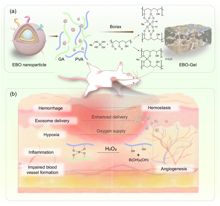
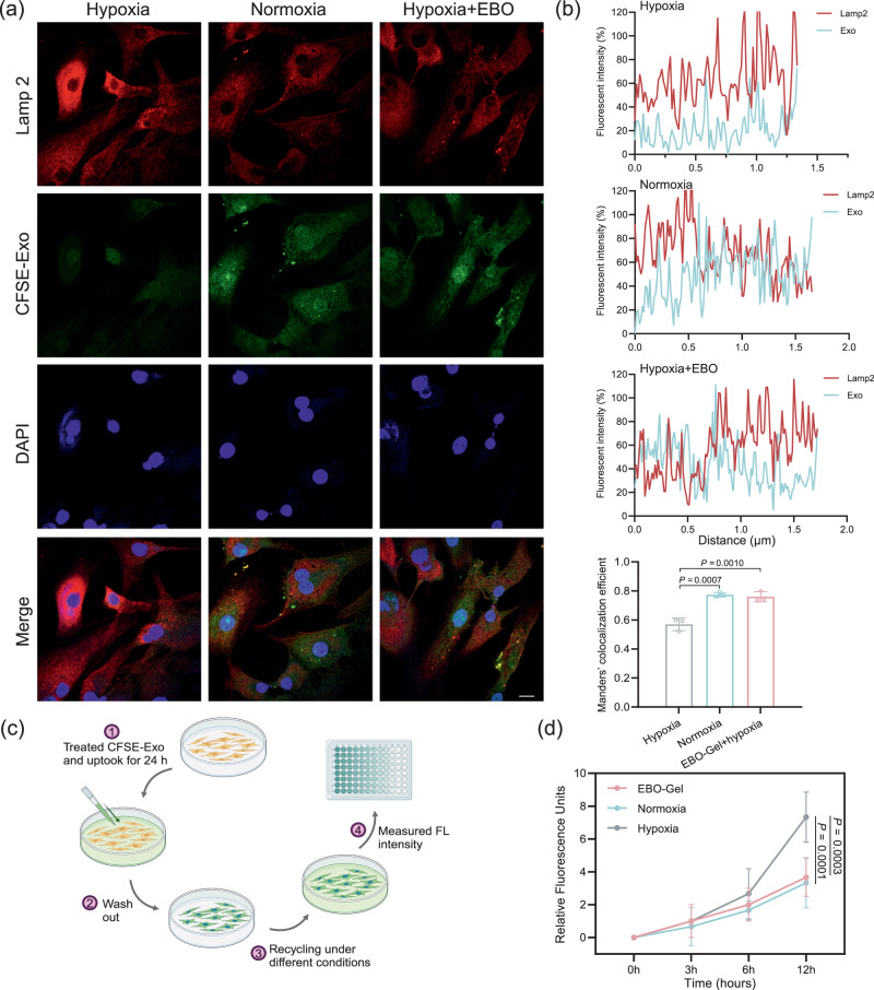
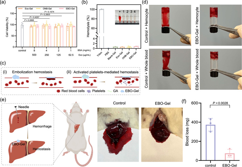
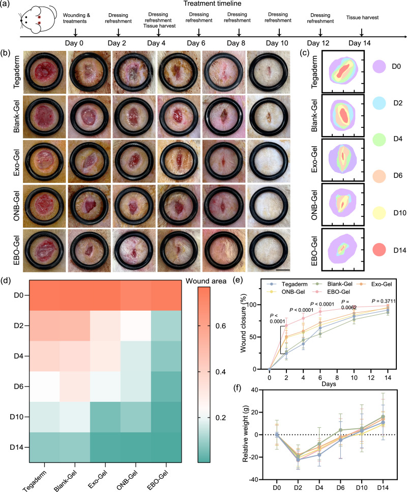
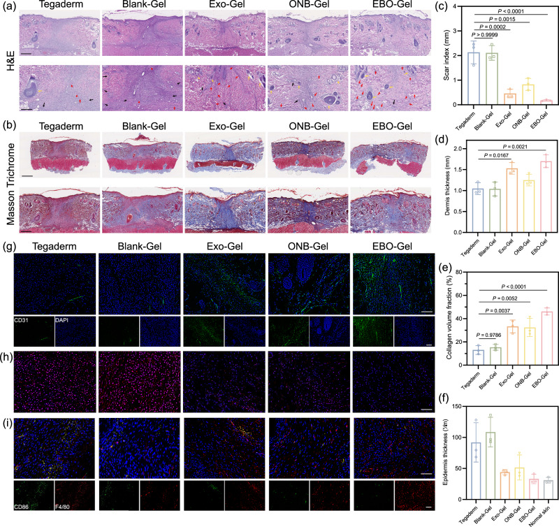
Wound healing is an obvious clinical concern that can be hindered by inadequate angiogenesis, inflammation, and chronic hypoxia. While exosomes derived from adipose tissue-derived stem cells have shown promise in accelerating healing by carrying therapeutic growth factors and microRNAs, intracellular cargo delivery is compromised in hypoxic tissues due to activated hypoxia-induced endocytic recycling. To address this challenge, we have developed a strategy to coat oxygen nanobubbles with exosomes and incorporate them into a polyvinyl alcohol/gelatin hybrid hydrogel. This approach not only alleviates wound hypoxia but also offers an efficient means of delivering exosome-coated nanoparticles in hypoxic conditions. The self-healing properties of the hydrogel, along with its component, gelatin, aids in hemostasis, while its crosslinking bonds facilitate hydrogen peroxide decomposition, to ameliorate wound inflammation. Here, we show the potential of this multifunctional hydrogel for enhanced healing, promoting angiogenesis, facilitating exosome delivery, mitigating hypoxia, and inhibiting inflammation in a male rat full-thickness wound model.


Conflict of interest statement

J.I. and X.H. filed a provisional patent application for the exosome-coated oxygen nanobubble-laden hydrogel (U.S. Patent Application No.: 63/631,255). The remaining authors declare no competing interests.

Figures

Fig. 1
Fig. 1. Schematic illustration of EBO-Gel-mediated wound healing.
a Crosslinking mechanisms and structure of EBO-Gel. b Enhanced wound healing programmed by hemostasis, promoted exosome delivery, oxygen supply, angiogenesis, and antioxidant properties offered by EBO-Gel. PVA polyvinyl alcohol, GA gelatin, EBO ADSC-derived exosome coated BSA-based oxygen nanobubbles, EBO-Gel EBO nanoparticles-embedded hydrogel.
Fig. 2
Fig. 2. Synthesis and characterization of EBO.
a Schematic of the preparation of ONB and EBO. b Concentration distribution and scattered images (background) of exosomes. Diameter distribution (c) and zeta potential values (d) of ONB and EBO (n  =  3 independent samples). e TEM images of (i) exosomes; (ii) ONB; (iii) EBO. Scale bar: 50 nm. f (i) EBO uptake and (ii) Z-stack slice with orthogonal views of EBO (Green) internalization in HDF-a cells after 6 h of incubation. Scale bar: 10 μm. g The browning intensity of early (A294) and late (A420) MRP (n =  3 independent samples). h Infrared spectra of BSA and ONB. Pink areas indicate the spectral regions of group vibrations (Left) and dextran (Right). (i) SDS-PAGE of different formulations. Lanes: 1. Natural BSA; 2. Mixture of BSA and dextran sulfate; 3. Ultrasonicated BSA; 4. Shell; 5. ONB; 6. Exosomes; 7. EBO. Data are presented as mean ± SD (b, d, g). Statistical analysis was performed by two-way ANOVA with Dunnett’s multiple comparisons (g). Representative images are shown from two independent experiments with similar results (e, f, i). Source data are provided as a Source Data file. BSA bovine serum albumin, ONB oxygen nanobubble, Exo adipose-derived stem cell (ADSC)-derived exosomes, EBO ADSC-derived exosome coated BSA-based oxygen nanobubbles, Dex dextran sulfate, Mix mixture of BSA and dextran sulfate.
Fig. 3
Fig. 3. Characterizations of EBO-Gel.
a Formation of EBO-Gel. b Injectability of EBO-Gel through the syringe. c Shape remodeling and adaptability of EBO-Gel. d Macroscopic self-healing property of EBO-Gel over time. Scale bar (in the 2nd row): 2 mm. e Storage modulus (G) and loss modulus (G) of EBO-Gel after cyclic cutting and reassembling process. f Adhesive capacity on different substrates. g Adhesion to finger with different bending angles. h Linear viscoelastic rheological characterization of Blank-Gel and EBO-Gel. The transition from the blue region to the yellow region signifies the shift from liquid-like to solid-like behavior during the gelation process. i SEM images of Blank-Gel and EBO-Gel. The red area indicates the magnified region. Scale bars are shown on each image respectively. j Adhesion to rat organs (from left to right: heart, liver, spleen, lung, and kidney). k Adhesion to skin tissue from different species (from left to right: rat, porcine, mouse, and human). Data are presented as mean ± SD (h). Representative images are shown from two independent experiments with similar results (i). Source data are provided as a Source Data file. PVA polyvinyl alcohol, GA gelatin, Blank-Gel hydrogel scaffold without nanoparticles, EBO-Gel EBO nanoparticles-embedded hydrogel.
Fig. 4
Fig. 4. Oxygen supply and antioxidant properties of EBO-Gel.
a Illustration of oxygen supply and anti-inflammation mechanisms offered by EBO-Gel. b Oxygen release curve monitored within 10 h. c Intracellular hypoxia conditions of different treatments. Scale bar: 50 μm. d H2O2 consumption capacity of EBO-Gel (n  =  3 independent samples). e ROS/SOD detection in HDF-a cells. Scale bar: 100 μm. f Quantification of ROS/SOD fluorescence (n  =  3 biologically independent samples). g Evaluation of H2DCFDA signal after treated with Blank-Gel, ONB-Gel, Exo-Gel, and EBO-Gel. Data are presented as mean ± SD (d, f). Statistical analysis was performed by one-way ANOVA with Tukey’s multiple comparisons (f). Representative images are shown from three independent experiments with similar results (c, e). Source data are provided as a Source Data file. Exo adipose-derived stem cell (ADSC)-derived exosomes, ONB oxygen nanobubble, NT no treatments, Blank-Gel hydrogel scaffold without nanoparticles, Exo-Gel adipose-derived stem cell (ADSC)-derived exosomes-embedded hydrogel, ONB-Gel oxygen nanobubble-embedded hydrogel, EBO-Gel EBO nanoparticles-embedded hydrogel.
Fig. 5
Fig. 5. Enhanced intracellular exosome delivery.
a Representative immunofluorescent images of CFSE-Exo and Lamp2 under different treatments. Scale bar: 10 μm. b Fluorescence intensity and colocalization efficiency of different treatments (n  =  3 biologically independent samples). c Illustrations of the procedures for the evaluation of exosome recycling. d Quantification of exosome recycling in the culture medium (n  =  3 biologically independent samples). Data are presented as mean ± SD (b, d). Statistical analysis was performed by one-way ANOVA with Tukey’s multiple comparisons (b) or two-way ANOVA with Dunnett’s multiple comparisons (d). Representative images are shown from two independent experiments with similar results (a). Source data are provided as a Source Data file. EBO-Gel EBO nanoparticles-embedded hydrogel.
Fig. 6
Fig. 6. Biocompatibility and hemostatic properties assessment.
a Cell viability of HDF-a cells incubated with different concentrations of Exo-Gel, ONB-Gel, and EBO-Gel (n  =  5 biologically independent samples). b Blood compatibility evaluated by hemolysis assay (n  =  3 biologically independent samples). Inserted image: +: Positive control (Triton); 1: Blank-Gel; 2: Exo-Gel; 3: ONB-Gel; 4: EBO-Gel. c Mechanisms of hemostasis capacity: (i) Embolization hemostasis offered by remodeling, adhesive, and self-healing properties of EBO-Gel; (ii) Activated platelet-mediated hemostasis offered by GA. d In vitro procoagulant effects of EBO-Gel. e Illustration (Left) and Digital photos (Right) of hemostasis evaluation on rat liver hemorrhage model. f Quantitation of blood loss in e (n  =  3 biologically independent experiments). Data are presented as mean ± SD (a, b, f). Statistical analysis was performed by two-way ANOVA with Dunnett’s multiple comparisons (a) or two-tailed Student’s t test (f). Representative images are shown from three independent experiments with similar results (b, d, e). Source data are provided as a Source Data file. GA gelatin, Exo-Gel adipose-derived stem cell (ADSC)-derived exosomes-embedded hydrogel, ONB-Gel oxygen nanobubble-embedded hydrogel, EBO-Gel EBO nanoparticles-embedded hydrogel.
Fig. 7
Fig. 7. Evaluation of enhanced cell proliferation, migration, and angiogenesis.
a Immunofluorescence images of BrdU staining in HDF-a cells. Scale bar: 50 μm. b Cell proliferation assay of HDF-a cells with different treatments (n  =  3 biologically independent samples). c Calcein AM/PI fluorescence staining to examine live/dead cells. Scale bar: 50 μm. d Scratch wound healing assay conducted on HDF-a cells with different treatments subjected to 0 h, 12 h, or 24 h of hypoxia. Scale bar: 200 μm. e In vitro wound closure rate followed by scratching assay in d (n  =  3 biologically independent samples). f, g Brightfield images and quantitative result of transwell migration of HDF-a cells (n  =  3 biologically independent samples). Scale bar: 200 μm. h Tube formation ability of HUVECs with different treatments. Scale bar: 200 μm. Quantitative results of (i) number of branches and (j) total branches length (n  =  3 biologically independent samples in i and j. k Cell cycle analysis of HDF-a cells with different treatments. Data are presented as mean ± SD (b, e, g, I, j). Statistical analysis was performed by two-way ANOVA with Dunnett’s multiple comparisons (b, e), one-way ANOVA with Tukey’s multiple comparisons (g, i, j). Representative images are shown from two (a, c) or three (d, f, h) independent experiments with similar results. Source data are provided as a Source Data file. NT no treatments, Blank-Gel hydrogel scaffold without nanoparticles, Exo-Gel adipose-derived stem cell (ADSC)-derived exosomes-embedded hydrogel, ONB-Gel oxygen nanobubble-embedded hydrogel, EBO-Gel EBO nanoparticles-embedded hydrogel.
Fig. 8
Fig. 8. In vivo efficacy of EBO-Gel in a rat full-thickness wound model.
a Illustration of wound creation and treatment timeline. b Representative digital photos of wounds with varied treatments at different time points. Scale bar: 7 mm. c Traces of wound closure from day 0 to day 14. Quantitative analysis of (d) wound area (cm2) and (e) wound closure rate (n  =  6 biologically independent wounds) over time. f Relative body weight of rats after varied treatments (n  =  4 animals per group). Data are presented as mean ± SD (e, f). Statistical analysis was performed by two-way ANOVA with Dunnett’s multiple comparisons (e). Source data are provided as a Source Data file. Tegaderm no treatments, Blank-Gel hydrogel scaffold without nanoparticles, Exo-Gel adipose-derived stem cell (ADSC)-derived exosomes-embedded hydrogel, ONB-Gel oxygen nanobubble-embedded hydrogel, EBO-Gel EBO nanoparticles-embedded hydrogel, D0-D14 days post-surgery.
Fig. 9
Fig. 9. histological analysis of wounds that underwent treatments with different hydrogels.
Representative images (top) and magnified images (bottom) of (a) H&E and (b) Masson’s trichrome staining of the wound tissue on Day 14. Red arrows: new blood vessel formation; Black arrows: inflammation area; Yellow arrows: newly generated-hair follicles. Scale bar: 500 μm (a) and 1 mm (b) for the normal-sized image; 250 μm (a) and 500 μm (b) for the magnified image. Various parameters for wound healing evaluation, including: (c) Scar index; (d) Dermis thickness; (e) Collagen volume fraction; and (f) Epidermis thickness (n  =  3 biologically independent samples in cf). Representative fluorescence images of (g) CD31 immunostaining, (h) DHE (red) staining, and (i) CD86 and F4/80 immunostaining of wound tissues on Day 14 post-treatment. Cell nuclei were stained with DAPI (blue). Scale bar: 100 μm (CD31) and 50 μm (DHE and CD86). Data in are presented as mean ± SD (cf). Statistical analysis was performed by one-way ANOVA with Tukey’s multiple comparisons (cf). Representative images are shown from three independent experiments with similar results (a, b, g, h, i). Source data are provided as a Source Data file. Tegaderm no treatments, Blank-Gel hydrogel scaffold without nanoparticles, Exo-Gel adipose-derived stem cell (ADSC)-derived exosomes-embedded hydrogel, ONB-Gel oxygen nanobubble-embedded hydrogel, EBO-Gel EBO nanoparticles-embedded hydrogel.

References

    1. Eming SA, Martin P, Tomic-Canic M. Wound repair and regeneration: Mechanisms, signaling, and translation. Sci. Transl. Med. 2014;6:265sr266–265sr266. doi: 10.1126/scitranslmed.3009337. - DOI - PMC - PubMed
    1. Menke NB, Ward KR, Witten TM, Bonchev DG, Diegelmann RF. Impaired wound healing. Clin. Dermatol. 2007;25:19–25. doi: 10.1016/j.clindermatol.2006.12.005. - DOI - PubMed
    1. Nussbaum SR, et al. An Economic Evaluation of the Impact, Cost, and Medicare Policy Implications of Chronic Nonhealing Wounds. Value Health. 2018;21:27–32. doi: 10.1016/j.jval.2017.07.007. - DOI - PubMed
    1. Singer AJ, Clark RAF. Cutaneous Wound Healing. N. Engl. J. Med. 1999;341:738–746. doi: 10.1056/NEJM199909023411006. - DOI - PubMed
    1. Gurtner GC, Werner S, Barrandon Y, Longaker MT. Wound repair and regeneration. Nature. 2008;453:314–321. doi: 10.1038/nature07039. - DOI - PubMed

Publication types