

Loss of AMPK activity induces organelle dysfunction and oxidative stress during oocyte aging
- PMID: 38654312
- PMCID: PMC11036640
- DOI: 10.1186/s13062-024-00471-4
Loss of AMPK activity induces organelle dysfunction and oxidative stress during oocyte aging
Abstract
Background: Oocyte quality is critical for the mammalian reproduction due to its necessity on fertilization and early development. During aging, the declined oocytes showing with organelle dysfunction and oxidative stress lead to infertility. AMP-activated protein kinase (AMPK) is a serine/threonine protein kinase which is important for energy homeostasis for metabolism. Little is known about the potential relationship between AMPK with oocyte aging.
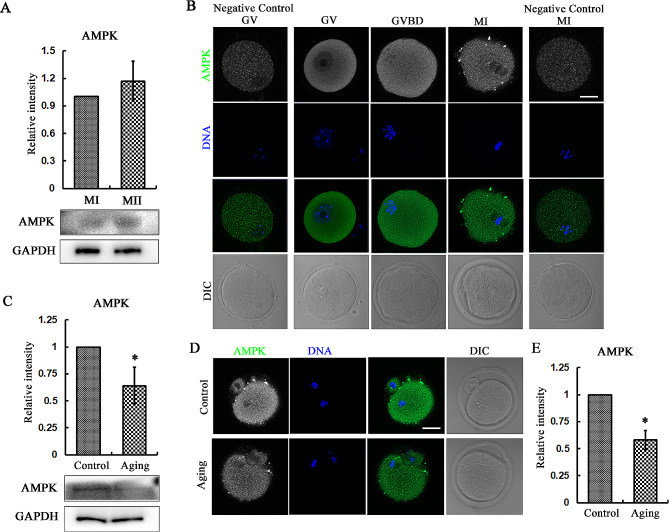
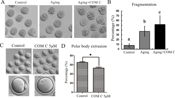
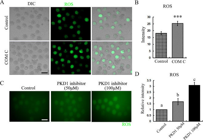
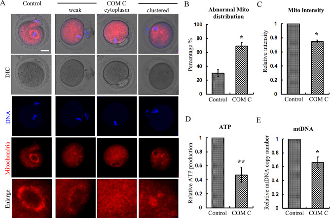
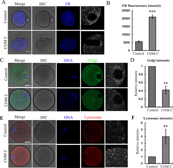
Results: In present study we reported that AMPK was related with low quality of oocytes under post ovulatory aging and the potential mechanism. We showed the altered AMPK level during aging and inhibition of AMPK activity induced mouse oocyte maturation defect. Further analysis indicated that similar with its upstream regulator PKD1, AMPK could reduce ROS level to avoid oxidative stress in oocytes, and this might be due to its regulation on mitochondria function, since loss of AMPK activity induced abnormal distribution, reduced ATP production and mtDNA copy number of mitochondria. Besides, we also found that the ER and Golgi apparatus distribution was aberrant after AMPK inhibition, and enhanced lysosome function was also observed.
Conclusions: Taken together, these data indicated that AMPK is important for the organelle function to reduce oxidative stress during oocyte meiotic maturation.
Keywords: AMPK; Meiosis; Mitochondria; Oocyte; Oxidative stress.
© 2024. The Author(s).
Conflict of interest statement
The authors declare that they have no competing interests.
Figures
Similar articles
-
FOXM1 affects oxidative stress, mitochondrial function, and the DNA damage response by regulating p21 in aging oocytes.Theriogenology. 2024 Nov;229:66-74. doi: 10.1016/j.theriogenology.2024.08.010. Epub 2024 Aug 10. Theriogenology. 2024. PMID: 39163804
-
Rescue in vitro maturation of germinal vesicle oocytes after ovarian stimulation: the importance of the culture media.Hum Reprod. 2025 Aug 1;40(8):1504-1515. doi: 10.1093/humrep/deaf099. Hum Reprod. 2025. PMID: 40447125 Free PMC article.
-
Epigallocatechin-3-gallate mitigates postovulatory oocyte aging by reducing oxidative stress and promoting embryonic development.Reproduction. 2025 Jun 25;170(1):e250125. doi: 10.1530/REP-25-0125. Print 2025 Jul 1. Reproduction. 2025. PMID: 40492572
-
Oocyte activation for women following intracytoplasmic sperm injection (ICSI).Cochrane Database Syst Rev. 2024 Dec 20;12(12):CD014040. doi: 10.1002/14651858.CD014040.pub2. Cochrane Database Syst Rev. 2024. PMID: 39704318
-
In vitro maturation in subfertile women with polycystic ovarian syndrome undergoing assisted reproduction.Cochrane Database Syst Rev. 2025 Feb 6;2(2):CD006606. doi: 10.1002/14651858.CD006606.pub5. Cochrane Database Syst Rev. 2025. PMID: 39912435
Cited by
-
Proteomic analysis reveals the alleviation of follicular development defects in offspring mice under DEHP exposure by melatonin.BMC Biol. 2025 Feb 28;23(1):65. doi: 10.1186/s12915-025-02165-3. BMC Biol. 2025. PMID: 40022026 Free PMC article.
-
NAMPT regulates mitochondria and oxidative stress level for mouse early embryo development.Biol Res. 2025 May 4;58(1):25. doi: 10.1186/s40659-025-00608-3. Biol Res. 2025. PMID: 40320561 Free PMC article.
-
Significance of Mitochondrial Dynamics in Reproductive Physiology: Current and Emerging Horizons in Mitochondrial Therapy for Assisted Reproductive Technologies.Reprod Med Biol. 2025 Aug 17;24(1):e12672. doi: 10.1002/rmb2.12672. eCollection 2025 Jan-Dec. Reprod Med Biol. 2025. PMID: 40832453 Free PMC article. Review.
-
The Importance of Mitochondrial Processes in the Maturation and Acquisition of Competences of Oocytes and Embryo Culture.Int J Mol Sci. 2025 Apr 25;26(9):4098. doi: 10.3390/ijms26094098. Int J Mol Sci. 2025. PMID: 40362337 Free PMC article. Review.
-
Early-stage administration of hydroxytyrosol extends lifespan and delays aging in C. elegans.Biol Direct. 2025 May 21;20(1):62. doi: 10.1186/s13062-025-00634-x. Biol Direct. 2025. PMID: 40399944 Free PMC article.
References
-
- Barone E, Di Domenico F, Perluigi M, Butterfield DA. The interplay among oxidative stress, brain insulin resistance and AMPK dysfunction contribute to neurodegeneration in type 2 diabetes and Alzheimer disease. Free Radic Biol Med. 2021;176:16–33. doi: 10.1016/j.freeradbiomed.2021.09.006. - DOI - PMC - PubMed
-
- Bertoldo MJ, Guibert E, Faure M, Rame C, Foretz M, Viollet B, Froment P. Specific deletion of AMP-activated protein kinase (alpha1AMPK) in murine oocytes alters junctional protein expression and mitochondrial physiology. PLoS ONE. 2015;10(3):e0119680. doi: 10.1371/journal.pone.0119680. - DOI - PMC - PubMed
Publication types
MeSH terms
Substances
LinkOut - more resources
Full Text Sources
Molecular Biology Databases
