. 2024 Apr 23;43(1):122.
doi: 10.1186/s13046-024-03049-0.

CircCDYL2 bolsters radiotherapy resistance in nasopharyngeal carcinoma by promoting RAD51 translation initiation for enhanced homologous recombination repair

Affiliations

CircCDYL2 bolsters radiotherapy resistance in nasopharyngeal carcinoma by promoting RAD51 translation initiation for enhanced homologous recombination repair

Hongke Qu et al. J Exp Clin Cancer Res. .

Abstract

Background: Radiation therapy stands to be one of the primary approaches in the clinical treatment of malignant tumors. Nasopharyngeal Carcinoma, a malignancy predominantly treated with radiation therapy, provides an invaluable model for investigating the mechanisms underlying radiation therapy resistance in cancer. While some reports have suggested the involvement of circRNAs in modulating resistance to radiation therapy, the underpinning mechanisms remain unclear.

Methods: RT-qPCR and in situ hybridization were used to detect the expression level of circCDYL2 in nasopharyngeal carcinoma tissue samples. The effect of circCDYL2 on radiotherapy resistance in nasopharyngeal carcinoma was demonstrated by in vitro and in vivo functional experiments. The HR-GFP reporter assay determined that circCDYL2 affected homologous recombination repair. RNA pull down, RIP, western blotting, IF, and polysome profiling assays were used to verify that circCDYL2 promoted the translation of RAD51 by binding to EIF3D protein.

Results: We have identified circCDYL2 as highly expressed in nasopharyngeal carcinoma tissues, and it was closely associated with poor prognosis. In vitro and in vivo experiments demonstrate that circCDYL2 plays a pivotal role in promoting radiotherapy resistance in nasopharyngeal carcinoma. Our investigation unveils a specific mechanism by which circCDYL2, acting as a scaffold molecule, recruits eukaryotic translation initiation factor 3 subunit D protein (EIF3D) to the 5'-UTR of RAD51 mRNA, a crucial component of the DNA damage repair pathway to facilitate the initiation of RAD51 translation and enhance homologous recombination repair capability, and ultimately leads to radiotherapy resistance in nasopharyngeal carcinoma.

Conclusions: These findings establish a novel role of the circCDYL2/EIF3D/RAD51 axis in nasopharyngeal carcinoma radiotherapy resistance. Our work not only sheds light on the underlying molecular mechanism but also highlights the potential of circCDYL2 as a therapeutic sensitization target and a promising prognostic molecular marker for nasopharyngeal carcinoma.

Keywords: Homologous recombination repair; Nasopharyngeal carcinoma; RAD51; Radiotherapy resistance; circCDYL2.


Conflict of interest statement

No conflict of interest exists in the submission of this manuscript, and the manuscript is approved by all authors for publication.

The authors declare that they have no conflict of interest.

Figures
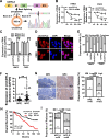
Fig. 1
Fig. 1
circCDYL2 is highly expressed in nasopharyngeal carcinoma and associated with poor prognosis. A. Schematic diagram of the structure of circCDYL2. Sanger sequencing confirmed that circCDYL2 is formed by reverse splicing of exon 2 of CDYL2 pre-mRNA, with a length of 592 nt. B. RT-qPCR was used to detect the relative abundance of circCDYL2 and CDYL2 mRNA in HNE2 and CNE2 cells after actinomycin D (1 μg/ml) treatment for 0, 8, 16, or 24 hours. C. RT-qPCR was performed to examine the expression of circCDYL2 and CDYL2 mRNA in HNE2 and CNE2 cells after RNase R treatment. D. Fluorescence in situ hybridization was used to determine the cellular localization of circCDYL2, showing distribution in both the cytoplasm and the nucleus. Scale bar = 20 μm. E. Cellular localization of circCDYL2 was assessed using nuclear-cytoplasmic fractionation followed by RT-qPCR, confirming the presence of circCDYL2 in both cytoplasm and nucleus. F. The expression of circCDYL2 was detected by RT-qPCR in fresh nasopharyngeal carcinoma (NPC) biopsy tissues (n = 45) and control nasopharyngeal epithelium (NPE) samples (n = 23). The expression of circCDYL2 was significantly upregulated in NPC tissues. G. In situ hybridization was used to detect circCDYL2 expression levels in 203 archived paraffin-embedded tissues, including 56 adjacent nasopharyngeal epithelium tissues. Results showed that circCDYL2 was highly expressed in 67% (136/203) of NPC tissues compared to 12.5% (7/56) in nasopharyngeal epithelium (NPE) tissues. Representative in situ hybridization results for nasopharyngeal epithelium and NPC tissues are shown; 20×, scale bar = 50 μm; 40×, scale bar = 20 μm. H. The overall survival time of NPC patients with high circCDYL2 expression is shorter than that of patients with low circCDYL2 expression, with a five-year survival rate of 48.55% vs 71.32%. HR = 2.38, p < 0.001. I. The expression of circCDYL2 was significantly higher in the radiotherapy resistance group (NPC_R, n = 48), compared with that in the radiotherapy-sensitive group (NPC_S, n = 38). All these NPC patients underwent conventional radiotherapy. Patients who did not experience recurrence or metastasis within 5 years after radiotherapy were defined as the radiotherapy-sensitive group (NPC_S, n = 38), while those who experienced recurrence or metastasis within the first 2 years after initial radiotherapy were considered the radiotherapy resistance group (NPC_R, n = 48). Data were represented as mean ± SD. ns, not significant; *, p < 0.05; **, p < 0.01; ***, p < 0.001
Fig. 2
Fig. 2
circCDYL2 promotes radiotherapy resistance in vitro and in vivo. A. Colony formation assay was performed to measure the survival rates after ionizing radiation in NPC cells. Cells were irradiated with X-rays at doses of 0, 2, 4, 6, or 8 Gy, respectively, after overexpression or knockdown of circCDYL2. The cell survival curve was obtained by using the irradiation dose as the abscissa (arithmetic coordinates), and the survival fraction as the ordinate (logarithmic coordinates). The results were analyzed and plotted using a linear-quadratic model. B. In vivo experiments were performed by subcutaneously injecting 3 × 106 CNE2 cells after overexpression or knockdown for circCDYL2 (n = 5). Tumor sites were irradiated with 6 Gy on day 18. Tumor volumes were measured every 2 days until day 28. C. Representative images of subcutaneous tumors. D & E. Tumor volumes (D) and weights (E) were measured in each group. F. Immunofluorescence was performed to measure the level of γ-H2AX in HNE2 cells after overexpression or knockdown of circCDYL2 with 6 Gy irradiation for 2, 6, 12, or 24 hours. The results showed that overexpression of circCDYL2 reduced the accumulation of radiation-induced foci (IRIF) of γ-H2AX in HNE2 cells post-irradiation, while knockdown of circCDYL2 had the opposite effect. Scale bar = 10 μm. The right graph represents the number of radiation-induced foci (IRIF) in 30 cells. G. Western blotting revealed that circCDYL2 promoted the disappearance of γ-H2AX foci in HNE2 cells with circCDYL2 overexpression or knockdown after irradiation. H. Comet assays showed that circCDYL2 promoted DNA damage repair in NPC cells with circCDYL2 overexpression or knockdown after radiation. Representative comet images were shown above, and the scale bar = 20 μm. The lower panel represented statistical analysis. Data were represented as mean ± SD. ns, not significant; *, p < 0.05; **, p < 0.01; ***, p < 0.001
Fig. 3
Fig. 3
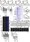
circCDYL2 enhances radiotherapy resistance in nasopharyngeal carcinoma by promoting DNA homologous recombination repair. A. Flow cytometry was performed to measure the GFP expression in DR-GFP-U2OS and EJ5-GFP-U2OS cells. Cells were transfected with the overexpression plasmid or ASO of circCDYL2. SiRNAs of BRCA1 or 53BP1 were selected as positive controls. Each group was co-transfected with the HA-Isce1 overexpression plasmid. B. Immunofluorescence showed that circCDYL2 enhanced the number of RAD51 irradiation-induced foci (IRIF) in HNE2 and CNE2 cell nuclei after irradiation. Scale bar = 10 μm. On the right, the quantification of IRIF per 30 cells was presented. C. The expression of RAD51 in the nucleus after irradiation was detected by western blotting. The results showed that circCDYL2 promoted the accumulation of RAD51 in the nucleus following irradiation. D. Western blotting showed that circCDYL2 promoted the expression of RAD51 protein in NPC cells after overexpression or knockdown of circCDYL2. Data were represented as mean ± SD. ns, not significant; *, p < 0.05; **, p < 0.01; ***, p < 0.001
Fig. 4
Fig. 4
circCDYL2 enhances RAD51 translation to promote DNA homologous recombination repair in nasopharyngeal carcinoma. A. Polysome profiling demonstrated that circCDYL2 enhances RAD51 mRNA expression on polysomes in NPC cells after overexpression or knockdown of circCDYL2. B. Flow cytometry showed that RAD51 could partially reverse the effect of circCDYL2 on homologous recombination repair in DR-GFP U2OS cells after simultaneous overexpression of circCDYL2 combined with knockdown of RAD51 or knockdown of circCDYL2 with overexpression of RAD51, respectively. Each group was co-transfected with the HA-Isce1 overexpression plasmid. C. Clonogenic assays showed that RAD51 partially reversed the effect of circCDYL2 on HNE2 cells survival post-irradiation. Cells were exposed to 0, 2, 4, 6, and 8 Gy X-ray irradiation, respectively after simultaneous overexpression of circCDYL2 combined with knockdown of RAD51 or knockdown of circCDYL2 with overexpression of RAD51. D. Immunofluorescence showed that RAD51 could partially reverse the regulation of γ-H2AX radiation-induced foci (IRIF) by circCDYL2 in HNE2 cells These cells were irradiated with 6 Gy X-rays for 6 hours after simultaneous overexpression of circCDYL2 combined with knockdown of RAD51 or knockdown of circCDYL2 with overexpression of RAD51. Scale bar = 10 μm. E. Western blotting demonstrated that RAD51 could partially reverse the regulation of γ-H2AX expression levels by circCDYL2 in HNE2 cells. Knockdown of circCDYL2, overexpression of RAD51, and simultaneous knockdown of circCDYL2 with overexpression of RAD51 in HNE2 cells, respectively. These cells were irradiated with 6 Gy X-rays for 0, 2, 6, 12, or 24 hours, respectively. F. Comet assays showed that RAD51 mediated the effect of circCDYL2 on DNA damage repair in HNE2 cells after simultaneous overexpression of circCDYL2 combined with knockdown of RAD51 or knockdown of circCDYL2 with overexpression of RAD51, respectively. These cells were irradiated with 6 Gy X-rays, and c Scale bar = 20 μm. Data were represented as mean ± SD. ns, not significant; *, p < 0.05; **, p < 0.01; ***, p < 0.001
Fig. 5
Fig. 5
RAD51 is highly expressed in NPC tissues and positively correlated with the expression of circCDYL2. A. Representative images of H&E staining, in situ hybridization of circCDYL2, and immunohistochemistry of RAD51 protein using the subcutaneous xenograft tumor tissues. 40×, scale bar = 20 μm. B. Statistical results of the relative expression levels of circCDYL2 and RAD51 in the subcutaneous xenograft tumor tissues according to Fig. 5A. C. Correlation between circCDYL2 and RAD51 expression was analyzed in the subcutaneous xenograft tumor tissues according to Fig. 5A. R = 0.8353. D. Representative immunohistochemistry results of RAD51 protein in 102 paraffin-embedded NPC tissues, compared with 28 adjacent nasopharyngeal epitheliums. 20×, scale bar = 50 μm, and 40×, scale bar = 20 μm. E. Overall survival analysis of NPC patients with low and high RAD51 expression using a Kaplan-Meier curve. F. Correlation between circCDYL2 and RAD51 expression was analyzed in 102 nasopharyngeal carcinoma tissues using the Pearson correlation analysis. R = 0.7026. G. The expression of RAD51 was analyzed in NPC and NPE tissues using four NPC gene expression profile data (GSE12452, GSE53819, GSE64634, and GSE61218). Data were represented as mean ± SD. ns, not significant; *, p < 0.05; **, p < 0.01; ***, p < 0.001
Fig. 6
Fig. 6
circCDYL2 recruits EIF3D protein to promote RAD51 translation initiation. A. RNA pulldown showed the interaction between circCDYL2 and EIF3D protein in HNE and CNE2 cells. B. Immunofluorescence demonstrated co-localization of circCDYL2 and EIF3D proteins in HNE and CNE2 cells. Blue, DAPI; Red, Dig-labeled circCDYL2; Green, anti-EIF3D antibody; Yellow, the co-localization of circCDYL2 and EIF3D protein. Scale bar = 10 μm. C. RNA immunoprecipitation (RIP) assay showed EIF3D proteins interacted with circCDYL2 in HNE2 and CNE2 cells. D. RIP experiments showed that EIF3D protein interacted with RAD51 mRNA in HNE2 and CNE2 cells. E. CircRIP experiments showed that circCDYL2 bound to RAD51 mRNA in HNE2 and CNE2 cells. F. RIP assays showed circCDYL2 enhanced the binding of EIF3D protein with RAD51 mRNA in NPC cells after overexpression or knockdown of circCDYL2. G. Polysome profiling was used to analyze the expression level of RAD51 mRNA in NPC cells after simultaneous knockdown of circCDYL2 with overexpression of EIF3D or overexpression of circCDYL2 with knockdown of EIF3D, respectively, using GAPDH mRNA as a control. H. Western blotting showed that EIF3D partially reverses the regulation of circCDYL2 on the expression of RAD51 protein in NPC cells after simultaneous knockdown of circCDYL2 with overexpression of EIF3D or overexpression of circCDYL2 with knockdown of EIF3D, respectively. Data were represented as mean ± SD. ns, not significant; *, p < 0.05; **, p < 0.01; ***, p < 0.001
Fig. 7
Fig. 7
circCDYL2 serves as a scaffold to promote EIF3D protein binding to RAD51 mRNA. A. The binding site of circCDYL2 to EIF3D protein was determined by RNA pull down in HNE2 and CNE2 cells after overexpression of circCDYL2 or its deletion mutants (DEL1 and DEL2). B. The binding site of circCDYL2 to EIF3D protein was determined by RIP assay in HNE2 and CNE2 cells after overexpression of circCDYL2 or its deletion mutants (DEL1 and DEL2). C. The binding site of circCDYL2 to RAD51 mRNA was determined by circRIP in HNE2 and CNE2 cells after overexpression of circCDYL2 or its deletion mutants (DEL1 and DEL2). D. The binding between EIF3D and RAD51 mRNA was determined by RIP in HNE2 and CNE2 cells after overexpression of circCDYL2 or its deletion mutants (DEL1 and DEL2). E. The expression of RAD51 was detected by western blotting in HNE2 and CNE2 cells after overexpression of circCDYL2 or its deletion mutants (DEL1 and DEL2). F. Schematic diagram of circCDYL2 promoting radiotherapy resistance in nasopharyngeal carcinoma via the EIF3D/RAD51 axis. circCDYL2 is generated by reverse splicing of exon 2 of CDYL2 pre-mRNA. It interacts with EIF3D protein and RAD51 5′-UTR and acts as a scaffold to recruit EIF3D protein to bind with RAD51 mRNA, facilitating the translation of RAD51 and accelerating homologous recombination repair, which ultimately leads to radiotherapy resistance in nasopharyngeal carcinoma. Data were represented as mean ± SD. ns, not significant; *, p < 0.05; **, p < 0.01; ***, p < 0.001

References

    1. Lo KW, To KF. Huang DP. Focus on nasopharyngeal carcinoma. Cancer Cell. 2004;5(5):423–428. doi: 10.1016/S1535-6108(04)00119-9. - DOI - PubMed
    1. Chang ET, Adami HO. The enigmatic epidemiology of nasopharyngeal carcinoma. Cancer epidemiology, biomarkers & prevention : a publication of the American Association for Cancer Research, cosponsored by the American Society of Preventive Oncology. 2006;15(10):1765–1777. doi: 10.1158/1055-9965.EPI-06-0353. - DOI - PubMed
    1. Chen Y, Zhao Y, Yang X, Ren X, Huang S, Gong S, et al. USP44 regulates irradiation-induced DNA double-strand break repair and suppresses tumorigenesis in nasopharyngeal carcinoma. Nat Commun. 2022;13(1):501. doi: 10.1038/s41467-022-28158-2. - DOI - PMC - PubMed
    1. Wei F, Wu Y, Tang L, He Y, Shi L, Xiong F, et al. BPIFB1 (LPLUNC1) inhibits migration and invasion of nasopharyngeal carcinoma by interacting with VTN and VIM. Br J Cancer. 2018;118(2):233–247. doi: 10.1038/bjc.2017.385. - DOI - PMC - PubMed
    1. Wang WJ, Wu SP, Liu JB, Shi YS, Huang X, Zhang QB, et al. MYC regulation of CHK1 and CHK2 promotes radioresistance in a stem cell-like population of nasopharyngeal carcinoma cells. Cancer Res. 2013;73(3):1219–1231. doi: 10.1158/0008-5472.CAN-12-1408. - DOI - PubMed

MeSH terms