. 2024 Apr 23;43(1):123.
doi: 10.1186/s13046-024-03048-1.

Tumor-suppressive miR-4732-3p is sorted into fucosylated exosome by hnRNPK to avoid the inhibition of lung cancer progression

Affiliations

Tumor-suppressive miR-4732-3p is sorted into fucosylated exosome by hnRNPK to avoid the inhibition of lung cancer progression

Wanzhen Zhuang et al. J Exp Clin Cancer Res. .

Abstract

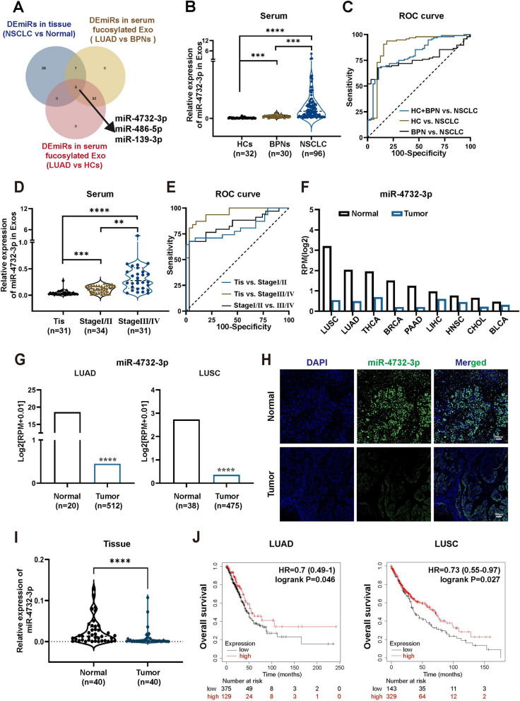
Background: Aberrant fucosylation observed in cancer cells contributes to an augmented release of fucosylated exosomes into the bloodstream, where miRNAs including miR-4732-3p hold promise as potential tumor biomarkers in our pilot study. However, the mechanisms underlying the sorting of miR-4732-3p into fucosylated exosomes during lung cancer progression remain poorly understood.

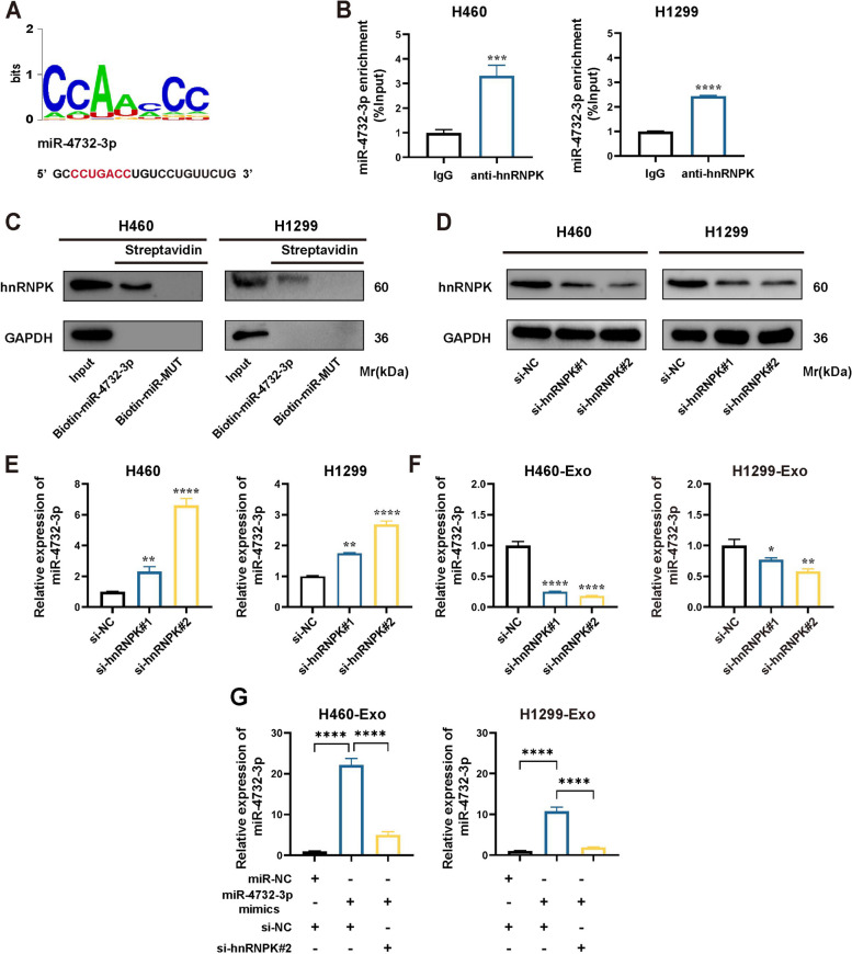
Methods: A fucose-captured strategy based on lentil lectin-magnetic beads was utilized to isolate fucosylated exosomes and evaluate the efficiency for capturing tumor-derived exosomes using nanoparticle tracking analysis (NTA). Fluorescence in situ hybridization (FISH) and qRT-PCR were performed to determine the levels of miR-4732-3p in non-small cell lung cancer (NSCLC) tissue samples. A co-culture system was established to assess the release of miRNA via exosomes from NSCLC cells. RNA immunoprecipitation (RIP) and miRNA pull-down were applied to validate the interaction between miR-4732-3p and heterogeneous nuclear ribonucleoprotein K (hnRNPK) protein. Cell functional assays, cell derived xenograft, dual-luciferase reporter experiments, and western blot were applied to examine the effects of miR-4732-3p on MFSD12 and its downstream signaling pathways, and the impact of hnRNPK in NSCLC.

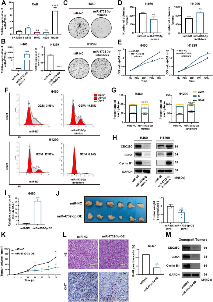
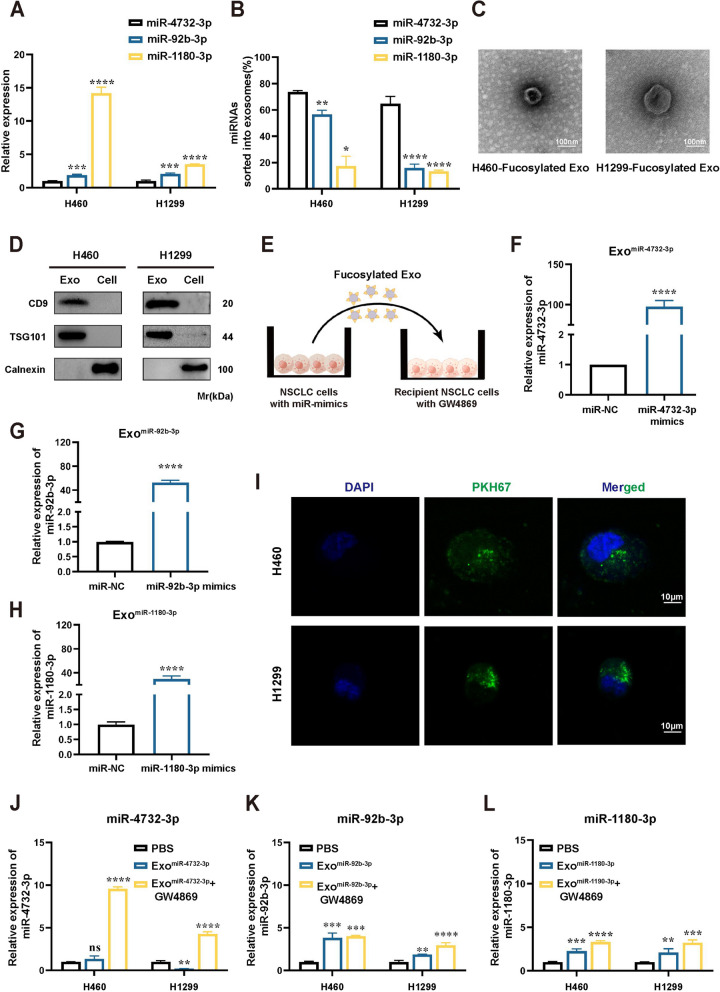
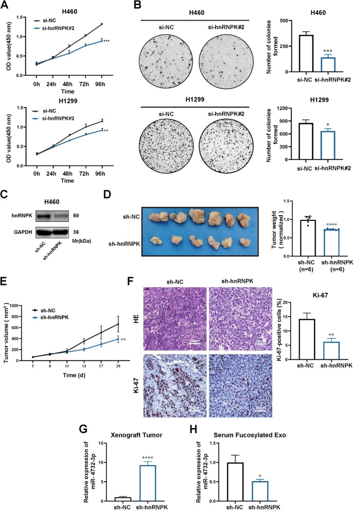
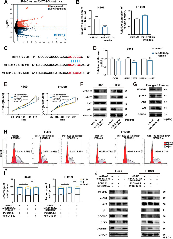
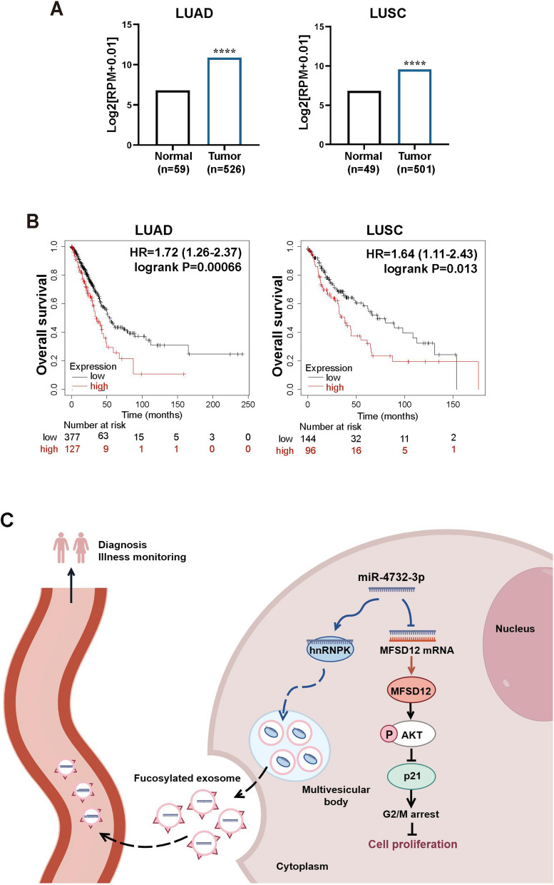
Results: We enriched exosomes derived from NSCLC cells using the fucose-captured strategy and detected a significant upregulation of miR-4732-3p in fucosylated exosomes present in the serum, while its expression declined in NSCLC tissues. miR-4732-3p functioned as a tumor suppressor in NSCLC by targeting 3'UTR of MFSD12, thereby inhibiting AKT/p21 signaling pathway to induce cell cycle arrest in G2/M phase. NSCLC cells preferentially released miR-4732-3p via exosomes instead of retaining them intracellularly, which was facilitated by the interaction of miR-4732-3p with hnRNPK protein for selective sorting into fucosylated exosomes. Moreover, knockdown of hnRNPK suppressed NSCLC cell proliferation, with the elevated levels of miR-4732-3p in NSCLC tissues but the decreased expression in serum fucosylated exosomes.

Conclusions: NSCLC cells escape suppressive effects of miR-4732-3p through hnRNPK-mediated sorting of them into fucosylated exosomes, thus supporting cell malignant properties and promoting NSCLC progression. Our study provides a promising biomarker for NSCLC and opens a novel avenue for NSCLC therapy by targeting hnRNPK to prevent the "exosome escape" of tumor-suppressive miR-4732-3p from NSCLC cells.

Keywords: Exosome escape; Fucosylated exosome; MFSD12; hnRNPK; miR-4732-3p.


Conflict of interest statement

The authors have declared that no conflict of interest exists.

Figures

Fig. 1
Fig. 1
miR-4732-3p is highly expressed in serum fucosylated exosomes but downregulated in NSCLC tissues. A Venn diagram exhibiting the overlap of DEmiRs in serum fucosylated exosomes from early LUAD patients compared with both BPNs and HCs groups, together with DEmiRs in NSCLC tissues. B qRT-PCR was performed to determine miR-4732-3p levels in serum fucosylated exosomes from NSCLC patients (n = 96), BPNs (n = 30), and HCs (n = 32). C ROC curve analysis of serum fucosylated exosomal miR-4732-3p for diagnosing NSCLC patients form BPNs and HCs. D Levels of miR-4732-3p were detected via qRT-PCR in serum fucosylated exosomes from NSCLC patients at diverse stages: Tis (n = 31), Stage I/II (n = 34), and Stage III/IV (n = 31). E The ability of serum fucosylated exosomal miR-4732-3p to discriminate NSCLC patients at diverse stages was evaluated using ROC curve analysis. F-G Expression of miR-4732-3p in cancerous tissues according to the GEDs (F) and ENCORI (G) databases. H-I miR-4732-3p levels in NSCLC tissues were evaluated by the fluorescence intensity via FISH (H) and relative expression via qRT-PCR (I) analysis. J Kaplan–Meier survival analysis of NSCLC patients according to hsa-miR-4732 expression. Data are shown as the mean ± SD from at least three independent experiments. **p < 0.01; ***p < 0.001; ****p < 0.0001; ns, not significant
Fig. 2
Fig. 2
miR-4732-3p inhibits the proliferation of NSCLC in vitro and in vivo. A qRT-PCR analysis of miR-4732-3p expression in NSCLC cell lines. B qRT-PCR was performed to examine miR-4732-3p expression in NSCLC cells transfected with miR-4732-3p mimics and inhibitors. C-E Colony formation assays (C-D), and CCK8 assays (E) were performed to evaluate the proliferation ability of NSCLC cells transfected with miR-4732-3p mimics and inhibitors. F-G Cell cycle assays were conducted to examine the effect of miR-4732-3p mimics and inhibitors on NSCLC cell cycle progression. H Western blot analysis was used to detect the expression of G2/M phase-related protein in NSCLC cells transfected with miR-4732-3p mimics and inhibitors. I qRT-PCR was performed to assess the expression of miR-4732-3p in H460 cells following infection with lentivirus engineered to overexpress miR-4732-3p (miR-4732-3p OE) and negative control (miR-NC). J The indicated H460 cells (miR-NC and miR-4732-3p OE) were injected into nude mice and xenograft tumors were harvested. Representative images and measurement of tumor weight in xenograft tumors were shown (n = 6). K Measurement and analysis of tumor volume in xenograft tumors with H460 cells stably overexpressing miR-4732-3p and miR-NC. L Histological examination of xenograft tumors by hematoxylin and eosin (HE) staining and Ki-67 expression levels examined by immunohistochemistry (IHC) (Scale bars = 50 μm). M Western blot analysis investigating the impact of miR-4732-3p on G2/M phase-related protein in vivo. Data are shown as the mean ± SD from at least three independent experiments. *p < 0.05; **p < 0.01; ***p < 0.001; ****p < 0.0001; ns, not significant
Fig. 3
Fig. 3
NSCLC cells preferentially release miR-4732-3p into exosomes. A Relative expression of miR-4732-3p, miR-92b-3p, and miR-1180-3p in NSCLC cells. B NSCLC cells was treated with GW4869 (10 μM) to assess the effects of exosome secretion on intracellular miRNA expression via qRT-PCR analysis and the proportion of each miRNA sorted into exosomes was calculated. C Images of fucosylated exosomes derived from NSCLC cells were photographed by transmission electron microscopy (TEM). Scale bar = 100 nm. D Western blot analysis was performed to detect typical exosome markers, including TSG101, CD9, and Calnexin (negative control). E Graphic illustration of co-culture system, in which we isolated fucosylated exosomes enriched with miRNA and added them into conditioned medium (CM) of NSCLC cells. F–H Fucosylated exosomes enriched with miR-4732-3p (F), miR-92b-3p (G), and miR-1180-3p (H) were obtained following transfecting NSCLC cells with miRNA mimics. I Internalization of PKH67-labeled fucosylated exosomes (green) by NSCLC cells. Scale bar = 10 μm. J-L NSCLC cells were co-cultured with blank control (PBS), or exosomes rich in miR-4732-3p, miR-92b-3p, and miR-1180-3p. We further added GW4869 (10 μM) to the co-culture system and analyzed the expression of intracellular miRNA in NSCLC cells co-cultured with exosomes under conditions where exosome secretion was suppressed, but exosome uptake was allowed. qRT-PCR was applied to verify the expression of miR-4732-3p (J), miR-92b-3p (K), and miR-1180-3p (L) in NSCLC cells after co-culturing. Data are shown as the mean ± SD from at least three independent experiments. *p < 0.05; **p < 0.01; ***p < 0.001; ****p < 0.0001; ns, not significant
Fig. 4
Fig. 4
miR-4732-3p is sorted into fucosylated exosomes via hnRNPK. A Sequence motifs of hnRNPK binding site predicted by RBPsuite. B RIP assay using hnRNPK antibody and qRT-PCR were performed, anti-IgG as the negative control. C miRNA pull-down and western blot analysis were conducted to confirm interaction between miR-4732-3p and hnRNPK protein. D Western blot analysis was applied to define knockdown efficiency of hnRNPK in NSCLC cells transfected with si-hnRNPK. E–F qRT-PCR analysis of miR-4732-3p levels in NSCLC cells (E), and their corresponding fucosylated exosomes (F) following hnRNPK knockdown. G qRT-PCR analysis was performed to determine the effects of miR-4732-3p mimics and si-hnRNPK on the levels of fucosylated exosomal miR-4732-3p. Data are shown as the mean ± SD from at least three independent experiments. *p < 0.05; **p < 0.01; ***p < 0.001; ****p < 0.0001; ns, not significant
Fig. 5
Fig. 5
Knockdown of hnRNPK suppresses NSCLC in vitro and in vivo. A-B The proliferation ability of NSCLC cells transfected with hnRNPK siRNAs was evaluated through CCK8 (A) and colony formation assays (B). C H460 cells were infected with lentivirus engineered to knock down hnRNPK (sh-hnRNPK) and negative control (sh-NC). The protein expressions were analyzed by western blot. D The indicated H460 cells (sh-NC and sh-hnRNPK) were injected into nude mice and xenograft tumors were harvested. Representative images and measurement of tumor weight in xenograft tumors were shown (n = 6). E Measurement and analysis of tumor volume in xenograft tumors of sh-NC and sh-hnRNPK groups. F Histological examination of xenograft tumors through HE staining and analysis of Ki-67 expression levels by IHC (Scale bars = 50 μm). G qRT-PCR analysis of miR-4732-3p expression in xenograft tumor tissues with stably hnRNPK knockdown. H qRT-PCR analysis of fucosylated exosomal miR-4732-3p expression in serum from nude mice with stably hnRNPK knockdown. Data are shown as the mean ± SD from at least three independent experiments. *p < 0.05; **p < 0.01; ***p < 0.001; ****p < 0.0001; ns, not significant
Fig. 6
Fig. 6
miR-4732-3p targets 3'UTR of MFSD12 in NSCLC cells. A Volcano plot presenting significantly differentially expressed genes (DEGs) (|log2FC|>1 and p < 0.05) between two groups: H460 cells transfected with miR-4732-3p mimics and miR-NC. B qRT-PCR analysis of MFSD12 mRNA expression in NSCLC cells following transfection of miR-4732-3p mimics and inhibitors. C Predicted binding sites between miR-4732-3p and the 3'UTR of MFSD12 mRNA. D Luciferase assay performed in 293T cells to validate the binding interaction between miR-4732-3p and MFSD12. E CCK8 assays examining the proliferation of NSCLC cells with overexpression or suppression of miR-4732-3p and MFSD12. F-G Western blot analysis of MFSD12 protein and the AKT/p21 signaling pathway in NSCLC cells after treatment as described above (F), and in xenograft tumors with miR-4732-3p overexpression (G). H-I Cell cycle analysis performed to assess the combined effects of miR-4732-3p and MFSD12 in NSCLC cells. J Western blot analysis conducted to investigate the AKT/p21 signaling pathway and G2/M phase-related protein after the indicated treatment. Data are shown as the mean ± SD from at least three independent experiments. *p < 0.05; **p < 0.01; ***p < 0.001; ****p < 0.0001; ns, not significant
Fig. 7
Fig. 7
MFSD12 is upregulated in NSCLC and associated with unfavorable prognosis. A ENCORI database analysis depicting the MFSD12 mRNA expression in LUAD and LUSC tissues. B Kaplan–Meier analysis illustrating the negative relationship between MFSD12 expression and overall survival (OS) rates of LUAD and LUSC patients. C Proposed working model depicting the mechanism by which tumor-suppressive miR-4732-3p is sorted into fucosylated exosomes through its interaction with hnRNPK protein, thereby promoting NSCLC progression. Data are shown as the mean ± SD from at least three independent experiments. ****p < 0.0001

Similar articles

Cited by

References

    1. Sung H, Ferlay J, Siegel RL, Laversanne M, Soerjomataram I, Jemal A, Bray F. Global Cancer Statistics 2020: GLOBOCAN Estimates of Incidence and Mortality Worldwide for 36 Cancers in 185 Countries. CA Cancer J Clin. 2021;71(3):209–249. doi: 10.3322/caac.21660. - DOI - PubMed
    1. Herbst RS, Morgensztern D, Boshoff C. The biology and management of non-small cell lung cancer. Nature. 2018;553(7689):446–454. doi: 10.1038/nature25183. - DOI - PubMed
    1. Siegel RL, Miller KD, Fuchs HE, Jemal A. Cancer statistics, 2022. CA Cancer J Clin. 2022;72(1):7–33. doi: 10.3322/caac.21708. - DOI - PubMed
    1. Kalluri R, LeBleu VS. The biology, function, and biomedical applications of exosomes. Science. 2020;367(6478):eaau6977. doi: 10.1126/science.aau6977. - DOI - PMC - PubMed
    1. Hu W, Liu C, Bi ZY, Zhou Q, Zhang H, Li LL, Zhang J, Zhu W, Song YYY, Zhang F, et al. Comprehensive landscape of extracellular vesicle-derived RNAs in cancer initiation, progression, metastasis and cancer immunology. Mol Cancer. 2020;19(1):102. doi: 10.1186/s12943-020-01199-1. - DOI - PMC - PubMed

MeSH terms