

Artificial intelligence in chorioretinal pathology through fundoscopy: a comprehensive review
- PMID: 38654344
- PMCID: PMC11036694
- DOI: 10.1186/s40942-024-00554-4
Artificial intelligence in chorioretinal pathology through fundoscopy: a comprehensive review
Abstract
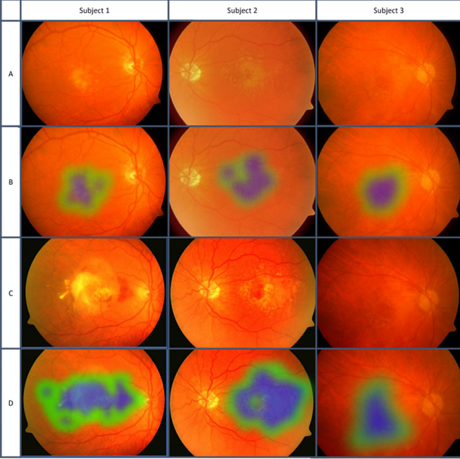
Background: Applications for artificial intelligence (AI) in ophthalmology are continually evolving. Fundoscopy is one of the oldest ocular imaging techniques but remains a mainstay in posterior segment imaging due to its prevalence, ease of use, and ongoing technological advancement. AI has been leveraged for fundoscopy to accomplish core tasks including segmentation, classification, and prediction.
Main body: In this article we provide a review of AI in fundoscopy applied to representative chorioretinal pathologies, including diabetic retinopathy and age-related macular degeneration, among others. We conclude with a discussion of future directions and current limitations.
Short conclusion: As AI evolves, it will become increasingly essential for the modern ophthalmologist to understand its applications and limitations to improve patient outcomes and continue to innovate.
Keywords: Age-related macular degeneration; Artificial intelligence; Choroid; Deep learning; Diabetic retinopathy; Fundoscopy; Fundus; Machine learning.
© 2024. The Author(s).
Conflict of interest statement
JC: Allergan, Salutaris, Biogen, Erasca. All else: None.
Figures
Similar articles
-
Artificial Intelligence in Ophthalmology - Status Quo and Future Perspectives.Semin Ophthalmol. 2023 Apr;38(3):226-237. doi: 10.1080/08820538.2022.2139625. Epub 2022 Nov 10. Semin Ophthalmol. 2023. PMID: 36356300 Review.
-
Artificial Intelligence (AI) Applications for Age-Related Macular Degeneration (AMD) and Other Retinal Dystrophies.Semin Ophthalmol. 2021 May 19;36(4):304-309. doi: 10.1080/08820538.2021.1896756. Epub 2021 Mar 25. Semin Ophthalmol. 2021. PMID: 33764255
-
An overview of artificial intelligence in diabetic retinopathy and other ocular diseases.Front Public Health. 2022 Oct 28;10:971943. doi: 10.3389/fpubh.2022.971943. eCollection 2022. Front Public Health. 2022. PMID: 36388304 Free PMC article. Review.
-
Artificial intelligence and deep learning in ophthalmology: Current status and future perspectives.Adv Ophthalmol Pract Res. 2022 Aug 24;2(3):100078. doi: 10.1016/j.aopr.2022.100078. eCollection 2022 Nov-Dec. Adv Ophthalmol Pract Res. 2022. PMID: 37846285 Free PMC article. Review.
-
Promising Artificial Intelligence-Machine Learning-Deep Learning Algorithms in Ophthalmology.Asia Pac J Ophthalmol (Phila). 2019 May-Jun;8(3):264-272. doi: 10.22608/APO.2018479. Epub 2019 May 31. Asia Pac J Ophthalmol (Phila). 2019. PMID: 31149787 Review.
Cited by
-
Advancing healthcare with artificial intelligence: diagnostic accuracy of machine learning algorithm in diagnosis of diabetic retinopathy in the Brazilian population.Diabetol Metab Syndr. 2024 Aug 29;16(1):209. doi: 10.1186/s13098-024-01447-0. Diabetol Metab Syndr. 2024. PMID: 39210394 Free PMC article.
References
-
- The Philadelphia photographer [Internet]. Philadelphia: Benerman & Wilson; 1864 [cited 2024 Mar 24]. 794 p. http://archive.org/details/philadelphiaphot18861phil.
-
- Retinal Atlas. The - ClinicalKey [Internet]. [cited 2024 Mar 24]. https://www-clinicalkey-com.my.wvsom.edu:2443/#!/browse/book/3-s2.0-C201....
Publication types
LinkOut - more resources
Full Text Sources
Research Materials
