

Management, risk factors and treatment outcomes of rhegmatogenous retinal detachment associated with giant retinal tears: scoping review
- PMID: 38654369
- PMCID: PMC11036595
- DOI: 10.1186/s40942-024-00552-6
Management, risk factors and treatment outcomes of rhegmatogenous retinal detachment associated with giant retinal tears: scoping review
Abstract
Background: Rhegmatogenous retinal detachment (RRD) is a serious condition that occurs when the retina detaches from its underlying retinal pigment epithelium. RRDs associated with giant retinal tears (GRTs) are caused by retinal tears at least 90° or one-quarter of the circumferential extent. This scoping review systematically identifies and summarizes clinical studies evaluating surgical techniques for the management of GRT-related RRDs, discusses functional and visual outcomes and the risk factors affecting treatment outcomes.
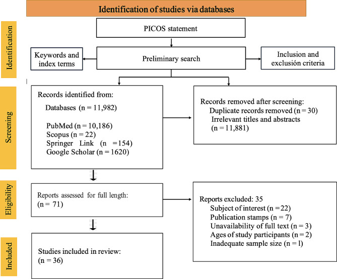
Methods: This study was conducted in accordance with the Preferred Reporting Items for Systematic Reviews and Meta-Analyses (PRISMA) guidelines. PubMed, Scopus, Google Scholar, and Springer Link databases were searched for relevant papers (from January 2001 to March 2023). Studies that were published in the English language and reported the risk factors, management, and treatment outcomes of GRT-related RRDs were included in the review. The outcome measures included anatomic success rates, changes in BCVA (logMAR) from baseline to the final follow-up, and adverse events.
Results: A total of 11,982 articles were identified. After the title and abstract review, 71 studies were deemed eligible for full-text review. Thirty-six studies that met the eligibility criteria were included in the final review. Four surgical techniques were identified: pars plana vitrectomy (PPV), combined PPV and scleral buckling, scleral buckling alone, and pneumatic retinopexy. Various types of tamponades, including gas, silicone oil, and air, have been used. PPV was the most commonly used surgical technique in 33.1-100% of patients. Among the 20 studies that used PPV alone, 17 were associated with preoperative PVR. In addition, scleral buckling alone or in combination with PPV was reported as a treatment option in 10 studies, with 2-100% of patients experiencing scleral buckling alone and 13.6-100% experiencing combined PPV and complementary scleral buckling. Primary anatomic success (PAS) was achieved with retinal reattachment via a single operation with no residual tamponade, whereas final anatomic success (FAS) was achieved via more than one operation with no residual tamponade. Reported single surgery anatomic success (SSAS) rates range from 65.51 to 100%. The preoperative best-corrected visual acuity (BCVA) ranged from 0.067 to 2.47 logMAR, whereas the postoperative BCVA ranged from 0.08 to 2.3 logMAR. An improvement in visual acuity was observed in 29 studies. Cataracts (3.9-28.3%) were the most common postoperative complication, followed by high IOP (0.01-51.2%) and PVR (0.8-31.57%).
Conclusion: PPV is the most common surgical technique, and currently microincision vitrectomy surgery (MIVS) systems are commonly employed. Silicone oil is the most frequently used tamponade in RRD repair. Risk factors for GRT-related RRD include age, sex, lens status, high myopia status, proliferative vitreoretinopathy (PVR), presenting visual acuity, the extent of the GRT and retinal detachment, and macular involvement. Future research areas include guidelines to reduce variability in the reporting of surgical methodology, choice of tamponades, and reporting of functional and visual outcomes to inform the best therapeutic interventions in GRT-related RRD.
Keywords: Giant retinal tear; High myopia; Primary vitrectomy; Proliferative vitreoretinopathy; Rhegmatogenous retinal detachment; Scleral buckling.
© 2024. The Author(s).
Conflict of interest statement
The authors declare no conflicts of interest.
Figures
Similar articles
-
Pars plana vitrectomy versus scleral buckling for repairing simple rhegmatogenous retinal detachments.Cochrane Database Syst Rev. 2019 Mar 8;3(3):CD009562. doi: 10.1002/14651858.CD009562.pub2. Cochrane Database Syst Rev. 2019. PMID: 30848830 Free PMC article.
-
Outcomes of Pars Plana Vitrectomy Alone versus Combined Scleral Buckling plus Pars Plana Vitrectomy for Primary Retinal Detachment.Ophthalmol Retina. 2021 Feb;5(2):169-175. doi: 10.1016/j.oret.2020.09.013. Epub 2020 Sep 25. Ophthalmol Retina. 2021. PMID: 32980532
-
Contemporary Management of Complex and Non-Complex Rhegmatogenous Retinal Detachment Due to Giant Retinal Tears.Clin Ophthalmol. 2021 Mar 8;15:1013-1022. doi: 10.2147/OPTH.S299762. eCollection 2021. Clin Ophthalmol. 2021. PMID: 33727784 Free PMC article.
-
Retinal Detachment Associated With Giant Retinal Tears: Surgical Management and Outcomes in the Past 2 Decades.J Vitreoretin Dis. 2023 Apr 29;7(4):293-298. doi: 10.1177/24741264231171458. eCollection 2023 Jul-Aug. J Vitreoretin Dis. 2023. PMID: 37927327 Free PMC article.
-
Scleral Buckling Alone or in Combination with Pars Plana Vitrectomy for Rhegmatogenous Retinal Detachment Repair: A Meta-Analysis of 7,212 Eyes.Ophthalmologica. 2022;245(4):296-314. doi: 10.1159/000524888. Epub 2022 May 9. Ophthalmologica. 2022. PMID: 35533652
Cited by
-
Updates on Treatment Modalities for Primary Rhegmatogenous Retinal Detachment Repair.Diagnostics (Basel). 2024 Jul 11;14(14):1493. doi: 10.3390/diagnostics14141493. Diagnostics (Basel). 2024. PMID: 39061630 Free PMC article. Review.
-
Therapeutic effect of suprachoroidal viscoelastic injection combined with 532 laser photocoagulation in treating rhegmatogenous retinal detachment.BMC Ophthalmol. 2025 May 30;25(1):322. doi: 10.1186/s12886-025-04121-9. BMC Ophthalmol. 2025. PMID: 40448059 Free PMC article.
-
Characteristics and surgical outcomes of giant retinal tear associated rhegmatogenous retinal detachment.Sci Rep. 2024 Aug 27;14(1):19943. doi: 10.1038/s41598-024-70898-2. Sci Rep. 2024. PMID: 39198536 Free PMC article.
-
Incidence and risk factors for recurrence after surgical treatment of rhegmatogenous retinal detachment: a retrospective cohort study.Int J Retina Vitreous. 2025 May 22;11(1):59. doi: 10.1186/s40942-025-00680-7. Int J Retina Vitreous. 2025. PMID: 40405309 Free PMC article.
References
-
- Gariano RF, Kim CH. Evaluation and management of suspected retinal detachment. Am Fam Physician. 2004;69(7):1691–8. - PubMed
-
- Koh JEW, Raghavendra U, Gudigar A, Ping OC, Molinari F, Mishra S et al. A novel hybrid approach for automated detection of retinal detachment using ultrasound images. Comput Biol Med 2020;120. 10.1016/j.compbiomed.2020.103704. - PubMed
Publication types
LinkOut - more resources
Full Text Sources
Research Materials
Miscellaneous
