[Preprint]. 2024 Mar 31:2024.03.28.587272.
doi: 10.1101/2024.03.28.587272.

IL-10 dependent adaptation allows macrophages to adjust inflammatory responses to TLR4 stimulation history

Affiliations

IL-10 dependent adaptation allows macrophages to adjust inflammatory responses to TLR4 stimulation history

H Bongartz et al. bioRxiv. .

Abstract

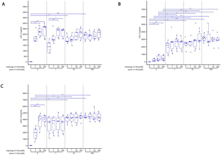
During an infection, innate immune cells must adjust nature and strength of their responses to changing pathogen abundances. To determine how stimulation of the pathogen sensing TLR4 shapes subsequent macrophage responses, we systematically varied priming and restimulation concentrations of its ligand KLA. We find that different priming strengths have very distinct effects at multiple stages of the signaling response, including receptor internalization, MAPK activation, cytokine and chemokine production, and nuclear translocation and chromatin association of NFκB and IκB members. In particular, restimulation-induced TNF-α production required KLA doses equal to or greater than those used for prior exposure, indicating that macrophages can detect and adaptively respond to changing TLR4 stimuli. Interestingly, while such adaptation was dependent on the anti-inflammatory cytokine IL-10, exogenous concentrations of IL-10 corresponding to those secreted after strong priming did not exert suppressive effects on TNF-α without such prior priming, confirming the critical role of TLR4 stimulation history.

Keywords: IL-10; adaptation; inflammation; innate immunity; macrophages; pattern recognition receptor (PRR); temporal gradients; toll-like receptor 4 (TLR4).


Figures

Fig. 1:
Fig. 1:. (A,B) Basic experimental protocol used for BMDM generation, priming and restimulation.
(A) Bone marrow derived macrophages (BMDMs) were derived from isolated bone marrow cells of wildtype C57BL/6J mice by M-CSF differentiation. BMDMs were treated with varying doses of KLA (prime) for 4 hrs. Then, after replacing the medium, cells are rested for 1 hr and are restimulated (challenge) again with KLA. Depending on the experimental read-out, challenge times differ. (C) TLR4 internalization becomes stronger as priming concentrations increase. BMDMs were incubated with 5% FBS DMEM. During priming, cells were stimulated with 0, 0.03, 0.1, 0.3, 1, 10 or 100 nM KLA . After 4 hrs, cells were washed and medium was replaced with 5% FBS DMEM and cells were incubated for 1 hr. Subsequently, cells were washed and stained for TLR4 surface expression. Cell surface expression was determined by using a LSRII flow cytometer (BDBiosciences). Raw data was further processed with FlowJo. Median fluorescence intensities were normalized to the median fluorescence intensity of the TLR4 detecting antibody from a TLR4 KO IMM cellline and is represented in percent of non-primed control sample (set as 100%). (D-G) Dose dependent activation and adaptation of MAP kinases and IκBa. BMDMs were incubated with 5% FBS DMEM. During priming, cells were stimulated with 0, 0.03, 0.1, 0.3, 1 or 100 nM KLA . After 4 hrs, cells were washed and medium was replaced with 5% FBS DMEM and cells were incubated for 1 hr. Subsequently, restimulation was performed using 0, 1 or 100 nM KLA. After 20 mins, cells were washed, fixed, permeabilized and intracellularly stained for (D) Erk1/2, (E) p38, (F) JNK1/2, and (G) IkBa expression. Median fluorescence intensity of samples was determined by using a LSRII flow cytometer (BDBiosciences). Raw data was further processed with FlowJo. Data is given in % of maximal median fluorescence intensity within each replicate (set as 100%) and was normalized by subtracting median fluorescence intensity of the sample with the detected minimal median fluorescence intensity (set as 0%). Each replicate was performed with BMDMs from different mice and data sets include at least 4 replicates (n=4). For (C): Kruskal–Wallis test with post hoc Dunn-Bonferroni comparisons of primed samples with untreated sample (planned comparisons) and for (D-G): Kruskal–Wallis test with post hoc Dunn-Bonferroni comparisons: *p < .05, **p < .01, ***p < .001, ****p < .0001.
Fig. 2:
Fig. 2:. KLA concentration during priming dictates hyper- and hyporesponsive behavior of cyto- and chemokine release in response to restimulation
BMDMs were incubated with 5% FBS DMEM. During priming, cells were stimulated with 0, 1, 10 or 100 nM KLA. After 4 hrs, cells were washed and medium was replaced with 5% FBS DMEM and cells were incubated for 1 hr. Subsequently, re-stimulation was performed using 0, 1, 10 or 100 nM KLA. After 3 hrs, supernatants were collected, processed with the LegendPlex Multiplex Assay Kits and cyto- and chemokine levels for (A) TNF-α, (B) IL-6, (C) CXCL-1, (D) IFN-β, (E) IL-10 and (F) CCL-2 were determined by using a LSRII flow cytometer (BDBiosciences). Raw data was further processed with LegendPlex Desktop software. Each replicate was performed with BMDMs from different mice and data sets include 6 replicates (n=6). Kruskal– Wallis test with post hoc Dunn-Bonferroni comparisons: *p < .05, **p < .01, ***p < .001, ****p < .0001. (G) Correlation analysis of TNF-α and IL-10 secretion in BMDMs. Spearman correlation coefficients for different prime and re-stimulation (challenge) KLA doses were calculated from samples shown in (A) and (E).
Fig. 3:
Fig. 3:. KLA concentration during priming dictates hyper- and hyporesponsive behavior of cyto- and chemokine release in response to restimulation while blocking IL-10 reverses hyporesponsiveness.
BMDMs were incubated with 5% FBS DMEM containing either IL-10R blocking antibody or it’s isotype control. During priming, cells were stimulated with 0, 0.03, 0.1, 0.3, 1 or 100 nM KLA . After 4 hrs, cells were washed and medium was replaced with 5% FBS DMEM containing either IL-10R blocking antibody or it’s isotype control and cells were incubated for 1 hr. Subsequently, re-stimulation was performed using 0, 1 or 100 nM KLA. After 3 hrs, supernatants were collected, processed with the LegendPlex Multiplex Assay Kits and cyto- and chemokine levels for (A) TNF-α, (B) IL-6, (C) CXCL-1, (D) IFN-β, (E) IL-10 and (F) CCL-2 were determined by using a LSRII flow cytometer (BDBiosciences). Raw data was further processed with LegendPlex Desktop software. Each replicate was performed with BMDMs from different mice and data sets include at least 5 replicates (n=5). Kruskal–Wallis test with post hoc Dunn-Bonferroni comparisons: *p < .05, **p < .01, ***p < .001, ****p < .0001.
Fig. 4:
Fig. 4:. Expression of negative feedback inhibitor SOCS-3 and BCL-3 induced by TLR4 activation depends on priming concentration and is differently affected by IL-10.
(A,B) BMDMs were incubated with 5% FBS DMEM containing either IL-10R blocking antibody or it’s isotype control. During priming, cells were stimulated with 0, 0.1, 1 or 100 nM KLA . After 4 hrs, cells were washed and medium was replaced with 5% FBS DMEM containing either IL-10R blocking antibody or it’s isotype control and cells were incubated for 1 hr. Subsequently, restimulation was performed using 0 or 100 nM KLA. After 1 hr, total RNA was isolated and subjected to qRT-PCR analysis to monitor (A) SOCS-3 and (B) BCL-3 mRNA expression. The expression of aforementioned mRNAs was normalized to SDHA mRNA expression. Relative expression of mRNA is given in fold of mRNA expression in naïve and unstimulated control cells. Each replicate was performed with BMDMs from different mice and data sets include at least 5 replicates (n=5). (C-F) BMDMs were incubated with 5% FBS DMEM containing either IL-10R blocking antibody or it’s isotype control. During priming, cells were stimulated with 0, 0.03, 0.1, 0.3, 1 or 100 nM KLA . After 4 hrs, cells were washed and medium was replaced with 5% FBS DMEM containing either IL-10R blocking antibody or it’s isotype control and cells were incubated for 1 hr. Subsequently, re-stimulation was performed using 0 or 100 nM for up to 2 hrs. After depicted times, cells were washed, fixed, permeabilized and intracellularly stained for (C,D) BCL-3 expression as well as (E,F) p50. Expression was determined by using a LSRII flow cytometer (BDBiosciences). Raw data was further processed with FlowJo. Data is given in % of maximal median fluorescence intensity within each replicate (set as 100%). Each replicate was performed with BMDMs from different mice and data sets include at least 3 replicates (n=3). Kruskal–Wallis test with post hoc Dunn-Bonferroni comparisons: *p < .05, **p < .01, ***p < .001, ****p < .0001.
Fig. 5:
Fig. 5:. Restimulation induced nuclear localization of p65, p50 and BCL-3 are differently affected by varying priming concentrations and depend on IL-10 signaling.
BMDMs were incubated with 5% FBS DMEM containing either IL-10R blocking antibody or it’s isotype control. During priming, cells were stimulated with 0, 0.03, 0.1, 0.3, 1 or 100 nM KLA . After 4 hrs, cells were washed and medium was replaced with 5% FBS DMEM containing either IL-10R blocking antibody or it’s isotype control and cells were incubated for 1 hr. Subsequently, re-stimulation was performed using 0, 1 or 100 nM KLA. After 20 mins, cells were washed, fixed, permeabilized and intracellularly stained for (A,B) p65, (C,D) p50, and (E,F) BCL-3 expression as well as chromatin. Expression and localization were determined by using a CellInsight CX7 Pro HCS Platform (Thermo Fisher Scientific) equipped with an 40x objective lens. Raw data was further processed with CellProfiler. Data is given in relative mean nuclear intensity (A,C,E) and nuclear to cytoplasmic ratio (B,D,F) of the corresponding fluorophore-coupled antibody for p65, p50 or BCL-3. Each replicate was performed with BMDMs from different mice and data sets include 6 replicates (n=6). Kruskal–Wallis test with post hoc Dunn-Bonferroni comparisons: *p < .05, **p < .01, ***p < .001, ****p < .0001.
Fig. 6:
Fig. 6:. IL-10 dependent adaptation of BCL-3, p50 and p65 association with κB sites in TNF-α, IL-6 and IL-10 genes.
BMDMs were incubated with 5% FBS DMEM containing either IL-10R blocking antibody or it’s isotype control. During priming, cells were stimulated with 100 nM KLA . After 4 hrs, cells were washed and medium was replaced with 5% FBS DMEM containing either IL-10R blocking antibody or it’s isotype control and cells were incubated for 1 hr. Subsequently, re-stimulation was performed using 0 or 100 nM KLA. After 20 mins, cells were washed and fixed to cross-link chromatin and protein interactions and chromatin was sheared and further processed. Chromatin immuno-precipitation (ChIP) of either, cross-linked (A-C) BCL-3, (D-F) p50 or (E-G) p65 – chromatin complexes was conducted with BCL-3, p50 or p65 targeting antibodies and, in parallel, with an Isotype control antibody. Protein – chromatin complexes were isolated and reverse cross-linked. DNA was purified and quantified with qPCR using specific primers for kB sites in TNF-α (A,D,G), IL-6 (B,E,FH and IL-10 (C,F,I) genes. The amount of aforementioned chromatin DNA in anti-p65 antibody immunoprecipitated samples was normalized to DNA amounts in their respective anti-Isotype-control antibody immuno-precipitated samples (fold enrichment). Each replicate was performed with BMDMs from different mice and data sets include 4 replicates (n=4). Kruskal–Wallis test with post hoc Dunn-Bonferroni comparisons in (A),(B),(C) and Wilcoxon- test in (D): *p < .05, **p < .01, ***p < .001, ****p < .0001 .
Fig. 7:
Fig. 7:. Hypo-responsiveness (Adaptation) induced not only by IL-10
BMDMs were incubated with 5% FBS DMEM. During priming, cells were stimulated with 0, 0.1 or 100 nM KLA. 20 minutes later, recombinant murine IL-10 was added in depicted concentrations. After 4 hrs priming, cells were washed, medium was replaced with 5% FBS DMEM and cells were incubated for 1 hr. Subsequently, re-stimulation was performed using 0 or 100 nM KLA. 20 minutes later, recombinant murine IL-10 was added in depicted concentrations. After 3 hrs re-stimulation, supernatants were collected, processed with the LegendPlex Multiplex Assay Kits and cyto- and chemokine levels for (A) TNF-α, (B) IL-6, (C) CXCL-1, (D) IFN-β, (E) IL-10 and (F) CCL-2 were determined by using a LSRII flow cytometer (BDBiosciences). Raw data was further processed with LegendPlex Desktop software. Each replicate was performed with BMDMs from different mice and data sets include 6 replicates (n=6). Kruskal–Wallis test with post hoc Dunn-Bonferroni comparisons: *p < .05, **p < .01, ***p < .001, ****p < .0001.

Similar articles

References

    1. Medzhitov R., Toll-like receptors and innate immunity. Nat Rev Immunol, 2001. 1(2): p. 135–45. - PubMed
    1. Lu Y.C., Yeh W.C., and Ohashi P.S., LPS/TLR4 signal transduction pathway. Cytokine, 2008. 42(2): p. 145–151. - PubMed
    1. Gustafson G.L., Rhodes M.J., and Hegel T., Monophosphoryl lipid A as a prophylactic for sepsis and septic shock. Prog Clin Biol Res, 1995. 392: p. 567–79. - PubMed
    1. Salkowski C.A., et al., Pulmonary and hepatic gene expression following cecal ligation and puncture: monophosphoryl lipid A prophylaxis attenuates sepsis-induced cytokine and chemokine expression and neutrophil infiltration. Infect Immun, 1998. 66(8): p. 3569–78. - PMC - PubMed
    1. Butcher S.K., et al., Toll-Like Receptors Drive Specific Patterns of Tolerance and Training on Restimulation of Macrophages. Front Immunol, 2018. 9: p. 933. - PMC - PubMed

Publication types