. 2024 Mar;28(1):1-12.
doi: 10.12717/DR.2024.28.1.1. Epub 2024 Mar 31.

Gonadotropins Regulate the mRNA Expression of Gonadotropin-Releasing Hormone and Its Receptors in the Mouse Ovary and Uterus

[Article in Korean]
Affiliations

Gonadotropins Regulate the mRNA Expression of Gonadotropin-Releasing Hormone and Its Receptors in the Mouse Ovary and Uterus

[Article in Korean]
Soeun Moon et al. Dev Reprod. 2024 Mar.

Abstract

Gonadotropin-releasing hormone (GnRH), a critical hormone produced in the hypothalamus, is essential for regulating reproductive processes. It has also been demonstrated the presence of GnRH and its receptors (GnRHR) in ovarian and uterine tissues, but little was known about the regulation mechanism of their expression in these organs and ovarian aging. Therefore, the aim of this study was to investigate the expression of GnRHR in the ovary and uterus of mice, particularly after high-dose gonadotropin treatments and in relation to aging. Quantitative real-time-PCR (qRT-PCR) revealed that pituitary gland had the highest GnRHR expression in both young and aged mice. In addition, liver expression was higher in young mice, whereas thymus expression was higher in aged mice. GnRHR mRNA was present in the ovaries of both young and aged mice but nearly undetectable in the uterus of aged mice. We next examined the expression of GnRHR in the ovary and uterus in response to high-dose administration of pregnant mare serum gonadotropin (PMSG). After PMSG administration, GnRH mRNA levels were significantly decreased in the ovary but increased in the uterus. The expression of GnRH mRNA in these organs showed opposite trends to that of GnRHR expression. These results suggest the involvement of GnRH in age-related reproductive decline and the potential effects of high-dose gonadotropin treatments on reproductive organ function.


Conflict of interest statement

The authors declare no potential conflict of interest.

Figures

Fig. 1.
Fig. 1.. Conventional RT-PCR analysis of GnRHR mRNA expression in various organs of young and aged female mice.
(A) Conventional RT-PCR analysis was conducted to examine the expression of GnRHR mRNA in the various organs of 6-week-old female mice. GnRHR mRNA is expressed in all the tested organs. Notably, GnRHR mRNA exhibits higher expression in the pituitary gland, heart, liver, kidney, and spleen. (B) At 12 months of age in female mice, GnRHR mRNA exhibits pervasive expression throughout all assessed organs, with the elevated levels in the pituitary gland, thymus, lung, small intestine, kidney, and spleen. RT-PCR, reverse transcription PCR; GnRHR, GnRH and its receptors; GnRH, gonadotropin-releasing hormone.
Fig. 2.
Fig. 2.. GnRHR expression in the ovary and uterus of young and aged female mice.
Conventional RT-PCR analysis shows GnRHR mRNA expression in the ovary and uterus of young and aged female mice. GnRHR mRNA is highly expressed in both the ovary and uterus of young mice, whereas its expression is notably reduced in the uterus compared to the ovary of aged mice. GnRH, gonadotropin-releasing hormone; GnRHR, GnRH and its receptors; RT-PCR, real-time PCR.
Fig. 3.
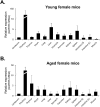
Fig. 3.. Quantitative RT-PCR analysis of GnRHR mRNA expression in various organs of young and aged female mice.
(A) GnRHR mRNA expression in various organs of 6-week-old female mice measured by qRT-PCR shows higher in pituitary, liver and spleen compared to other organs. In particular, the pituitary gland shows a 20-fold higher level of expression compared to the next highest levels in the liver or spleen. (B) The expression of GnRHR mRNA in various organs of 12-month-old female mice, as measured by qRT-PCR, shows higher levels in the pituitary gland, lung, and thymus compared to the other organs. Significantly, the pituitary gland shows a 5-fold higher level of expression compared to the next highest levels in the lung and the thymus. GnRH, gonadotropin-releasing hormone; GnRHR, GnRH and its receptors; qRT-PCR, quantitative real-time PCR.
Fig. 4.
Fig. 4.. GnRHR expression in the ovary and uterus of young and aged female mice.
(A) qRT-PCR analysis exhibits that GnRHR mRNA is highly expressed in the ovary and uterus of young mice compared to aged mice. Interestingly, its expression in the uterus is much lower in aged mice than in young mice. (B) Localization of GnRH receptor in the ovary by immunohistochemical staining. GnRH receptor is presented in the granulosa cells of an ovarian follicle. Magnification, ×1,000. GnRH, gonadotropin-releasing hormone; GnRHR, GnRH and its receptors; qRT-PCR, quantitative real-time PCR.
Fig. 5.
Fig. 5.. GnRHR mRNA expression in the ovary and uterus after PMSG injection.
(A) GnRHR mRNA expression in the ovary and uterus shows a dose-dependent response to PMSG injection. GnRHR mRNA expression levels show a significant decrease in the ovary after 10 and 30 IU administration compared to the saline control. (B) GnRHR mRNA expression in the uterus exhibits a significant increase in the uterus after 10 and 30 IU administration compared to the saline control. All data are presented as mean±SEM (n=3). * p<0.05. GnRHR, GnRH and its receptors; GnRH, gonadotropin-releasing hormone.
Fig. 6.
Fig. 6.. GnRH mRNA expression in the ovary and uterus after PMSG injection.
(A) GnRH mRNA expression in the ovary and uterus shows a dose-dependent response to PMSG injection. GnRH mRNA expression levels show a significant increase in the ovary after 10 and 30 IU administration compared to the saline control. (B) GnRH mRNA expression in the uterus exhibits a significant decrease in the uterus after 10 and 30 IU administration compared to the saline control. All data are presented as mean±SEM (n=3). * p<0.05. GnRH, gonadotropin-releasing hormone.

References

    1. Acevedo B, Gomez-Palomares JL, Ricciarelli E, Hernández ER. Triggering ovulation with gonadotropin-releasing hormone agonists does not compromise embryo implantation rates. Fertil Steril. 2006;86:1682–1687. doi: 10.1016/j.fertnstert.2006.05.049. - DOI - PubMed
    1. Aguilar-Rojas A, Huerta-Reyes M. Human gonadotropin-releasing hormone receptor-activated cellular functions and signaling pathways in extra-pituitary tissues and cancer cells. Oncol Rep. 2009;22:981–990. doi: 10.3892/or_00000525. - DOI - PubMed
    1. Caligara C, Navarro J, Vargas G, Simón C, Pellicer A, Remohí J. The effect of repeated controlled ovarian stimulation in donors. Hum Reprod. 2001;16:2320–2323. doi: 10.1093/humrep/16.11.2320. - DOI - PubMed
    1. Cao N, Cao L, Gao M, Wang H, Zhang L, Yang L. Changes in mRNA and protein levels of gonadotropin releasing hormone and receptor in ovine thymus, lymph node, spleen, and liver during early pregnancy. Domest Anim Endocrinol. 2021;76:106607. doi: 10.1016/j.domaniend.2021.106607. - DOI - PubMed
    1. Catt KJ, Loumaye E, Wynn PC, Iwashita M, Hirota K, Morgan RO, Chang JP. GnRH receptors and actions in the control of reproductive function. J Steroid Biochem. 1985;23:677–689. doi: 10.1016/S0022-4731(85)80003-0. - DOI - PubMed