

Brain Fluid Clearance After Traumatic Brain Injury Measured Using Dynamic Positron Emission Tomography
- PMID: 38655117
- PMCID: PMC11035850
- DOI: 10.1089/neur.2024.0010
Brain Fluid Clearance After Traumatic Brain Injury Measured Using Dynamic Positron Emission Tomography
Abstract
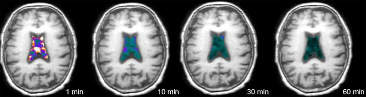
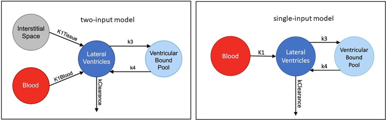
Brain fluid clearance by pathways including the recently described paravascular glymphatic system is a critical homeostatic mechanism by which metabolic products, toxins, and other wastes are removed from the brain. Brain fluid clearance may be especially important after traumatic brain injury (TBI), when blood, neuronal debris, inflammatory cells, and other substances can be released and/or deposited. Using a non-invasive dynamic positron emission tomography (PET) method that models the rate at which an intravenously injected radiolabeled molecule (in this case 11C-flumazenil) is cleared from ventricular cerebrospinal fluid (CSF), we estimated the overall efficiency of brain fluid clearance in humans who had experienced complicated-mild or moderate TBI 3-6 months before neuroimaging (n = 7) as compared to healthy controls (n = 9). While there was no significant difference in ventricular clearance between TBI subjects and controls, there was a significant group difference in dependence of ventricular clearance upon tracer delivery/blood flow to the ventricles. Specifically, in controls, ventricular clearance was highly, linearly dependent upon blood flow to the ventricle, but this relation was disrupted in TBI subjects. When accounting for blood flow and group-specific alterations in blood flow, ventricular clearance was slightly (non-significantly) increased in TBI subjects as compared to controls. Current results contrast with past studies showing reduced glymphatic function after TBI and are consistent with possible differential effects of TBI on glymphatic versus non-glymphatic clearance mechanisms. Further study using multi-modal methods capable of assessing and disentangling blood flow and different aspects of fluid clearance is needed to clarify clearance alterations after TBI.
Keywords: PET scanning; adult brain injury; cerebrospinal fluid; clearance; glymphatic system.
© Tracy Butler et al., 2024; Published by Mary Ann Liebert, Inc.
Conflict of interest statement
No competing financial interests exist.
Figures

References
Grants and funding
LinkOut - more resources
Full Text Sources
Miscellaneous