. 2024 May 16;9(5):e0017924.
doi: 10.1128/msystems.00179-24. Epub 2024 Apr 24.

Deciphering Staphylococcus aureus- host dynamics using dual activity-based protein profiling of ATP-interacting proteins

Affiliations

Deciphering Staphylococcus aureus- host dynamics using dual activity-based protein profiling of ATP-interacting proteins

Stephen Dela Ahator et al. mSystems. .

Abstract

The utilization of ATP within cells plays a fundamental role in cellular processes that are essential for the regulation of host-pathogen dynamics and the subsequent immune response. This study focuses on ATP-binding proteins to dissect the complex interplay between Staphylococcus aureus and human cells, particularly macrophages (THP-1) and keratinocytes (HaCaT), during an intracellular infection. A snapshot of the various protein activity and function is provided using a desthiobiotin-ATP probe, which targets ATP-interacting proteins. In S. aureus, we observe enrichment in pathways required for nutrient acquisition, biosynthesis and metabolism of amino acids, and energy metabolism when located inside human cells. Additionally, the direct profiling of the protein activity revealed specific adaptations of S. aureus to the keratinocytes and macrophages. Mapping the differentially activated proteins to biochemical pathways in the human cells with intracellular bacteria revealed cell-type-specific adaptations to bacterial challenges where THP-1 cells prioritized immune defenses, autophagic cell death, and inflammation. In contrast, HaCaT cells emphasized barrier integrity and immune activation. We also observe bacterial modulation of host processes and metabolic shifts. These findings offer valuable insights into the dynamics of S. aureus-host cell interactions, shedding light on modulating host immune responses to S. aureus, which could involve developing immunomodulatory therapies.

Importance: This study uses a chemoproteomic approach to target active ATP-interacting proteins and examines the dynamic proteomic interactions between Staphylococcus aureus and human cell lines THP-1 and HaCaT. It uncovers the distinct responses of macrophages and keratinocytes during bacterial infection. S. aureus demonstrated a tailored response to the intracellular environment of each cell type and adaptation during exposure to professional and non-professional phagocytes. It also highlights strategies employed by S. aureus to persist within host cells. This study offers significant insights into the human cell response to S. aureus infection, illuminating the complex proteomic shifts that underlie the defense mechanisms of macrophages and keratinocytes. Notably, the study underscores the nuanced interplay between the host's metabolic reprogramming and immune strategy, suggesting potential therapeutic targets for enhancing host defense and inhibiting bacterial survival. The findings enhance our understanding of host-pathogen interactions and can inform the development of targeted therapies against S. aureus infections.

Keywords: ATP-interacting proteins; HaCaT cells; Staphylococcus aureus; THP-1 cells; activity-based protein profiling (ABPP); bacterial metabolism; host immune response; host–pathogen interactions.


Conflict of interest statement

The authors declare no conflict of interest.

Figures
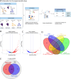
Fig 1
Fig 1
(A) Schematic representation of the experimental setup including infection, ATP-interacting protein profiling, and data analysis. The diagram gives an overview of the S. aureus infection of THP-1 and HaCaT cells and the control cultures of the human cells and bacteria grown in isolation. Probe labeling of the protein extracts from the samples was performed using the desthiobiotin-ATP probe and the modified proteins identified using mass spectrometry. After infection, the differentially activated proteins in the S. aureus and host cells were identified by comparing them with the control samples, which are bacteria and human cell lines grown in isolation. (B and C) Activated and repressed proteins in S. aureus following infection in the HaCaT and THP-1 cells and chemoproteomic enrichment, respectively. The abundance ratio is expressed as a log2 value of the relative activity of the proteins. (D) Differentially activated and repressed S. aureus protein distribution following infection in the HaCaT and THP-1 cells. (E) The general protein profile derived from the ATP probe in the S. aureus cell following infection in the THP-1 and HaCaT cells and their overlap with the total proteome in the S. aureus USA300 genome using the hidden Markov model (HMM) profile derived from the UniProt database.
Fig 2
Fig 2
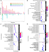
(A) COG analysis of the activated and repressed S. aureus proteins following infection in the THP-1 and HaCaT cells and chemoproteomic enrichment. The length of the up and down bars represents the number of activated and repressed proteins, respectively. The bar chart illustrates the differential activation of S. aureus proteins categorized by COG during infection in the THP-1 and HaCaT cells. Each bar represents the degree to which proteins within a specific COG category are either activated or repressed. The x-axis labels, denoted by single-letter codes, correspond to distinct functional COG categories. The GO analysis of the (B) bacterial activated and (C) repressed proteins following infection in the HaCaT cells and (D) the bacterial activated proteins following infection in the THP-1 cells.
Fig 3
Fig 3
(A) The metabolic map and heatmap show S. aureus proteins involved in energy metabolism and carbon source utilization after infection in HaCaT and THP-1 cells. The heatmap shows the relative activity of the bacterial proteins. The heatmap scale shows the log2 value for the protein activity. The map renders the pathways derived from the KEGG and EggNOG databases. (B and C) The time-course HaCaT and THP-1 infection and cell viability assays following infection with the S. aureus strains; the WT, transposon mutant of dhaK, and its in-trans complemented strain [dhaK(dhaK)]. The data represent the mean ± SD of three independent experiments. A P-value >0.05 is considered significant (*P < 0.01; **P < 0.001).
Fig 4
Fig 4
(A) A diagrammatic rendering of the amino acid synthesis and transport pathway involving bacterial proteins activated and repressed following infection in the THP-1 and HaCaT cells. (B) The heatmap shows the relative activity (log2 value) of bacterial proteins involved in the amino acid synthesis, metabolism, and transport pathway. (C and D) The time-course HaCaT and THP-1 infection and cell viability assay following infection with the WT S. aureus strains; transposon mutants of brnQ, IlvN, and leuC; and their corresponding complemented strains brnQ(brnQ), ilvN(ilvN), and leuC(leuC). The data represent the mean ± SD of three independent experiments. A P-value >0.05 is considered significant. *P < 0.01; **P < 0.001.
Fig 5
Fig 5
Relative activity of S. aureus histidine kinases and serine/threonine kinases following infection in (A) HaCaT cells and (B) THP-1 cells. (C) Galleria melonella survival following infection by the wild-type S. aureus USA300_JE2 and the transposon mutants of the TCSs SrrAB and ArlSR. PBS was used as a control. (D and E) The time-course HaCaT and THP-1 infection and cell viability assay following infection with the WT S. aureus strains; transposon mutants of srrA, srrB, arlR, and arlS; and their corresponding complemented strains srrA(srrA), srrB(srrB), arlS(arlR), and arlR(arlR). The data represent the mean ± SD of three independent experiments. A P-value >0.05 is considered significant. *P < 0.01; **P < 0.001.
Fig 6
Fig 6
(A) Distribution of activated and repressed human proteins from HaCaT and THP-1 cells following S. aureus infection. (B) This bar chart illustrates the differential activation of human proteins categorized by COG in THP-1 and HaCaT cells in response to S. aureus infection. Each bar represents the degree to which proteins within a specific COG category are either activated or repressed. The x-axis labels, denoted by single-letter codes, correspond to distinct functional COG categories.
Fig 7
Fig 7
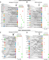
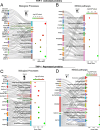
Sankey dot plot showing the flow of activated proteins to their respective (A) biological processes and (B) KEGG pathway enrichment in HaCaT cells following S. aureus infection. (C) Sankey dot plots depicting the repressed proteins and their corresponding biological processes and (D) KEGG pathway enrichment in HaCaT cells following S. aureus infection. The dot size indicates gene count, and color intensity represents the −log10(P-value).
Fig 8
Fig 8
Sankey dot plot illustrating the activated proteins and their related (A) biological processes and (B) KEGG pathway enrichment in THP-1 cells following S. aureus infection. (C) Sankey dot plots of repressed proteins and their biological processes and (D) KEGG pathway in THP-1 cells following S. aureus infection. The dot size indicates gene count, and color intensity represents the −log10(P-value).
Fig 9
Fig 9
(A) List of human immune response pathways. Heatmap and pathway mapping of differentially activated human proteins from the HaCaT and THP-1 cells following S. aureus infection. The heatmap scale shows the log2 value for the protein activity.

Similar articles

References

    1. Brown AF, Leech JM, Rogers TR, McLoughlin RM. 2014. Staphylococcus aureus colonization: modulation of host immune response and impact on human vaccine design. Front Immunol 4:507. doi:10.3389/fimmu.2013.00507 - DOI - PMC - PubMed
    1. Bien J, Sokolova O, Bozko P. 2011. Characterization of virulence factors of Staphylococcus aureus: novel function of known virulence factors that are implicated in activation of airway epithelial proinflammatory response. J Pathog 2011:601905. doi:10.4061/2011/601905 - DOI - PMC - PubMed
    1. Foster TJ. 2005. Immune evasion by staphylococci. Nat Rev Microbiol 3:948–958. doi:10.1038/nrmicro1289 - DOI - PubMed
    1. De Jong NWM, Van Kessel KPM, Van Strijp JAG. 2019. Immune evasion by Staphylococcus aureus. Microbiol Spectr 7:2–7. doi:10.1128/microbiolspec.GPP3-0061-2019 - DOI - PMC - PubMed
    1. Wójcik-Bojek U, Różalska B, Sadowska B. 2022. Staphylococcus aureus—a known opponent against host defense mechanisms and vaccine development—do we still have a chance to win? Int J Mol Sci 23:948. doi:10.3390/ijms23020948 - DOI - PMC - PubMed

MeSH terms

LinkOut - more resources