. 2024 Jun 1;131(6):1101-1111.
doi: 10.1152/jn.00077.2024. Epub 2024 Apr 24.

Postsynaptic potentials of soleus motor neurons produced by transspinal stimulation: a human single-motor unit study

Affiliations

Postsynaptic potentials of soleus motor neurons produced by transspinal stimulation: a human single-motor unit study

Nilgün Yildiz et al. J Neurophysiol. .

Abstract

Transspinal (or transcutaneous spinal cord) stimulation is a noninvasive, cost-effective, easily applied method with great potential as a therapeutic modality for recovering somatic and nonsomatic functions in upper motor neuron disorders. However, how transspinal stimulation affects motor neuron depolarization is poorly understood, limiting the development of effective transspinal stimulation protocols for rehabilitation. In this study, we characterized the responses of soleus α motor neurons to single-pulse transspinal stimulation using single-motor unit (SMU) discharges as a proxy given the 1:1 discharge activation between the motor neuron and the motor unit. Peristimulus time histogram, peristimulus frequencygram, and surface electromyography (sEMG) were used to characterize the postsynaptic potentials of soleus motor neurons. Transspinal stimulation produced short-latency excitatory postsynaptic potentials (EPSPs) followed by two distinct phases of inhibitory postsynaptic potentials (IPSPs) in most soleus motor neurons and only IPSPs in others. Transspinal stimulation generated double discharges at short interspike intervals in a few motor units. The short-latency EPSPs were likely mediated by muscle spindle group Ia and II afferents, and the IPSPs via excitation of group Ib afferents and recurrent collaterals of motor neurons leading to activation of diverse spinal inhibitory interneuronal circuits. Further studies are warranted to understand better how transspinal stimulation affects depolarization of α motor neurons over multiple spinal segments. This knowledge will be seminal for developing effective transspinal stimulation protocols in upper motor neuron lesions.NEW & NOTEWORTHY Transspinal stimulation produces distinct actions on soleus motor neurons: an early short-latency excitation followed by two inhibitions or only inhibition and doublets. These results show how transspinal stimulation affects depolarization of soleus α motor neurons in healthy humans.

Keywords: motoneurons; motor units; postsynaptic potentials; transcutaneous spinal cord stimulation; transspinal stimulation.


Conflict of interest statement

No conflicts of interest, financial or otherwise, are declared by the authors.

Figures

None
Graphical abstract
Figure 1.
Figure 1.
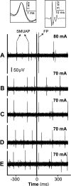
Transspinal stimulation during single-motor unit (SMU) activation. Transspinal stimulation was delivered during voluntary ankle plantar flexion and when a motor unit was repeatedly discharged, as evident by the similar single-motor unit action potential (SMUAP; top left box) recorded with intramuscular fine wire electrodes. When transspinal stimulation simultaneously recruited several motor unit action potentials known as field potentials (FPs; top right box) at a time consistent with the transspinal-evoked potential (TEP) latency in the single-motor unit channel (trace A: stimulus intensity = 80 mA), the intensity was reduced so that the field potential was no longer elicited (traces B and D). However, the same stimulus intensity (70 mA) was sufficient to occasionally recruit SMUAP at the TEP latency (traces C and E). SMUAPs were followed regularly on a template matching program to ensure that the spikes recruited at the TEP latency were the original SMUAP spikes. The top box on the left shows the shapes of the superimposed SMUAPs that were followed during this experiment. The timing of transspinal stimulation is indicated by a thick vertical line at 0 ms. SMUAPs and FPs are identified with arrows.
Figure 2.
Figure 2.
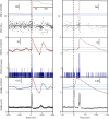
Net excitation followed by net inhibition on single-motor units during transspinal stimulation. From top to bottom: peristimulus frequencygram (PSF)-cumulative sum (CUSUM), PSF, peristimulus time histogram (PSTH)-CUSUM, PSTH, sEMG-CUSUM, surface electromyography (sEMG). In these recordings, 130 stimuli were delivered while the motor unit was discharging at around 6.5 Hz (± 3.3 Hz). Transspinal stimulation of 60.0 mA generated excitation followed by inhibition. The left figure shows the stimulus-induced responses for 800 ms (400 ms before and after the onset of transspinal stimulation). In contrast, the figure on the right highlights the events on an expanded scale. The horizontal lines indicate the maximum prestimulus CUSUM variations, i.e., the error box. EPSP, excitatory postsynaptic potential; IPSP, inhibitory postsynaptic potential. PSF-CUSUM identifies first and second inhibitory responses as 1st and 2nd double-headed arrows. The motor unit shown here is number 1 in Table 1.
Figure 3.
Figure 3.
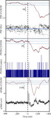
Typical net inhibition on a single-motor unit (SMU) during transspinal stimulation. From top to bottom: peristimulus frequencygram (PSF)-cumulative sum (CUSUM), PSF, peristimulus time histogram (PSTH)-CUSUM, PSTH (counts), surface electromyography (EMG)-CUSUM, and sEMG. One hundred fifty-eight (158) transspinal stimuli were delivered while the motor unit was discharging at around 6 Hz (± 1.4 Hz). The stimulus intensity was 43.3 mA. The inhibition shows two stages in the PSF analysis: an early stage immediately after the short latency weak excitation and a longer latency (around 100 ms after the stimulus onset), more prolonged substantial inhibition. sEMG, on the other hand, indicates three significant peaks that do not correspond with the PSF-CUSUM. These peaks may designate that transspinal stimulation activates three different neuronal pathways.
Figure 4.
Figure 4.
A representative example of a doublet induced by transspinal stimulation (arrow) at latency similar to that of the soleus transspinal-evoked potential (TEP). Single-motor unit action potentials are shown superimposed on the upper right corner.
Figure 5.
Figure 5.
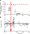
Motoneuron excitability following transspinal stimulation in healthy humans. A: amplitude of the soleus H-reflex conditioned by single-pulse transspinal stimulation at conditioning-test intervals of −50 to 150 ms [modified from Figure 3 of Knikou and Murray (17)]. The soleus H-reflex is presented as a percentage of the control H-reflex. B: peristimulus frequencygram (PSF) analysis of a motor unit from the current study represents changes in the membrane potential of the motoneuron following transspinal stimulation in an excitatory postsynaptic potential (EPSP) + inhibitory postsynaptic potential (IPSP) motor unit. Transspinal stimulation at time zero generates an early excitation indicated by an increased discharge rate at 20 ms followed by inhibition (low discharge rate relative to the prestimulus mean discharge rate shown as a horizontal line). Likely, the soleus H-reflex depression and IPSPs that last up to 150 ms are mediated by similar neuronal mechanisms and/or pathways triggered by transspinal stimulation.

Similar articles

Cited by

References

    1. Gerasimenko Y, Gorodnichev R, Moshonkina T, Sayenko D, Gad P, Edgerton RV. Transcutaneous electrical spinal-cord stimulation in humans. Ann Phys Rehabil Med 58: 225–231, 2015. doi:10.1016/j.rehab.2015.05.003. - DOI - PMC - PubMed
    1. Gorodnichev RM, Pivovarova EA, Puhov A, Moiseev SA, Savochin AA, Moshonkina TR, Chsherbakova NA, Kilimnik VA, Selionov VA, Kozlovskaya IB, Edgerton VR, Gerasimenko YP. Transcutaneous electrical stimulation of the spinal cord: a noninvasive tool for the activation of stepping pattern generators in humans. Hum Physiol 38: 158–167, 2012. doi:10.1134/S0362119712020065. - DOI - PubMed
    1. Hofstoetter US, McKay WB, Tansey KE, Mayr W, Kern H, Minassian K. Modification of spasticity by transcutaneous spinal cord stimulation in individuals with incomplete spinal cord injury. J Spinal Cord Med 37: 202–211, 2014. doi:10.1179/2045772313Y.0000000149. - DOI - PMC - PubMed
    1. Hofstoetter US, Krenn M, Danner SM, Hofer C, Kern H, McKay WB, Mayr W, Minassian K. Augmentation of voluntary locomotor activity by transcutaneous spinal cord stimulation in motor-incomplete spinal cord-injured individuals. Artif Organs 39: E176–E186, 2015. doi:10.1111/aor.12615. - DOI - PubMed
    1. Hofstoetter US, Danner SM, Freundl B, Binder H, Mayr W, Rattay F, Minassian K. Periodic modulation of repetitively elicited monosynaptic reflexes of the human lumbosacral spinal cord. J Neurophysiol 114: 400–410, 2015. doi:10.1152/jn.00136.2015. - DOI - PMC - PubMed

Publication types

LinkOut - more resources