. 2024 Apr 24:13:RP95514.
doi: 10.7554/eLife.95514.

Dependence of nucleosome mechanical stability on DNA mismatches

Affiliations

Dependence of nucleosome mechanical stability on DNA mismatches

Thuy T M Ngo et al. Elife. .

Abstract

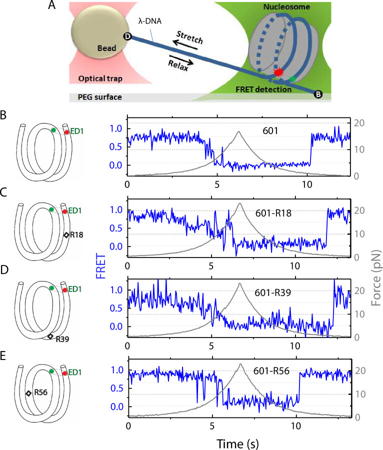
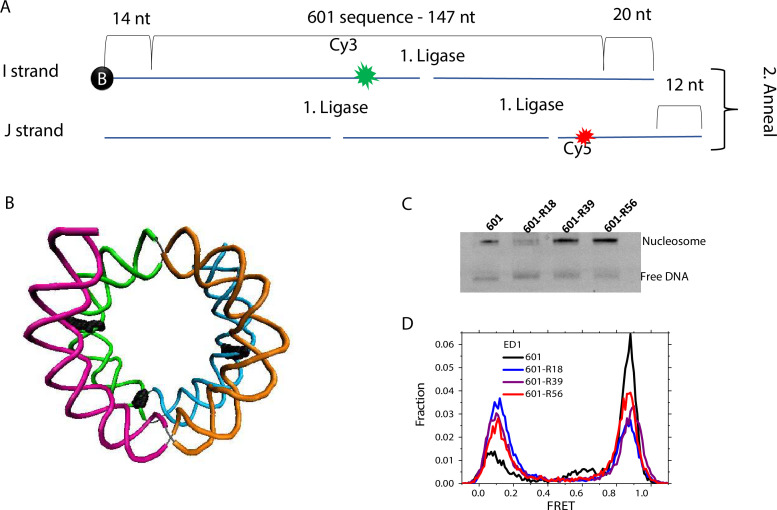
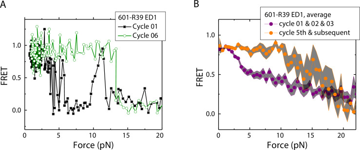
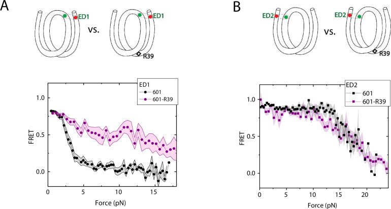
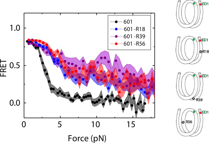
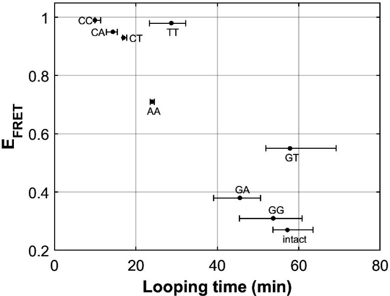
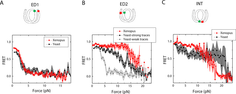
The organization of nucleosomes into chromatin and their accessibility are shaped by local DNA mechanics. Conversely, nucleosome positions shape genetic variations, which may originate from mismatches during replication and chemical modification of DNA. To investigate how DNA mismatches affect the mechanical stability and the exposure of nucleosomal DNA, we used an optical trap combined with single-molecule FRET and a single-molecule FRET cyclization assay. We found that a single base-pair C-C mismatch enhances DNA bendability and nucleosome mechanical stability for the 601-nucleosome positioning sequence. An increase in force required for DNA unwrapping from the histone core is observed for single base-pair C-C mismatches placed at three tested positions: at the inner turn, at the outer turn, or at the junction of the inner and outer turn of the nucleosome. The results support a model where nucleosomal DNA accessibility is reduced by mismatches, potentially explaining the preferred accumulation of single-nucleotide substitutions in the nucleosome core and serving as the source of genetic variation during evolution and cancer progression. Mechanical stability of an intact nucleosome, that is mismatch-free, is also dependent on the species as we find that yeast nucleosomes are mechanically less stable and more symmetrical in the outer turn unwrapping compared to Xenopus nucleosomes.

Keywords: DNA mismatch; DNA repair; S. cerevisiae; fluorescence resonance energy transfer; molecular biophysics; nucleosome; optical tweezers; single molecule biophysics; structural biology; xenopus.


Conflict of interest statement

TN, BL, FW, AB, CW, TH No competing interests declared

Figures

Figure 1.
Figure 1.. Nucleosome unwrapping measurement.
(A) Experimental scheme. The red and green stars represent labelled Cy5 (acceptor) and Cy3 (donor) fluorophores, respectively. Biotin, B, and digoxigenin, D, are used to tether the nucleosome-lambda DNA construct to the surface and the bead, respectively. (B, C, D, E): Representative stretching traces of the outer turn (ED1) for nucleosomes reconstituted from the 601 sequence (B) and from the 601 sequence with containing a mismatch at different positions: on the outer turn (C), at the junction of the outer turn and inner turn (D) and at the inner turn (E). The red and green dots on the DNA bends represent labelled Cy5 and Cy3 fluorophores. The elongated circles enclosing red and green dots represent the ED labeling position. The black diamonds on the DNA bends represent the mismatch position with R18 and R39 on histone = facing minor grooves and R56 on a histone-facing major groove.
Figure 1—figure supplement 1.
Figure 1—figure supplement 1.. Nucleosome preparation.
(A) Scheme of the DNA template prepared by ligation of short, labeled oligos. (B) DNA structure marking three sites of mismatch insertion (R56, R39, and R18, running from left to right). (C) Migration of the 601 nucleosome mismatch containing nucleosomes on 5% native PAGE. (D) FRET histogram of the 601 nucleosome mismatch containing nucleosomes with ED1 labeling scheme. The low FRET peak contains nucleosomes without a fluorescently active acceptor.
Figure 2.
Figure 2.. Unwrapping force of mismatch-containing nucleosomes is higher for subsequent stretching cycles.
(A) Representative single-molecule stretching traces at two stretching cycles from the sample molecule, probe by the ED1 FRET pair in the 601-R18 nucleosome. (B) Averaging FRET vs. Force for many molecules at the first three stretching cycles (purple) and the subsequent stretching cycles (orange). Histone proteins were expressed in Xenopus. The error bars represent S.D. of n=25 and 11 traces for the first three stretching cycles (purple) and for the cycle 5th and the subsequent stretching cycles (orange), respectively.
Figure 3.
Figure 3.. Enhancement of nucleosome mechanical stability by DNA mismatch.
Average of FRET vs. Force for ED1 probe (A) and ED2 probe (B) for the 601 nucleosome (black) and for the first stretching cycle of the mismatch containing nucleosome 601-R39 (purple). Histone proteins were expressed in Xenopus. The error bars represent S.D. of n=25 and 7 for the ED1 probe of the 601 and 601-R39 nucleosomes (A) and n=20 and 39 for the ED2 probe of the 601 and 601-R39 nucleosomes (B), respectively.
Figure 4.
Figure 4.. Mismatch position-dependence of nucleosome unwrapping.
Average of FRET vs. Force for ED1 probe for the 601 nucleosome (black) and the mismatch-containing nucleosome 601-R39 (purple), 601-R18 (blue) and 601-R56 (red). Histone proteins were expressed in Xenopus. The error bars represent S.D. of n=25, 11, 7, and 10 for the 601, 601-R18, 601-R39, and 601-R56 nucleosomes, respectively.
Figure 5.
Figure 5.. C-C mismatch enhances DNA flexibility.
(A) Single-molecule cyclization assay: The DNA construct with 10-nucleotide complementary sticky ends is immobilized on a PEG passivated imaging chamber. DNA looping is induced using the imaging buffer containing 1 M NaCl followed by time course TIRF imaging. To calculate the looping time, the fraction of looped molecules (high FRET) as a function of time is fitted to an exponential function, et/(loopingtime) (right panel for one run of experiments). (B, C) Fitted looping time for the right half of the 601 construct without and with mismatches (B) and with the biotin position being moved by 16 nt (C). Error bars represented the S.E.M with n=3 technical replicates.
Figure 5—figure supplement 1.
Figure 5—figure supplement 1.. Single-molecule cyclization time course quantification.
Each run has four times courses of looped fraction (percentage in high FRET population) vs time. Approximately 2500–3500 molecules were quantified at each timestamp during the experiment, and three independent experiments (run 1, run 2, and run 3) were performed for each sequence. (A - C) Fraction of DNA molecules in high FRET over time for the 601 sequences with a C-C mismatch. Run 1 in panel A is also shown in Figure 5. (D - F) Fraction of DNA molecules in high FRET over time for the 601 sequences with a C-C mismatch and biotin moved 16 nucleotides toward the center of the construct.
Figure 6.
Figure 6.. DNA flexibility enhancement is dependent on mismatch type.
Looping times for DNA containing a single mismatch (one of eight types each) and an intact DNA without a mismatch. Also shown are ensemble FRET efficiencies (EFRET) from Fields et al., 2013 as a measure of DNA buckling for the same type of mismatch.
Figure 7.
Figure 7.. Unwrapping of yeast vs. Xenopus reconstituted nucleosomes.
Average of FRET vs. Force for nucleosomes reconstituted from Xenopus (red) vs yeast (black and gray) histone proteins with DNA labeled by outer turn probes ED1 (A), ED2 (B) and inner turn probe INT (C). The error bars represent S.D. of n=17 (Xenopus) and 5 (Yeast) nucleosomes with the ED1 probe (A), n=20 (Xenopus), 6 (Yeast – strong) and 4 (Yeast-weak) nucleosomes with the ED2 probe (B), and n=22 (Xenopus) and 6 (Yeast) nucleosomes with the INT probe (C), respectively.

Update of

  • doi: 10.1101/2022.11.21.517409
  • doi: 10.7554/eLife.95514.1
  • doi: 10.7554/eLife.95514.2

Similar articles

References

    1. Basu A, Bobrovnikov DG, Qureshi Z, Kayikcioglu T, Ngo TTM, Ranjan A, Eustermann S, Cieza B, Morgan MT, Hejna M, Rube HT, Hopfner KP, Wolberger C, Song JS, Ha T. Measuring DNA mechanics on the genome scale. Nature. 2021;589:462–467. doi: 10.1038/s41586-020-03052-3. - DOI - PMC - PubMed
    1. Basu A, Bobrovnikov DG, Cieza B, Arcon JP, Qureshi Z, Orozco M, Ha T. Deciphering the mechanical code of the genome and epigenome. Nature Structural & Molecular Biology. 2022;29:1178–1187. doi: 10.1038/s41594-022-00877-6. - DOI - PMC - PubMed
    1. Blosser TR, Yang JG, Stone MD, Narlikar GJ, Zhuang X. Dynamics of nucleosome remodelling by individual ACF complexes. Nature. 2009;462:1022–1027. doi: 10.1038/nature08627. - DOI - PMC - PubMed
    1. Chen X, Chen Z, Chen H, Su Z, Yang J, Lin F, Shi S, He X. Nucleosomes suppress spontaneous mutations base-specifically in eukaryotes. Science. 2012;335:1235–1238. doi: 10.1126/science.1217580. - DOI - PubMed
    1. Chen P, Li W, Li G. Structures and functions of chromatin fibers. Annual Review of Biophysics. 2021;50:95–116. doi: 10.1146/annurev-biophys-062920-063639. - DOI - PubMed

Publication types