. 2024 May 1;11(Pt 3):395-404.
doi: 10.1107/S2052252524002549.

Crystal structure of human peptidylarginine deiminase type VI (PAD6) provides insights into its inactivity

Affiliations

Crystal structure of human peptidylarginine deiminase type VI (PAD6) provides insights into its inactivity

Fanomezana M Ranaivoson et al. IUCrJ. .

Abstract

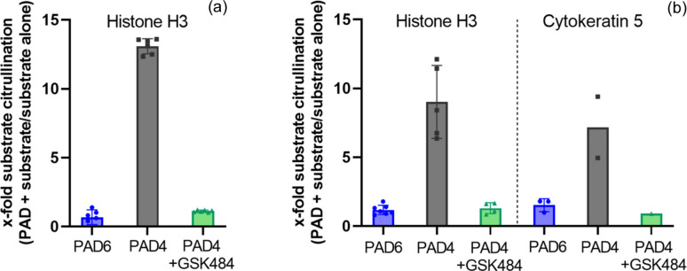
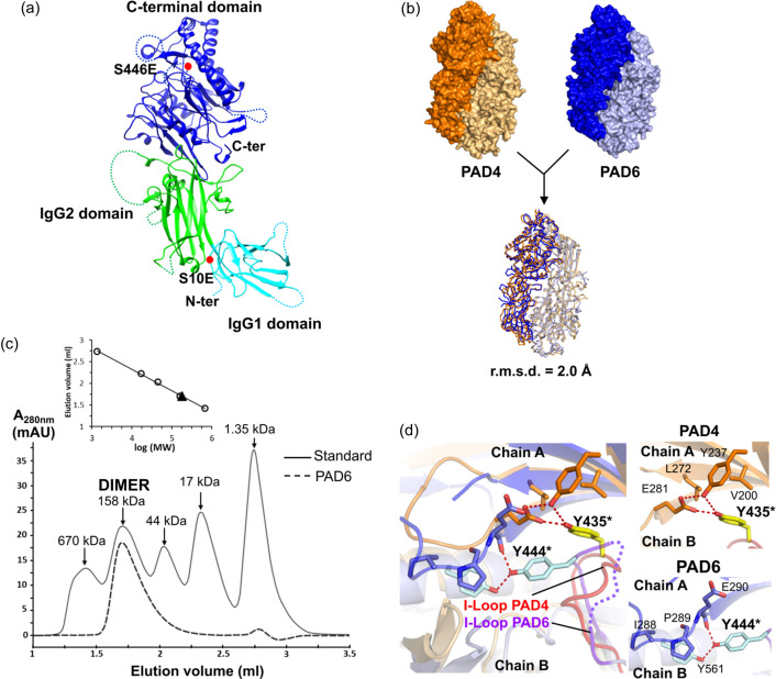
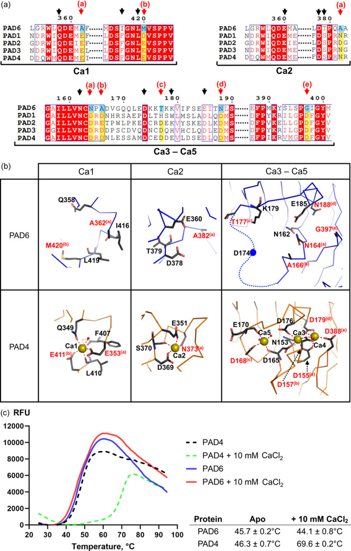
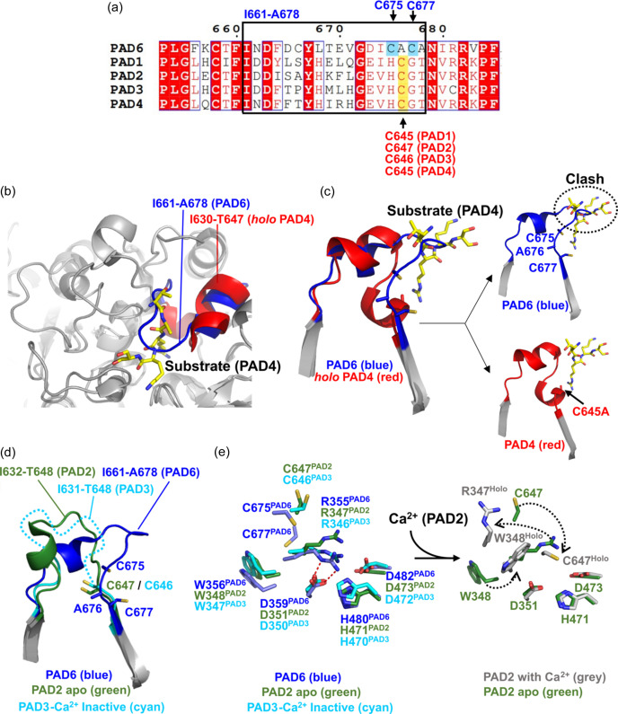
Human peptidylarginine deiminase isoform VI (PAD6), which is predominantly limited to cytoplasmic lattices in the mammalian oocytes in ovarian tissue, is essential for female fertility. It belongs to the peptidylarginine deiminase (PAD) enzyme family that catalyzes the conversion of arginine residues to citrulline in proteins. In contrast to other members of the family, recombinant PAD6 was previously found to be catalytically inactive. We sought to provide structural insight into the human homologue to shed light on this observation. We report here the first crystal structure of PAD6, determined at 1.7 Å resolution. PAD6 follows the same domain organization as other structurally known PAD isoenzymes. Further structural analysis and size-exclusion chromatography show that PAD6 behaves as a homodimer similar to PAD4. Differential scanning fluorimetry suggests that PAD6 does not coordinate Ca2+ which agrees with acidic residues found to coordinate Ca2+ in other PAD homologs not being conserved in PAD6. The crystal structure of PAD6 shows similarities with the inactive state of apo PAD2, in which the active site conformation is unsuitable for catalytic citrullination. The putative active site of PAD6 adopts a non-productive conformation that would not allow protein-substrate binding due to steric hindrance with rigid secondary structure elements. This observation is further supported by the lack of activity on the histone H3 and cytokeratin 5 substrates. These findings suggest a different mechanism for enzymatic activation compared with other PADs; alternatively, PAD6 may exert a non-enzymatic function in the cytoplasmic lattice of oocytes and early embryos.

Keywords: PAD6; cytoplasmic lattices; human peptidylarginine deiminase VI; mammalian fertilization; protein structures.


Figures

Figure 1
Figure 1
In vitro citrullination assays of different substrates. (a) ELISA assay for measuring citrullination activity of PAD4 and PAD6 on the histone H3 substrate. PAD6 (blue) and PAD4 (grey) were tested for histone H3 citrullination activity in the presence and absence of 1 µg ml−1 histone H3 (two independent experiments with n = 3). PAD4 citrullination activity was inhibited by 50 µM GSK484 inhibitor (green). All data were normalized to the citrullination signal of the substrate in the absence of PAD. Error bars indicate the standard deviation. (b) On-blot assay for measuring citrullination activity of PAD6 and PAD4 on different substrates. Citrullination of two substrates (histone H3 and cytokeratin 5) was evaluated in presence of PAD6 (blue, n = 7 for histone H3, n = 3 for cytokeratin 5) or PAD4 (grey, n = 5 for histone H3, n = 2 for cytokeratin 5) and normalized to the citrullination signal of the substrate in absence of PAD. PAD4 activity was inhibited by 50 µM GSK484 inhibitor (green, n = 4 for histone H3, n = 1 for cytokeratin 5).
Figure 2
Figure 2
Overall structure of PAD6. (a) Domain organization of PAD6. The N-terminal IgG1, N-terminal IgG2 and the C-terminal catalytic domains are coloured in cyan, green and blue, respectively. (b) Model of the PAD6 dimer constructed with the symmetry mate of the crystal lattice. Overlay and comparison with the PAD4 dimer and r.m.s.d. calculated from PDB entry 2dew. (c) Analytical size-exclusion chromatography of PAD6 using a Superdex 200 5/150GL column equilibrated with 20 mM Tris pH 7.5, 200 mM NaCl, 1 mM TCEP. The elution volumes for PAD6 and bovine gamma globulin standard (158 kDa) were 1.7 ml for both samples. Inlet: standards in white circles and PAD6 in the black triangle. The molecular weight for PAD6 is estimated at ∼182 kDa. (d) Comparison between interacting residues involving the I-loops of PAD4 (orange) or that of PAD6 (blue). The I-loop is coloured red in PAD4 and purple in PAD6. In PAD6, the I-loop contains a disordered fragment (Pro445–Gly449) represented as a dashed line. The equivalent residues Tyr435 (PAD4) and Tyr444 (PAD6) are indicated with a star. Left panel: an overlay between PAD4 and PAD6 focused on the I-loop, showing different interface interactions involving Tyr435 (PAD4) and Tyr444 (PAD6). Right panels: detailed descriptions of the interactions involving Tyr435 in PAD4 (top) and those involving Tyr444 in PAD6 (bottom).
Figure 3
Figure 3
Comparing calcium binding of PAD6 and PAD4. (a) Sequence alignment of human PAD6 and other PADs focused on regions containing PAD4 residues involved in Ca1, Ca2, Ca3–5. The arrows point to residues involved in Ca2+ coordination in PAD4. The red arrows, with associated letters, highlight residues not conserved and unable to coordinate Ca2+ in PAD6. (b) Close-up views of PAD6 regions equivalent to Ca2+-binding sites in PAD4. Below, PAD4 Ca2+-binding sites are shown for reference (PDB entry 2dew). The yellow spheres represent the Ca2+ ions in PAD4. Residues that differ between the two proteins for which the side chain cannot coordinate Ca2+ in PAD6 are highlighted in red (equivalent positions between PAD6 and PAD4 are indicated with letters in brackets). (c) Thermal stability profiles of PAD4 and PAD6 in the presence of 0 or 10 mM CaCl2. The table provides the average protein melting temperatures (determined as the inflection point of the thermal transition) and the standard deviation from triplicate measurements.
Figure 4
Figure 4
PAD6 structural analysis of the C-terminal domain. (a) Sequence alignment between five human PADs, focusing on the PAD6 loop I661–A678. Within this loop, the active or putative active cysteines are underlined and labelled: blue for PAD6, red for the other PADs. (b) Superimposition between structures of PAD6 and PAD4 in complex with substrate (PDB entry 2dew). The substrate is in yellow stick representation. (c) Same superimposition as in (b), showing a close-up view of the segment containing the I661–A678 loop and the β-strands connected by this loop, with the equivalent segment in PAD4 and the substrate. This view shows that the conformation of the PAD6 I661–A678 loop (blue) is different from the equivalent loop in PAD4 (I630–T647, red), and occludes the active site, so that the substrate would clash if positioned as in PAD4 (right panels). (d) PAD6 I661–A678 loop in blue superimposed with the equivalent segment of the apo PAD2 structure in green (PDB entry 4n20) and the inactive Ca2+-PAD3 in cyan (PDB entry 7d8n). Flexible loop I631–T648 (PAD3) represented by a dashed line. (e) Similarities in the active sites of the PAD6 structures and the apo PAD2 and non-productive form of Ca2+-bound PAD3, showing that equivalent Arg residues (Arg355 in PAD6, Arg347 in PAD2 and Arg346 in PAD3) occupy the active site, in lieu of the substrate. Ca2+ binding triggers the displacement of Arg347 in holo PAD2 (PDB entry 4n2c) together with other residues to shape a functional active site (right panel).

Similar articles

Cited by

References

    1. Arita, K., Hashimoto, H., Shimizu, T., Nakashima, K., Yamada, M. & Sato, M. (2004). Nat. Struct. Mol. Biol. 11, 777–783. - PubMed
    1. Arita, K., Shimizu, T., Hashimoto, H., Hidaka, Y., Yamada, M. & Sato, M. (2006). Proc. Natl Acad. Sci. USA, 103, 5291–5296. - PMC - PubMed
    1. Begemann, M., Rezwan, F. I., Beygo, J., Docherty, L. E., Kolarova, J., Schroeder, C., Buiting, K., Chokkalingam, K., Degenhardt, F., Wakeling, E. L., Kleinle, S., González Fassrainer, D., Oehl-Jaschkowitz, B., Turner, C. L. S., Patalan, M., Gizewska, M., Binder, G., Bich Ngoc, C. T., Chi Dung, V., Mehta, S. G., Baynam, G., Hamilton-Shield, J. P., Aljareh, S., Lokulo-Sodipe, O., Horton, R., Siebert, R., Elbracht, M., Temple, I. K., Eggermann, T. & Mackay, D. J. G. (2018). J. Med. Genet. 55, 497–504. - PMC - PubMed
    1. Bielecka, E., Scavenius, C., Kantyka, T., Jusko, M., Mizgalska, D., Szmigielski, B., Potempa, B., Enghild, J. J., Prossnitz, E. R., Blom, A. M. & Potempa, J. (2014). J. Biol. Chem. 289, 32481–32487. - PMC - PubMed
    1. Boggs, J. M., Rangaraj, G., Koshy, K. M., Ackerley, C., Wood, D. D. & Moscarello, M. A. (1999). J. Neurosci. Res. 57, 529–535. - PubMed

Publication types

MeSH terms