. 2024 May 1;11(Pt 3):384-394.
doi: 10.1107/S2052252524003075.

Structural insights into the molecular mechanism of phytoplasma immunodominant membrane protein

Affiliations

Structural insights into the molecular mechanism of phytoplasma immunodominant membrane protein

Chang Yi Liu et al. IUCrJ. .

Abstract

Immunodominant membrane protein (IMP) is a prevalent membrane protein in phytoplasma and has been confirmed to be an F-actin-binding protein. However, the intricate molecular mechanisms that govern the function of IMP require further elucidation. In this study, the X-ray crystallographic structure of IMP was determined and insights into its interaction with plant actin are provided. A comparative analysis with other proteins demonstrates that IMP shares structural homology with talin rod domain-containing protein 1 (TLNRD1), which also functions as an F-actin-binding protein. Subsequent molecular-docking studies of IMP and F-actin reveal that they possess complementary surfaces, suggesting a stable interaction. The low potential energy and high confidence score of the IMP-F-actin binding model indicate stable binding. Additionally, by employing immunoprecipitation and mass spectrometry, it was discovered that IMP serves as an interaction partner for the phytoplasmal effector causing phyllody 1 (PHYL1). It was then shown that both IMP and PHYL1 are highly expressed in the S2 stage of peanut witches' broom phytoplasma-infected Catharanthus roseus. The association between IMP and PHYL1 is substantiated through in vivo immunoprecipitation, an in vitro cross-linking assay and molecular-docking analysis. Collectively, these findings expand the current understanding of IMP interactions and enhance the comprehension of the interaction of IMP with plant F-actin. They also unveil a novel interaction pathway that may influence phytoplasma pathogenicity and host plant responses related to PHYL1. This discovery could pave the way for the development of new strategies to overcome phytoplasma-related plant diseases.

Keywords: X-ray crystallography; actin-binding proteins; immunodominant membrane proteins; phytoplasma; protein structure; α-helix bundles.


Figures

Figure 1
Figure 1
Purification and gel-filtration analysis of recombinant IMP. (a) SDS–PAGE of purified recombinant IMP on a 12% SDS–PAGE gel. The samples were loaded with 12 µl in each well. Lane M contains molecular-weight markers (labelled in kDa). (b) Gel-filtration standards for recombinant IMP. The calibration standard curve for size-exclusion chromatography was measured using the standard substances aprotinin (APR, 6.5 kDa), ribonuclease A (R, 13.7 kDa), carbonic anhydrase (CA, 29 kDa) and conalbumin (C, 75 kDa).
Figure 2
Figure 2
The crystal structure of recombinant IMP. (a) Refined electron-density map of the IMP crystal. (b) Ribbon diagram of the IMP dimer in the unit cell. (c) The secondary-structural elements of the IMP monomer. The red cylinders representing α-helices are labelled in parallel with the sequence.
Figure 3
Figure 3
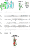
Structural analysis of the IMP–PHYL1 model and homologue comparison of the IMP structure. (a) The dimeric structure of TLNRD1; the two monomers of the rod domain are presented as green and cyan ribbons. (b) The structure of the TLNRD1_4H bundle (dark green ribbon; amino acids 148–270). (c) Alignment of the IMP and TLNRD1 protein sequences, which show relatively low sequence identity. (d) The models of IMP and TLNRD1_4H superimposed with an r.m.s.d. of 1.181 Å.
Figure 4
Figure 4
Calculated structural modelling of potent F-actin-binding proteins. (a) The proposed IMP–actin binding model from the HDOCK server with a docking score of −155.95 and a confidence score of 0.530. The F-actin model is coloured blue, while the IMP model is coloured red. (b) The proposed TLNRD1–actin binding model from the HDOCK server with a docking score of −262.13 and a confidence score of 0.904. The F-actin model is coloured blue, while the TLNRD1 model is coloured light green. (c) The proposed TLNRD1_4H–actin binding model from the HDOCK server with a docking score of −260.80 and a confidence score of 0.902. The F-actin model is coloured blue, while the TLNRD1_4H model is coloured dark green. (d) The alignment of all docking ligands (IMP, TLNRD1, TLNRD1_4H and IMP/PHYL1) to F-actin (solid ribbons) with the electron-density maps from the PDB structures.
Figure 5
Figure 5
Detection of the expression of PHYL1 and IMP in PnWB-infected C. roseus. (a) The various leafy flower symptoms of PnWB-infected C. roseus. (b, c) Expression levels of PHYL1 and IMP at various stages of leafy flower symptoms. Asterisks indicate Rubisco staining as a comparable quality loading control for leafy flower formation.
Figure 6
Figure 6
Confirmation of the PHYL1–IMP interaction. (a, b) The in vivo co-immunoprecipitation of PHYL1 (a) and IMP (b) from PnWB-infected C. roseus plants. α-PHYL1 was diluted 1:5000 for detection. α-IMP was diluted 1:40 000 for detection. (c) Cross-linking analysis of PHYL1 and IMP. The shifting band due to protein interactions is marked with an asterisk.
Figure 7
Figure 7
Schematic model of phytoplasma binding to plant F-actin.

References

    1. Bonnot, F., de Franqueville, H. & Lourenço, E. (2010). Phytopathology, 100, 300–312. - PubMed
    1. Boonrod, K., Kuaguim, L., Braun, M., Müller-Renno, C., Ziegler, C. & Krczal, G. (2023). Int. J. Mol. Sci. 24, 968. - PMC - PubMed
    1. Boonrod, K., Munteanu, B., Jarausch, B., Jarausch, W. & Krczal, G. (2012). Mol. Plant Microbe Interact. 25, 889–895. - PubMed
    1. Chichili, V. P. R., Kumar, V. & Sivaraman, J. (2013). Intrinsically Disord. Proteins, 1, e25464. - PMC - PubMed
    1. Constable, F. E. (2009). Phytoplasmas: Genomes, Plant Hosts and Vectors, edited by P. G. Weintraub & P. Jones, pp. 188–212. Wallingford: CABI.

Publication types