. 2024 Jun 1;35(6):ar88.
doi: 10.1091/mbc.E24-01-0046. Epub 2024 Apr 24.

Interplay of condensation and chromatin binding underlies BRD4 targeting

Affiliations

Interplay of condensation and chromatin binding underlies BRD4 targeting

Amy R Strom et al. Mol Biol Cell. .

Abstract

Nuclear compartments form via biomolecular phase separation, mediated through multivalent properties of biomolecules concentrated within condensates. Certain compartments are associated with specific chromatin regions, including transcriptional initiation condensates, which are composed of transcription factors and transcriptional machinery, and form at acetylated regions including enhancer and promoter loci. While protein self-interactions, especially within low-complexity and intrinsically disordered regions, are known to mediate condensation, the role of substrate-binding interactions in regulating the formation and function of biomolecular condensates is underexplored. Here, utilizing live-cell experiments in parallel with coarse-grained simulations, we investigate how chromatin interaction of the transcriptional activator BRD4 modulates its condensate formation. We find that both kinetic and thermodynamic properties of BRD4 condensation are affected by chromatin binding: nucleation rate is sensitive to BRD4-chromatin interactions, providing an explanation for the selective formation of BRD4 condensates at acetylated chromatin regions, and thermodynamically, multivalent acetylated chromatin sites provide a platform for BRD4 clustering below the concentration required for off-chromatin condensation. This provides a molecular and physical explanation of the relationship between nuclear condensates and epigenetically modified chromatin that results in their mutual spatiotemporal regulation, suggesting that epigenetic modulation is an important mechanism by which the cell targets transcriptional condensates to specific chromatin loci.


Figures
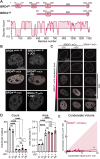
FIGURE 1:
FIGURE 1:
Chromatin binding influences BRD4 condensate distribution in living cells. (A) Schematic of BRD4 full-length (FL) and deltaN-terminus (∆N) constructs, as well as PONDR predicted disorder score. (B) From top: Immunofluorescence of endogenous BRD4 protein in cultured human U2OS cells without and with addition of 1 µM JQ1. Exogenous live expression of BRD4FL-mCherry and BRD4∆N-mCherry in the same cell before (−JQ1) and after (+JQ1) addition of 1 µM JQ1. (C) Panel of images of U2OS cells with increasing expression level of BRD4FL-mCherry (same cell −/+ JQ1) or BRD4∆N-mCherry at similar expression levels. (D) Quantification of number and size of BRD4 condensates from immunofluorescence of endogenous, or live expression of BRD4FL-mCherry or BRD4∆N-mCherry, −/+ JQ1. Points represent averages of three biological replicates of 25 cells each within the expression level gate defined in 1C, error bars SEM. Statistical test one-way ANOVA, ****p < 0.0001. (E) Estimated condensate volume measured in the same set of cells expressing BRD4FL-mCherry before (x-axis) and after (y-axis) disruption of chromatin binding through addition of 1 µM JQ1. If condensate volume is not affected, points should lie on the diagonal, indicated by shading.
FIGURE 2:
FIGURE 2:
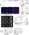
Thermodynamic effects of BRD4 chromatin binding are measured the Corelet synthetic oligomerization system. Representative images of BRD4FL Corelets before and after light activation of a nucleus without (A) or with (B) 1µM JQ1. (C) Quantification of the number and size of BRD4FL Corelet condensates per nucleus induced with light activation −/+ JQ1 in the same set of nuclei. Error bars represent SEM across four trials of 25 cells each, Student’s t test ***p = 0.0002 in Count, *** p = 0.001 in Area. (D) Condensate volume comparison in −/+JQ1 conditions in the same set of cells with BRD4FL (pink) or BRD4∆N (black) Corelets. BRD4∆N Corelet condensate volume is unaffected by the addition of JQ1, as can be seen from points largely along the diagonal (shaded pink), while BRD4FL Corelet condensate volume is much lower after addition of JQ1. Lines are linear fit. (E, F) Phase diagram and logistic regression of BRD4FL measured with the Corelet system in the absence (E) and presence (F) of JQ1. Phase diagrams are constructed from the same cells expressing BRD4FL −/+ JQ1 demonstrating the shifted valence between −JQ1 (gray) and +JQ1 (pink).
FIGURE 3:
FIGURE 3:
A coarse-grained model for simulating BRD4 Corelet condensation. (A) Coarse-grained simulations contain representations of the chromatin polymer (blue) and Corelet oligomerization platform (gold) with attached BRD4 molecules, each composed of two spheres representing the N-terminus (capable of interacting with chromatin) and C-terminus (capable of self-interaction). (B) The valence-dependent phase diagram in the absence of chromatin, obtained via direct-coexistence simulation (representative snapshot on left), shows a critical valence-1 of 1/6. C. A phase diagram showing the presence of chromatin-Corelet condensates in simulations with strong chromatin binding (40% acetylated histone tails) predicts an apparent critical valence-1 of 1/2. Representative snapshots are shown (on right) for the indicated simulation conditions. Related simulation data at lower acetylation levels are provided in Supplemental Figure S2F.
FIGURE 4:
FIGURE 4:
BRD4 condensate nucleation is seeded on chromatin. (A) Schematic of two modes of condensate nucleation: substrate-seeded (heterogeneous) and not seeded (homogeneous). Without seeding, the free energy of protein clustering increases until the cluster reaches its critical radius (rcrit), at which point it becomes energetically favorable to grow. A seed can lower the energetic barrier to reach rcrit. (B) Seeded nucleation is expected to have a shorter delay time before droplet formation, and increased nucleation rate (slope) compared with nonseeded nucleation. (C) Quantification of the number of condensates nucleated over time in the same cell before and after JQ1 treatment demonstrates the expected changes in delay time and slope. (D) Representative images of a 4 by 4 micron square nuclear area as light-induced BRD4FL-mCh-sspB Corelet condensates nucleate rapidly in untreated conditions (2 s, top), but are delayed in the same cell after JQ1 treatment (10 s, bottom). (E) Quantification of the nucleation rate (slope), delay time, and number density for 9 cells of similar expression level before and after JQ1 treatment. (F) Repeated activation-deactivation cycles of BRD4∆N and overlay of condensate positions in subsequent cycles shows whether nucleation occurs repeatedly in the same nuclear locations. (G) Overlay of condensate positions in subsequent cycles of activation for BRD4FL −/+ JQ1. (H) PCC quantification and difference in PCC (deltaPCC) of overlaid images of 33 cells in subsequent activation cycles. Negative controls are PCCs between images of different cells. ***p = 0.0015 by Mann–Whitney exact, two-tailed t test. (I) Short-term (2 min) 200 nM JQ1 treatment disperses BRD4 condensates (JQ1), yet they form again quickly after washing out JQ1 (Recovery). Segmented masks of identified BRD4 puncta within nuclear outline, overlay of JQ1 and recovery timepoints (green) with pretreatment condensate positions (magenta). (J) Quantification of condensate dissolution during JQ1 treatment (shaded area) and recovery after washout. Error bars represent SD of three biological trials of 10 cells each. (K) Overlay of images before/during JQ1 treatment and before treatment/after JQ1 washout (left). PCC of 30 cells before/during JQ1, and before treatment/after JQ1 washout.
FIGURE 5:
FIGURE 5:
Simulations of BRD4FL Corelet condensation quantify enhancement of chromatin-seeded nucleation. (A) Schematic of a typical nucleation event. τnucl is the time required for cluster size to reach the threshold to form a stable nucleus (red line). τdelay is the time required for the nucleated cluster to grow via diffusion-limited growth to reach a size that is observable under the microscope (green line), which is estimated to be 15 times larger than the stable nucleus size in our simulations. (B) Example of a heterogeneous (on-chromatin) nucleation event, in which the largest cluster (highlighted in red) interacts with the chromatin polymer. (C) Example of a homogeneous (off-chromatin) nucleation event. (D) Probability of on-chromatin (orange) or off-chromatin (blue) nucleation events for endogenous (top) and Corelet (bottom) simulations at two BRD4-histone tail interaction strengths (representing +JQ1 and −JQ1), and four acetylation fractions (0.1, 0.2, 0.3, 0.4). (E) Quantification of the nucleation rate in endogenous and Corelet simulations with (−JQ1) and without (+JQ1) strong BRD4-histone tail interactions. (F) Quantification of the delay time before observable condensates are formed in endogenous and Corelet simulation systems with (−JQ1) and without (+JQ1) strong BRD4-histone tail interactions. (E, F) Seven independent simulations for each measurement are run at an acetylation fraction of 0.4 for both +JQ1 and −JQ1. In the +JQ1 Corelet simulations, only two out of seven simulations achieved nucleation. Error bars represent the SE. ***p ≤ 0.001, ****p ≤ 0.0001 by Student’s t test.
FIGURE 6:
FIGURE 6:
Epigenetic acetylation level influences nucleation behaviors in endogenous and exogenous BRD4 systems. (A) Representative images and analysis examples of endogenous BRD4 puncta by immunofluorescence in cells treated with control media (Control), histone acetyltransferase inhibitor (A485), or histone deacetylase inhibitor (TSA). (B, C) Quantification of endogenous BRD4 condensate count per nucleus (B) and condensate average area (C) measured by immunofluorescence in drug-treated cells. n = 25 cells each, error is SD. ***p = 0.002, ****p = 0.0001 by one-way ANOVA. D. Representative images of BRD4FL-mCh puncta in living cells treated with control media, A485, or TSA at three timepoints: before addition of JQ1 (−JQ1), during JQ1 incubation (+JQ1) and after JQ1 washout and recovery. (E–F) Quantification of the number of condensates per nucleus in drug-treated cells before addition of JQ1 (E) and after washout and recovery (F). Expression range as defined in Supplemental Figure S4, D and E. n = 11, 11, 12 cells, error is SD, *p = 0.031, **p = 0.008 by one-way ANOVA. (G) Timecourse graph of the number of BRD4FL-mCh puncta per nucleus during JQ1 treatment (shaded area) and nucleation after washout. (H) Nucleation rate of BRD4FL-mCh puncta measured as slope after JQ1 washout. N = 11, 11, 12 for A485, control, TSA respectively. **p = 0.006, ****p = 0.001. (I) Model figure summarizing findings about on- and off-chromatin BRD4 nucleation.

Similar articles

Cited by

References

    1. Alekseyenko AA, Walsh EM, Zee BM, Pakozdi T, Hsi P, Lemieux ME, Cin PD, Ince TA, Kharchenko PV, Kuroda M, French CA (2017). Ectopic protein interactions within BRD4-chromatin complexes drive oncogenic megadomain formation in NUT midline carcinoma. Proc Natl Acad Sci USA 114, E4184–4192. - PMC - PubMed
    1. Banani SF, Lee HO, Hyman AA, Rosen MK (2017). Biomolecular condensates: organizers of cellular biochemistry. Nat Rev Mol Cell Biol 18, 285–298. - PMC - PubMed
    1. Bianchi E, Largo J, Tartaglia P, Zaccarelli E, Sciortino F (2006). Phase diagram of patchy colloids: towards empty liquids. Phys Rev Lett 97, 168301. - PubMed
    1. Boija A, Klein IA, Sabari BR, Dall’Agnese A, Coffey EL, Zamudio AV, Li CH, Shrinivas K, Manteiga JC, Hannett NM, et al. (2018). Transcription factors activate genes through the phase-separation capacity of their activation domains. Cell 175, 1842–1855.e16. - PMC - PubMed
    1. Boisvert F-M, van Koningsbruggen S, Navascués J, Lamond AI (2007). The multifunctional nucleolus. Nat Rev Mol Cell Biol 8, 1021–574– 585. - PubMed

LinkOut - more resources