

MRI of cardiac involvement in COVID-19
- PMID: 38656976
- PMCID: PMC11256941
- DOI: 10.1093/bjr/tqae086
MRI of cardiac involvement in COVID-19
Abstract
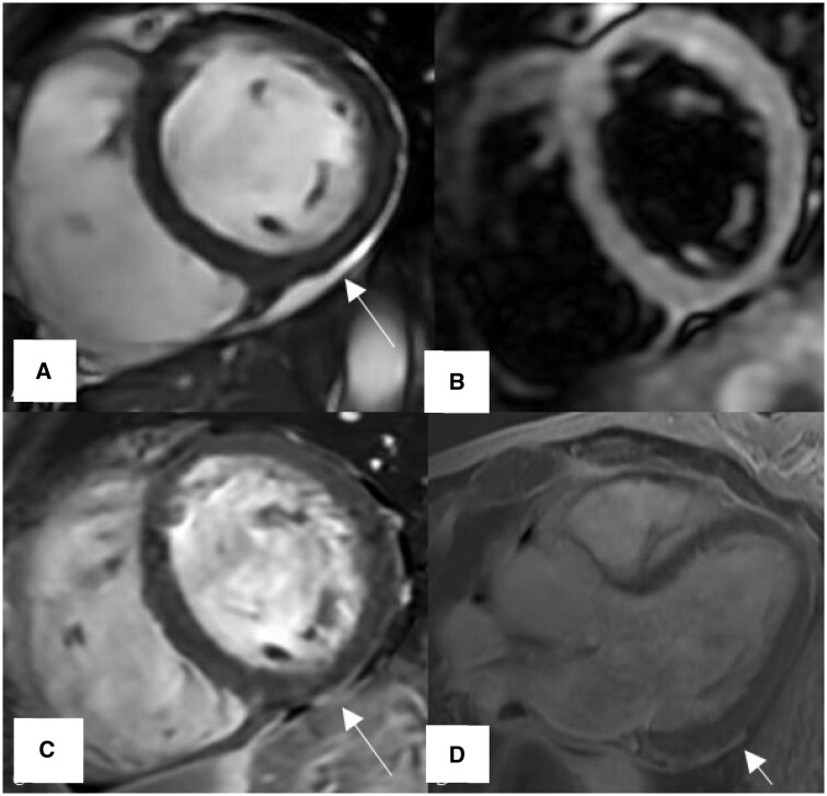
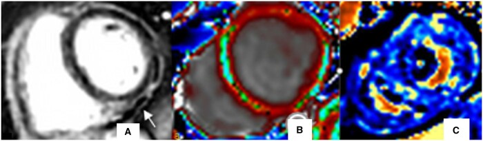
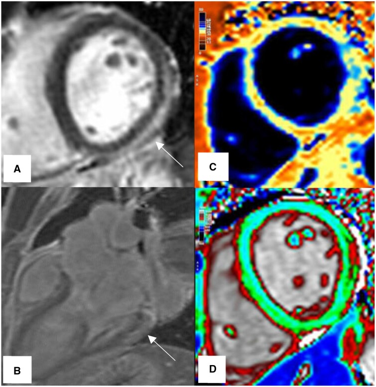
The COVID-19 pandemic, caused by the SARS-CoV-2 virus, has led to a diverse pattern of myocardial injuries, including myocarditis, which is linked to adverse outcomes in patients. Research indicates that myocardial injury is associated with higher mortality in hospitalized severe COVID-19 patients (75.8% vs 9.7%). Cardiovascular Magnetic Resonance (CMR) has emerged as a crucial tool in diagnosing both ischaemic and non-ischaemic myocardial injuries, providing detailed insights into the impact of COVID-19 on myocardial tissue and function. This review synthesizes existing studies on the histopathological findings and CMR imaging patterns of myocardial injuries in COVID-19 patients. CMR imaging has revealed a complex pattern of cardiac damage in these patients, including myocardial inflammation, oedema, fibrosis, and ischaemic injury, due to coronary microthrombi. This review also highlights the role of LLC criteria in diagnosis of COVID-related myocarditis and the importance of CMR in detecting cardiac complications of COVID-19 in specific groups, such as children, manifesting multisystem inflammatory syndrome in children (MIS-C) and athletes, as well as myocardial injuries post-COVID-19 infection or following COVID-19 vaccination. By summarizing existing studies on CMR in COVID-19 patients and highlighting ongoing research, this review contributes to a deeper understanding of the cardiac impacts of COVID-19. It emphasizes the effectiveness of CMR in assessing a broad spectrum of myocardial injuries, thereby enhancing the management and prognosis of patients with COVID-19 related cardiac complications.
Keywords: CMR; COVID-19; SARS-CoV-2; myocarditis.
© The Author(s) 2024. Published by Oxford University Press on behalf of the British Institute of Radiology.
Conflict of interest statement
None declared.
Figures
Similar articles
-
Myocardial involvement in children with post-COVID multisystem inflammatory syndrome: a cardiovascular magnetic resonance based multicenter international study-the CARDOVID registry.J Cardiovasc Magn Reson. 2021 Dec 30;23(1):140. doi: 10.1186/s12968-021-00841-1. J Cardiovasc Magn Reson. 2021. PMID: 34969397 Free PMC article.
-
Cardiovascular magnetic resonance for evaluation of cardiac involvement in COVID-19: recommendations by the Society for Cardiovascular Magnetic Resonance.J Cardiovasc Magn Reson. 2023 Mar 27;25(1):21. doi: 10.1186/s12968-023-00933-0. J Cardiovasc Magn Reson. 2023. PMID: 36973744 Free PMC article. Review.
-
Cardiovascular Magnetic Resonance for Patients With COVID-19.JACC Cardiovasc Imaging. 2022 Apr;15(4):685-699. doi: 10.1016/j.jcmg.2021.08.021. Epub 2021 Oct 13. JACC Cardiovasc Imaging. 2022. PMID: 34656482 Free PMC article. Review.
-
SARS-CoV-2 Cardiac Involvement in Young Competitive Athletes.Circulation. 2021 Jul 27;144(4):256-266. doi: 10.1161/CIRCULATIONAHA.121.054824. Epub 2021 Apr 17. Circulation. 2021. PMID: 33866822 Free PMC article.
-
Cardiac Magnetic Resonance Imaging in Coronavirus Disease 2019 (COVID-19): A Systematic Review of Cardiac Magnetic Resonance Imaging Findings in 199 Patients.J Thorac Imaging. 2021 Mar 1;36(2):73-83. doi: 10.1097/RTI.0000000000000574. J Thorac Imaging. 2021. PMID: 33306666
Cited by
-
Possible mechanisms of SARS-CoV-2-associated myocardial fibrosis: reflections in the post-pandemic era.Front Microbiol. 2024 Oct 8;15:1470953. doi: 10.3389/fmicb.2024.1470953. eCollection 2024. Front Microbiol. 2024. PMID: 39444690 Free PMC article. Review.
-
Symptom burden, coagulopathy and heart disease after acute SARS-CoV-2 infection in primary practice.Sci Rep. 2024 Sep 11;14(1):21229. doi: 10.1038/s41598-024-71535-8. Sci Rep. 2024. PMID: 39261512 Free PMC article.
-
Abnormal coronary vascular response in patients with long COVID syndrome - a case-control study using oxygenation-sensitive cardiovascular magnetic resonance.J Cardiovasc Magn Reson. 2025 Summer;27(1):101890. doi: 10.1016/j.jocmr.2025.101890. Epub 2025 Apr 2. J Cardiovasc Magn Reson. 2025. PMID: 40185235 Free PMC article.
-
Late Gadolinium Enhancement Magnetic Resonance Imaging (MRI) for Predicting Left Ventricular Reverse Remodeling in Non-Ischemic Cardiomyopathy: A Systematic Review and Meta-Analysis.J Clin Med. 2025 Jan 29;14(3):895. doi: 10.3390/jcm14030895. J Clin Med. 2025. PMID: 39941566 Free PMC article. Review.
References
-
- WHO Coronavirus (COVID-19) Dashboard [Internet]. Accessed October 15, 2023. https://covid19.who.int
Publication types
MeSH terms
LinkOut - more resources
Full Text Sources
Medical
Miscellaneous
