Review
. 2024 Sep 3;36(9):3036-3056.
doi: 10.1093/plcell/koae128.

Vacuolar degradation of plant organelles

Affiliations
Review

Vacuolar degradation of plant organelles

Marisa S Otegui et al. Plant Cell. .

Abstract

Plants continuously remodel and degrade their organelles due to damage from their metabolic activities and environmental stressors, as well as an integral part of their cell differentiation programs. Whereas certain organelles use local hydrolytic enzymes for limited remodeling, most of the pathways that control the partial or complete dismantling of organelles rely on vacuolar degradation. Specifically, selective autophagic pathways play a crucial role in recognizing and sorting plant organelle cargo for vacuolar clearance, especially under cellular stress conditions induced by factors like heat, drought, and damaging light. In these short reviews, we discuss the mechanisms that control the vacuolar degradation of chloroplasts, mitochondria, endoplasmic reticulum, Golgi, and peroxisomes, with an emphasis on autophagy, recently discovered selective autophagy receptors for plant organelles, and crosstalk with other catabolic pathways.


Conflict of interest statement

Conflict of interest statement. None declared.

Figures

Figure 1.
Figure 1.
Autophagy. A) Activation of canonical autophagy and downstream pathways depending on the ATG1 kinase complex, including the assembly of the VPS34 complex, the delivery of membrane to the phagophore mediated by ATG9, and the ATG12 and ATG8 conjugation pathways. ABA, abscisic acid; TOR (TARGET OF RAPAMYCIN); SnRK1 and 2 (SNF1-Related Protein Kinase1 and 2). B) Schematic representation of macroautophagy and microautophagy and their main molecular components. Selective autophagy of organelles, by either macroautophagy or microautophagy, is mediated by autophagy receptors.
Figure 2.
Figure 2.
Canonical and noncanonical chlorophagy modalities identified in Arabidopsis thaliana. The figure depicts 4 modalities of macrochlorophagy that depend on canonical autophagy. The 4 modalities are defined by their cargo and known receptors. Microchlorophagy of photodamaged chloroplasts proceeds through either canonical autophagy and ATG8 lipidation or a noncanonical pathway that uses NBR1 as receptor.
Figure 3.
Figure 3.
Mitophagy for recycling damaged mitochondria under stress conditions. A) Under stress conditions leading to mitochondrial damage, irreparable mitochondria lose their membrane potential (gray) and are eliminated via mitophagy. Mitochondria also use self-protection mechanisms, including isolation of disrupted segments through fission, formation of donut-like structures, and generation of MDVs. Damaged mitochondria are engulfed by autophagosomes. Such mitochondria are characterized by ruptured OMM and collapsed cristae (black spots), indicating loss of structural integrity. B) Molecular factors involved in mitophagosome formation in Arabidopsis include the OMM protein TRB1 that directly interacts with ATG8. FMT becomes associated with ATG8 on depolarized mitochondria, contributing to mitophagosome assembly. Ubiquitinated proteins accumulate in mitochondria when Arabidopsis cells are incubated with uncouplers. C) Etiolated seedlings are heterotrophic that consume nutrient reserves in seeds; mitochondria are required for catabolic mobilization of these reserves. During de-etiolation, seedlings transition to autotrophs capable of photosynthesis. Mitophagy facilitates the transition by recycling existing mitochondria to support the formation of new organelles, including chloroplast biogenesis.
Figure 4.
Figure 4.
ER-phagy pathways in plants. A) ER-phagy entails 4 main steps: (1) recognition of damaged or unwanted ER compartments; (2) fragmentation of the ER network; (3) recruitment into the growing phagophore via ER-phagy receptors; and (4) autophagic degradation in the vacuole. B) Known plant ER-phagy receptors that localize to different ER subcompartments and induce distinct autophagy mechanisms (see the text for details). The N and C termini, the ATG8 interaction motifs (AIM, in yellow), and known co-receptors are highlighted. The transmembrane domains are in red, the reticulon homology domains are shown in blue. The UBA domain of the ATI3 co-receptor UBAC2A is highlighted in purple.
Figure 5.
Figure 5.
Regulation of Golgi remodeling and Golgiphagy by the autophagy machinery. During nutrient starvation, a portion of the Golgi apparatus is captured and enclosed within double membrane-bound autophagosomes, whose formation depends on the conjugation of PE to ATG8 and its interaction with Golgiphagy receptors. Under acute heat stress conditions, the Golgi apparatus undergoes vacuolation, leading to the loss of cis-to-trans polarity and disruption of Golgi-mediated endomembrane trafficking. Lipidated ATG8, possibly in PS form, binds to the vacuolated Golgi cisternal membranes. ATG8 proteins recruit clathrin, enabling the formation of ATG8-positive vesicles on the vacuolated Golgi. These vesicles may subsequently fuse with the vacuole to eliminate denatured cargo molecules within the Golgi caused by heat stress. PM, plasma membrane; TGN, trans-Golgi network.
Figure 6.
Figure 6.
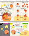
Schematic diagram of peroxisome degradation. A) ROS cause oxidative damage to peroxisomal proteins and membranes. Peroxisomal enzyme catalase degrades H2O2, and LON2 protease supports the folding and degradation of impaired proteins. Progressed damage in peroxisomes leads to the formation of protein aggregates, which are recognized by autophagy-related factors (e.g. PI3P, ATG18, and ATG8). The adaptor protein NBR1 is involved in pexophagy induced by cadmium (Cd) treatment. ARP2/3 complex also colocalizes with peroxisomes and ATG8. Pexophagy degrades individual peroxisomes through macropexophagy by enclosing them withing autophagosomes and transporting them to the vacuole. Under stressful conditions such as high light, peroxisomes form large aggregates, which can be degraded via micropexophagy. B) The role of pexophagy in the functional transition of peroxisomes. During germination, peroxisomes involved in lipid metabolism (glyoxysomes) transition into peroxisomes that perform photorespiration (leaf peroxisomes) as the seedling are exposed to light. While LON2 degrades enzymes of the old metabolic system inside the peroxisomes, pexophagy eliminates old peroxisomes; these 2 pathways work in coordination to enable an efficient functional transition.

Similar articles

Cited by

References

    1. Adriaenssens E, Ferrari L, Martens S. Orchestration of selective autophagy by cargo receptors. Curr Biol. 2022:32(24):R1357–R1371. 10.1016/j.cub.2022.11.002 - DOI - PubMed
    1. Anding AL, Wang C, Chang T-K, Sliter DA, Powers CM, Hofmann K, Youle RJ, Baehrecke EH. Vps13D encodes a ubiquitin-binding protein that is required for the regulation of mitochondrial size and clearance. Curr Biol. 2018:28(2):287–295.e6. 10.1016/j.cub.2017.11.064 - DOI - PMC - PubMed
    1. Barazzuol L, Giamogante F, Brini M, Calì T. PINK1/Parkin mediated mitophagy, ca2 + signalling, and ER–mitochondria contacts in Parkinson's disease. Int J Mol Sci. 2020:21(5):1772. 10.3390/ijms21051772 - DOI - PMC - PubMed
    1. Bar-On YM, Milo R. The global mass and average rate of rubisco. Proc Natl Acad Sci U S A. 2019:116(10):4738–4743. 10.1073/pnas.1816654116 - DOI - PMC - PubMed
    1. Barreto P, Okura VK, Neshich IAP, Maia IG, Arruda P. Overexpression of UCP1 in tobacco induces mitochondrial biogenesis and amplifies a broad stress response. BMC Plant Biol. 2014:14(1):144. 10.1186/1471-2229-14-144 - DOI - PMC - PubMed

LinkOut - more resources