. 2024 Aug;31(8):2433-2446.
doi: 10.1007/s43032-024-01567-y. Epub 2024 Apr 24.

The Effect of Thymoquinone on the TNF-α/OTULIN/NF-κB Axis Against Cisplatin-İnduced Testicular Tissue Damage

Affiliations

The Effect of Thymoquinone on the TNF-α/OTULIN/NF-κB Axis Against Cisplatin-İnduced Testicular Tissue Damage

Tuba Yalçın et al. Reprod Sci. 2024 Aug.

Abstract

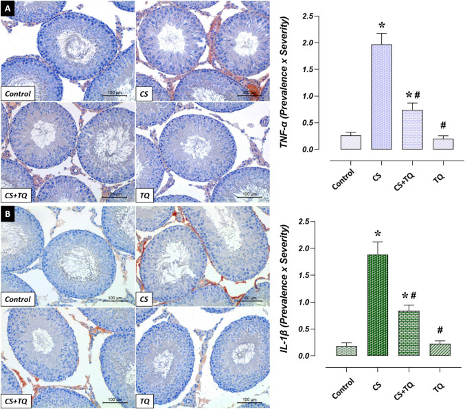
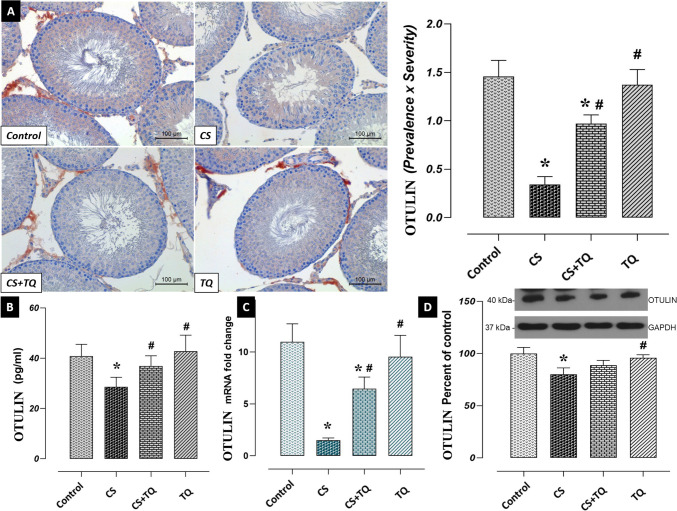
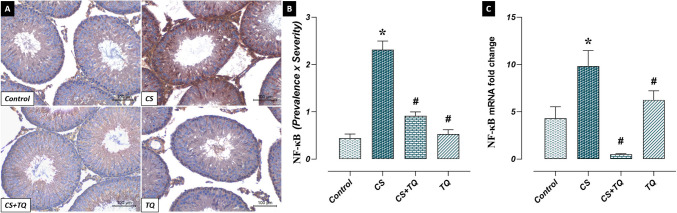
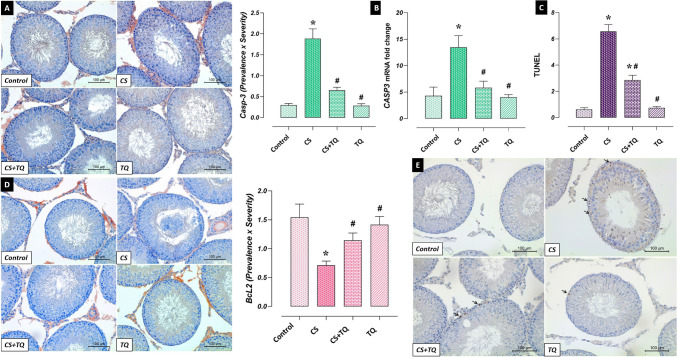
One of the adverse effects of the antineoplastic drug cisplatin (CS) is damage to testicular tissue. This study aimed to examine the potential therapeutic effect of thymoquinone (TQ), a strong antioxidant, against testicular damage caused by CS. In the experiment, 28 rats were used, and the rats were randomly divided into four groups: control (n = 7), CS (n = 7), CS + TQ (n = 7), and TQ (n = 7). The experiment was called off after all treatments were finished on day 15. Blood serum and testicular tissues were utilized for biochemical, histological, immunohistochemical, mRNA expression, and gene protein investigations. The testosterone level decreased and oxidative stress, histopathological damage, dysregulation in mitochondrial dynamics, inflammation and apoptotic cells increased in testicular tissue due to CS administration. TQ supplementation showed anti-inflammatory, antioxidant, and anti-apoptotic effects in response to CS-induced testicular damage. In addition, TQ contributed to the reduction of CS-induced toxic effects by regulating the TNF-α/OTULIN/NF-κB pathway. TQ supplementation may be a potential therapeutic strategy against CS-induced testicular damage by regulating the TNF-α/OTULIN/NF-κB axis, inhibiting inflammation, oxidative stress, and apoptosis.

Keywords: Cisplatin; Nuclear factor kappa B; OTULIN; Testicular damage; Thymoquinone; Tumour necrosis factor-alpha.


Conflict of interest statement

The authors have no conflicts of interest to declare regarding the content of this article.

Figures

Fig. 1
Fig. 1
Effect of CS and\or TQ applications on oxidant/antioxidant parameters in testicular tissue Oxidant/antioxidant parameters in testicular tissues of control and TQ groups were similar. When the CS group was compared with the control group, an increase in MDA level and a decrease in CAT and SOD levels were observed. In the CS + TQ group, it was determined that the oxidant/antioxidant parameters that changed due to CS were regulated. *; compared to the control group (p < 0.05), #; Compared with the CS group (p < 0.05). CS; Cisplatin, TQ; Thymoquinone, MDA; Malondialdehyde, CAT; Catalase, SOD; Superoxide dismutase
Fig. 2
Fig. 2
Effect of CS and\or TQ applications on reproductive hormones Serum testosterone and GnRH levels were similar in the control and TQ groups. GnRH levels increased while testosterone levels decreased in the CS group compared to the control group. In the CS + TQ group, it was determined that while testosterone levels increased compared to the CS group, GnRH levels decreased. *; compared to the control group (p < 0.05), #; Compared with the CS group (p < 0.05). CS; Cisplatin, TQ; Thymoquinone, GnRH; Gonadotropin-releasing hormone
Fig. 3
Fig. 3
Histopathological effects of CS and/or TQ applications on testicular tissues: A; JS graph, B; histopathological microphotographs, C; histopathological score graph. Testicular tissues of the control and TQ groups had similar JS and normal histological structure. While JS and HS decreased in the CS group compared to the control group, histopathological damage increased. Thin arrow; vascular congestion, thick arrow; seminiferous tubule degeneration, triangle; vacuolization, star; immature cells shed into the lumen. In the CS + TQ group, it was determined that there was an increase in JS and a decrease in histopathological damage compared to the CS group. *; compared to the control group (p < 0.05), #; Compared to CS group (p < 0.05). Hematoxylin and eosin staining, scale bar; 100 μm. JS; Johnsen score, HS; histopathological score, CS; Cisplatin, TQ; Thymoquinone, LC; Leyding cells, SC; Sertoli cells, SG; Spermatogonia, PS; Primary spermatocytes, S; Spermatids, SZ; Spermatozoa
Fig. 4
Fig. 4
Effect of CS and/or TQ applications on immunoreactivity of mitochondrial dynamics in testicular tissues: A; Drp1 immunoreactivity microphotographs and graph, B; Mfsn2 immunoreactivity microphotographs and graph. Drp1 and Mfsn2 immunoreactivities in testicular tissues of control and TQ groups were similar. While Drp1 immunoreactivity increased in the CS group compared to the control group, Mfsn2 immunoreactivity decreased. In the CS + TQ group, Drp1 immunoreactivity decreased while Mfsn2 immunoreactivity increased compared to the CS group. *; compared to the control group (p < 0.05), #; Compared to CS group (p < 0.05). Drp1 and Mfsn2 immunohistochemical staining, scale bar; 100 μm. CS; Cisplatin, TQ; Thymoquinone, Drp1; Dynamin related protein 1, Mfsn2; Mitofusin 2
Fig. 5
Fig. 5
Effect of CS and/or TQ applications on inflammatory cytokine immunoreactivities in testicular tissues: A; TNF-α immunoreactivity microphotographs and graph, B; IL-1β immunoreactivity microphotographs and graph. TNF-α and IL-1β immunoreactivities in testicular tissues of control and TQ groups were similar. TNF-α and IL-1β immunoreactivities were increased in the CS group compared to the control group. In the CS + TQ group, the immunoreactivities of pro-inflammatory cytokines were decreased compared to the CS group. *; compared to the control group (p < 0.05), #; Compared to CS group (p < 0.05). TNF-α and IL-1β immunohistochemical staining, scale bar; 100 μm. CS; Cisplatin, TQ; Thymoquinone, TNF-α; Tumor necrosis factor-alpha, IL-1β; Interleukin 1 Beta
Fig. 6
Fig. 6
Effect of CS and/or TQ applications on OTULIN levels in testicular tissues: A; OTULIN immunoreactivity microphotographs and graph, B; OTULIN ELISA levels, C; OTULIN mRNA expression, D; OTULIN gene protein level. OTULIN levels in testicular tissues of control and TQ groups were similar. OTULIN immunoreactivity, ELISA level, mRNA expression and gene protein level were decreased in the CS group compared to the control group. An increase in OTULIN levels was detected in the CS + TQ group compared to the CS group. *; compared to the control group (p < 0.05), #; Compared to CS group (p < 0.05). A; OTULIN immunohistochemical staining, scale bar; 100 μm. CS; Cisplatin, TQ; Thymoquinone
Fig. 7
Fig. 7
Effect of CS and/or TQ applications on NF-κB levels in testicular tissues: A; OTULIN immunoreactivity microphotographs, B; OTULIN immunoreactivity graph, C; NF-κB mRNA expression. NF-κB levels in testicular tissues of control and TQ groups were similar. NF-κB immunoreactivity and mRNA expression were increased in the CS group compared to the control group. A decrease in NF-κB levels was detected in the CS + TQ group compared to the CS group. *; compared to the control group (p < 0.05), #; Compared to CS group (p < 0.05). A; NF-κB immunohistochemical staining, scale bar; 100 μm. CS; Cisplatin, TQ; Thymoquinone
Fig. 8
Fig. 8
Effect of CS and/or TQ applications on apoptotic markers in testicular tissues: A; Casp3 immunoreactivity microphotographs and graph, B; Casp3 mRNA expression, C; TUNEL graph, D; BcL2 immunoreactivity microphotographs and graph, E; TUNEL microphotographs. Levels of apoptotic markers in testicular tissues of control and TQ groups were similar. It was determined that BcL2 immunoreactivity decreased in the CS group compared to the control group, while Casp3 levels and TUNEL-positive apoptotic cells increased. In the CS + TQ group, it was observed that BcL2 immunoreactivity increased compared to the CS group, while Casp3 levels and TUNEL-positive apoptotic cells decreased. *; compared to the control group (p < 0.05), #; Compared to CS group (p < 0.05). A; Casp3 immunohistochemical staining, D; BcL2 immunohistochemical staining, E; TUNEL staining, scale bar; 100 μm. CS; Cisplatin, TQ; Thymoquinone

Similar articles

Cited by

References

    1. Zoń A, Bednarek I. Cisplatin in ovarian cancer treatment—known limitations in therapy force new solutions. Int J Mol Sci. 2023;24(8):7585. - PMC - PubMed
    1. Keshta AT, Fathallah AM, Attia YA, Salem EA, Watad SH. Ameliorative effect of selenium nanoparticles on testicular toxicity induced by cisplatin in adult male rats. Food Chem Toxicol. 2023;179:113979. - PubMed
    1. Wang TE, Lai YH, Yang KC, Lin SJ, Chen CL, Tsai PS. Counteracting cisplatin-induced testicular damages by natural polyphenol constituent honokiol. Antioxidants. 2020;9(8):723. - PMC - PubMed
    1. Mesbahzadeh B, Hassanzadeh-Taheri M, Aliparast MS, Baniasadi P, Hosseini M. The protective effect of crocin on cisplatin-induced testicular impairment in rats. BMC Urol. 2021;21(1):117. - PMC - PubMed
    1. Jahan S, Munawar A, Razak S, Anam S, Ain QU, Ullah H, Almajwal A. Ameliorative effects of rutin against cisplatin-induced reproductive toxicity in male rats. BMC Urol. 2018;18(1):107. - PMC - PubMed

MeSH terms

LinkOut - more resources