Review
. 2024 Aug;42(8):801-819.
doi: 10.1007/s11604-024-01569-5. Epub 2024 Apr 25.

Endometriosis, a common but enigmatic disease with many faces: current concept of pathophysiology, and diagnostic strategy

Affiliations
Review

Endometriosis, a common but enigmatic disease with many faces: current concept of pathophysiology, and diagnostic strategy

Mayumi Takeuchi et al. Jpn J Radiol. 2024 Aug.

Abstract

Endometriosis is a benign, common, but controversial disease due to its enigmatic etiopathogenesis and biological behavior. Recent studies suggest multiple genetic, and environmental factors may affect its onset and development. Genomic analysis revealed the presence of cancer-associated gene mutations, which may reflect the neoplastic aspect of endometriosis. The management has changed dramatically with the development of fertility-preserving, minimally invasive therapies. Diagnostic strategies based on these recent basic and clinical findings are reviewed. With a focus on the presentation of clinical cases, we discuss the imaging manifestations of endometriomas, deep endometriosis, less common site and rare site endometriosis, various complications, endometriosis-associated tumor-like lesions, and malignant transformation, with pathophysiologic conditions.

Keywords: Deep endometriosis; Endometrioma; Endometriosis; Magnetic resonance imaging (MRI); Malignant transformation.


Figures

Fig. 1
Fig. 1
Endometrioma (typical) and hemorrhagic corpus luteum cyst. A 40-year-old with a history of pelvic pain and suspicion of bilateral ovarian cysts on ultrasonography examination. A T2-weighted image reveals bilateral ovarian cysts. The right cysts (arrowheads) show a faint, layered signal loss, whereas the left cyst (arrow) shows a partial signal loss. T2 dark spots are demonstrated in the right posterior cyst. B On T1-weighted image, the right cysts show high signal intensity (≥ fat) and the left cyst exhibits low signal intensity. No signal decrease is observed in the right ovarian cysts on C fat-saturated T1-weighted image. Multiple T1-high signal intensity cysts (multiplicity), the shading sign (T2-shortening in adnexal cyst exhibiting T1-high signal intensity), and the presence of T2 dark spot suggest right ovarian endometriomas. The left cyst is suggestive of the non-endometriotic hemorrhagic cyst (corpus luteum cyst). D Diffusion-weighted image (DWI) (b = 800 s/mm2) shows totally high signal intensity in the right cysts, and ring-like high signal intensity in the left cyst. E Susceptibility-weighted image (SWAN: susceptibility-weighted angiography) reveals dotty to curved linear signal voids in the walls of the right cysts. Signal voids are prominent within the left cysts but not in the cyst wall
Fig. 2
Fig. 2
Endometrioma. A 45-year-old with a history of dysmenorrhea and suspicion of a left ovarian cyst on ultrasonography examination. A T2-weighted image reveals a high signal intensity left ovarian cyst (arrow). B On fat-saturated T1-weighted image, the left ovarian cyst (arrow) shows high signal intensity. C SWAN reveals dotty to curved linear signal voids in the cyst wall (arrow), which suggest endometrioma
Fig. 3
Fig. 3
Deep endometriosis. A 48-year-old with a history of pelvic pain and suspicion of deep posterior endometriosis. A Sagittal and B oblique coronal T2-weighted images show the elevation of the posterior vaginal fornix, low signal intensity fibrotic plaque with stellate margins (arrow) on the serosal surface of the retroflexed uterus. Tethered appearance of the rectum to the uterus with low signal intensity faint fibrous strands is seen
Fig. 4
Fig. 4
Deep endometriosis (kissing ovaries /cloverleaf sign). A 27-year-old with a history of irregular menstruation and suspicion of bilateral ovarian cysts on ultrasonography examination. A On T2-weighted image and B fat-saturated T1-weighted image, bilateral endometriomas (short arrows) are observed posterior to the uterus (long arrow), appearing as “kissing ovaries” with extensive fibrous adhesions exhibit low signal intensity on T2-weighted image and intense contrast enhancement on C post-contrast fat-saturated T1-weighted image. A tethered appearance of the rectum (arrowhead) with T2-low signal intensity faint fibrous strands is observed. Small hemorrhagic foci within deep endometriosis are scattered as T1-high signal intensity spots and spotty signal voids on D SWI. Curved linear signal voids along the cyst wall are observed on SWI. A 36-year-old with deep endometriosis shows the cloverleaf sign as both ovaries, uterus, and rectum coming together in the center of the figure formed by low signal intensity constrictive adhesions (arrow) on E T2-weighted image
Fig. 5
Fig. 5
Deep endometriosis (rectosigmoid/round ligaments). A 29-year-old with a history of dysmenorrhea. A T2-weighted image and B fat-saturated T1-weighted image reveal a fibrotic plaque (arrow) on the right posterior serosal surface of the uterus to the right uterosacral ligament. Mottled T1-high signal intensity hemorrhagic foci are observed. C The right ureter is involved (arrow) resulting in right hydronephrosis on coronal T2-weighted image. A 46-year-old with right endometrioma. D On T2-weighted image, the right thickened round ligament (arrow) and a tethered appearance of the rectum to the uterus with low signal intensity faint fibrous strands (arrowhead) are revealed
Fig. 6
Fig. 6
Focal adenomyosis located in the outer myometrium (FAOM). A 39-year-old with a history of dysmenorrhea and hypermenorrhea. A On sagittal T2-weighted image ill-defined low signal intensity area is revealed in the thickened posterior myometrium separated from the junctional zone. Low signal intensity fibrous plaque with adhesive change (arrow) is seen posterior to the myometrial lesion. B On fat-saturated T1-weighted image high signal intensity hemorrhagic spots (arrows) are scattered
Fig. 7
Fig. 7
Bladder endometriosis. A 42-year-old with frequent urination during menstruation. A Sagittal T2-weighted image shows low signal intensity mass (arrow) at the posterior wall of the bladder. Small hemorrhagic foci are scattered in the mass (arrow) as high signal intensity spots on B sagittal fat-saturated T1-weighted image and dotty signal voids on C sagittal SWAN
Fig. 8
Fig. 8
Bowel endometriosis. A 47-year-old with hematochezia during menstruation. A On sagittal T2-weighted image, the anterior wall of the rectosigmoid colon is thickened exhibiting low signal intensity covered by high signal intensity mucosa and submucosa as "mushroom cap" shaped appearance (arrow). Low signal intensity fibrous strands between the uterus and rectosigmoid colon are observed. B DWI (b = 800 s/mm2), C SWAN. The mass-like thickened wall (arrow) shows no diffusion restriction on DWI. Small hemorrhagic foci are scattered as dotty signal voids on SWAN
Fig. 9
Fig. 9
Retroperitoneal endometriosis (lymph node involvement). A 72-year-old with suspicion of abdominal mass on ultrasonography examination. A Contrast-enhanced CT reveals a cystic mass (arrow) located in the retroperitoneal space. The mass shows high signal intensity on both B T2-weighted image and C T1-weighted image suggesting its hemorrhagic contents with clots
Fig. 10
Fig. 10
Abdominal wall endometriosis. A 34-year-old with lower abdominal discomfort during menstruation. A On T2-weighted image, a subcutaneous mass with irregular margins (arrow) exhibits heterogeneous signal intensity at the previous cesarean surgical scar. Small high signal intensity hemorrhagic foci are revealed on B fat-saturated T1-weighted image. Prominent signal voids are observed in and around the mass (arrow) on C SWI. A 37-year-old with lower abdominal discomfort during menstruation. D On T2-weighted image a subcutaneous low signal intensity mass (arrow) at the previous cesarean surgical scar is revealed. E On CT, linear infiltration irradiating peripherally from a central soft tissue mass (arrow) as the gorgon sign is observed. *: Concomitant mesenteric liposarcoma
Fig. 11
Fig. 11
Thoracic endometriosis. A 36-year-old with repeated catamenial right pneumothorax. A On CT, right pneumothorax (arrows) is observed. A 28-year-old with repeated catamenial hemoptosis. B On CT, patchy ground-glass opacity (arrow) at the left upper lobe reflecting pulmonary hemorrhage is observed
Fig. 12
Fig. 12
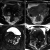
Ruptured endometrioma. A 26-year-old with acute abdomen. MRI is obtained at 3 days after the onset. A Fat-saturated T1-weighted image and B T2-weighted image show a left endometrioma (arrow) with a lack of tension. High signal intensity peritoneal fluid collection (arrowheads) is revealed on fat-saturated T1-weighted image. Diffuse intense contrast enhancement (arrows) is observed on C the post-contrast fat-saturated T1-weighted image reflecting chemical peritonitis
Fig. 13
Fig. 13
Peritoneal inclusion cyst associated with endometriosis. A 32-year-old with suspicion of a large ovarian cyst on ultrasonography examination. A T2-weighted image and, B T1-weighted image show a left small endometrioma (arrow) surrounded by a pseudocystic fluid collection (arrowheads) defined by the pelvic wall and pelvic organs. C SWAN revealed curved linear signal voids along the wall of the endometrioma
Fig. 14
Fig. 14
Endometriosis-associated ovarian carcinoma: clear cell carcinoma. A 52-year-old with suspicion of an ovarian cystic mass with mural nodules on ultrasonography examination. A T2-weighted image, B T1-weighted image, C DWI (b = 800 s/mm2), D ADC map, and E contrast-enhanced subtraction image show a left ovarian cystic mass with a mural nodule (arrowhead). The cyst contents show high signal intensity on both T1- and T2-weighted images reflecting hemorrhagic fluid. The mural nodule shows intermediate signal intensity on T2-weighted image and water diffusion restriction on DWI, and intense contrast-enhancement on post-contrast images clarified on the subtraction image
Fig. 15
Fig. 15
Endometriosis-associated ovarian carcinoma: endometrioid carcinoma. A 35-year-old with suspicion of an ovarian cystic mass with mural nodules on ultrasonography examination. A T2-weighted image, B fat-saturated T1-weighted image, C DWI (b = 800 s/mm2), D computed DWI with high b value (b = 1500 s/mm2), and E ADC map show a left ovarian cystic mass (arrow) with mural nodules. The cyst contents show high signal intensity on both T1- and T2-weighted images reflecting hemorrhagic fluid. The mural nodules show water diffusion restriction on DWI, however, high signal intensity hemorrhagic cyst contents mask the signal of mural nodules. High b-value computed DWI can reduce the signal of cyst contents and high signal intensity of mural nodules is clarified
Fig. 16
Fig. 16
Clots in endometrioma. A 34-year-old with suspicion of an ovarian cystic mass with mural nodules on ultrasonography examination. A T2-weighted image, B fat-saturated T1-weighted image, and C contrast-enhanced subtraction image show a left endometrioma (arrow) with a solid component. The solid component shows intermediate signal intensity on T2-weighted image. The high signal intensity of cyst contents masks the signal of the solid component on the post-contrast image, and no contrast enhancement of the clot is revealed on the contrast-enhanced subtraction image. A 27-year-old with a history of dysmenorrhea. D T2-weighted image, and E DWI (b = 800 s/mm2) show a left endometrioma (arrow) with a solid component. The solid component shows intermediate signal intensity on T2-weighted image and water diffusion restriction on DWI, however, appears as a signal void on F SWAN suggesting a clot
Fig. 17
Fig. 17
Endometriosis-associated ovarian carcinoma: clear cell carcinoma. A 47-year-old with suspicion of an ovarian cystic mass with mural nodules on ultrasonography examination. A On sagittal DWI (reduced field-of-view DWI, b = 800 s/mm2) a large left endometrioma with small mural nodules (arrowheads) is observed. The high signal intensity of the cyst contents masks the signal of small mural nodules and B computed DWI (b = 2000s/mm2) clarified the high signal intensities of the small mural nodules (arrowheads)
Fig. 18
Fig. 18
Endometriosis-related ovarian neoplasm: seromucinous borderline tumor. A 55-year-old with suspicion of an ovarian cystic mass with mural nodules on ultrasonography examination. A T2-weighted image, B fat-saturated T1-weighted image, C contrast-enhanced subtraction image, D DWI (b = 800 s/mm2), E ADC map show an endometrioma with a papillary mural nodule (arrow). The papillary mural nodule shows high signal intensity with low signal intensity dendritic fibrous core on T2-weighted image, relatively weak contrast-enhancement, and high signal intensity on DWI with high ADC (T2 shine-through)
Fig. 19
Fig. 19
Decidualized endometrioma. A 32-year-old pregnant (13 weeks) with suspicion of an ovarian cystic mass with mural nodules on ultrasonography examination. A T2-weighted image, B fat-saturated T1-weight image, C DWI (b = 800 s/mm2). D computed DWI (b = 2000s/mm2), and E ADC map show a right endometrioma with multiple, flat mural nodules (arrowheads) exhibiting T2-prominent high signal intensity, T1-low signal intensity, and high signal intensity on DWI with high ADC (T2 shine-through) similar to those of the placenta (*). On the high b-value (b = 2000s/mm2) computed DWI shows the signal decrease of the mural nodules
Fig. 20
Fig. 20
Polypoid endometriosis. A 47-year-old with lower abdominal pain associated with genital bleeding and suspicion of deep endometriosis. A T2-weighted image and B post-contrast T1-weighted image show polypoid masses (arrowheads) exhibiting T2-high signal intensity and intense contrast-enhancement protruding to the posterior wall of the uterine body with adenomyosis. The masses are surrounded by T2-low signal intensity adhesive fibrous tissue as “black rim sign”. The fibrous rim also shows intense contrast enhancement. A 33-year-old with a history of hypermenorrhea and dysmenorrhea and suspicion of deep endometriosis. C. Oblique coronal fat-saturated T2-weighted image shows a left endometrioma with high signal intensity mural nodule that extends to the Douglas' pouch (arrowheads). A 30-year-old with a history of dysmenorrhea and suspicion of deep endometriosis. D T2-weighted image shows polypoid masses infiltrating into the myometrium (arrowheads)

Similar articles

Cited by

References

    1. Zondervan KT, Becker CM, Missmer SA. Endometriosis. N Engl J Med. 2020;26(382):1244–56. - PubMed
    1. Shafrir AL, Farland LV, Shah DK, Harris HR, Kvaskoff M, Zondervan K, et al. Risk for and consequences of endometriosis: A critical epidemiologic review. Best Pract Res Clin Obstet Gynaecol. 2018;51:1–15. - PubMed
    1. Saunders PTK, Horne AW. Endometriosis: Etiology, pathobiology, and therapeutic prospects. Cell. 2021;27(184):2807–24. - PubMed
    1. Irving JA, Clement PB. Diseases of the peritoneum. In: Kurman RJ, editor. Blaustein’s pathology of the female genital tract. 7th ed. New York: Springer-Verlag; 2018. p. 771–840.
    1. Stewart CJR, Ayhan A, Fukunaga M, Huntsman DG (2020). Endometriosis and related conditions. In: WHO Classification of Tumours Editorial Board, ed. WHO classification of tumours female genital tumours, 5th ed. Lyon: IARC Library Cataloguing-in-Publication Data:169–74.

LinkOut - more resources