. 2024 Apr 24;10(1):31.
doi: 10.1038/s41523-024-00637-3.

Rapid autopsies to enhance metastatic research: the UPTIDER post-mortem tissue donation program

Affiliations

Rapid autopsies to enhance metastatic research: the UPTIDER post-mortem tissue donation program

Tatjana Geukens et al. NPJ Breast Cancer. .

Abstract

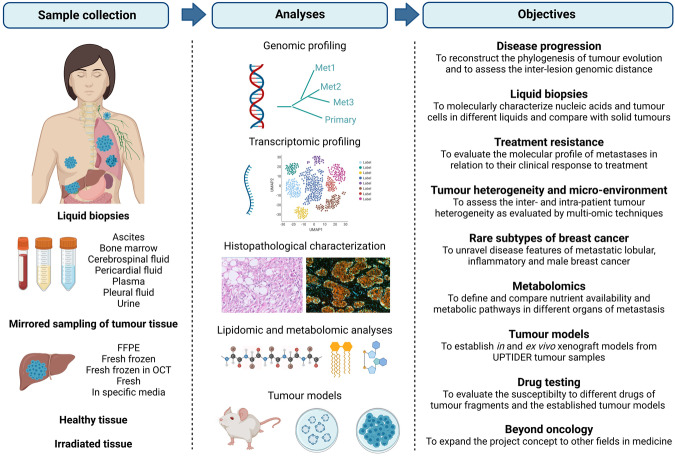
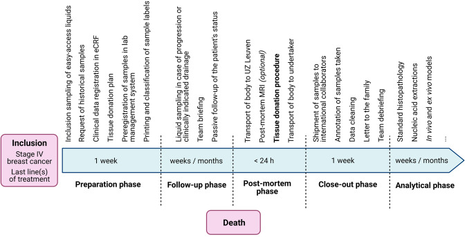
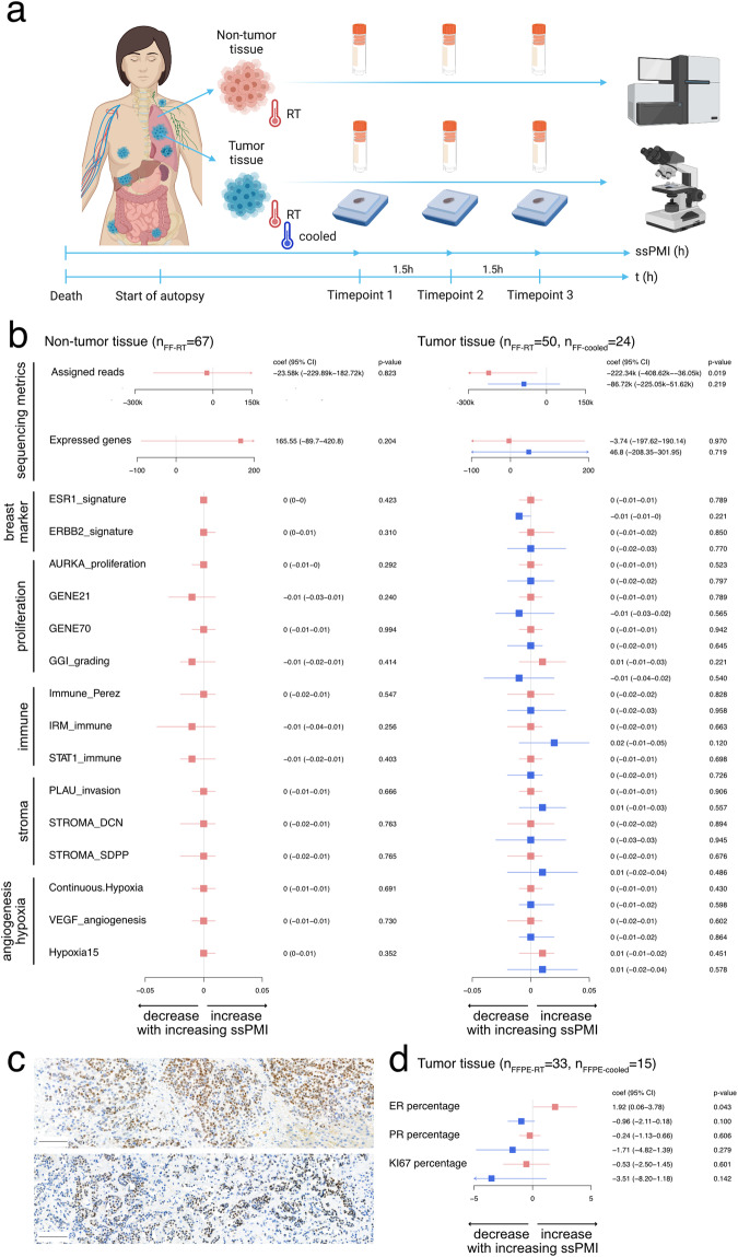
Research on metastatic cancer has been hampered by limited sample availability. Here we present the breast cancer post-mortem tissue donation program UPTIDER and show how it enabled sampling of a median of 31 (range: 5-90) metastases and 5-8 liquids per patient from its first 20 patients. In a dedicated experiment, we show the mild impact of increasing time after death on RNA quality, transcriptional profiles and immunohistochemical staining in tumor tissue samples. We show that this impact can be counteracted by organ cooling. We successfully generated ex vivo models from tissue and liquid biopsies from distinct histological subtypes of breast cancer. We anticipate these and future findings of UPTIDER to elucidate mechanisms of disease progression and treatment resistance and to provide tools for the exploration of precision medicine strategies in the metastatic setting.


Conflict of interest statement

T.G., M.D.S., W.V.D.B., K.V.B., M.M., A.P., A.M., S.L., E.I., H-L.N., I.B., M.H., G.Z., J.V.C., K.B., V.V., B.W., P.V., E.L., M.F.B., G.S., L.B., C.B., P.W.B.D., T.K., D.V., C.L.G.J.S., D.S.T., S.H., E.V., T.V.B., R.S., B.B., D.L., G.M., E.B., A.S., I.N., K.P., P.N., H.W., F.R., G.F., C.D.: no competing financial or non-financial interests to declare. S.M.F.: has received funding from Gilead, Black Belt Therapeutics and Alesta Therapeutics, has consulted for Fund+ and is in the advisory board of Alesta Therapeutics but declares no non-financial competing interests.

Figures

Fig. 1
Fig. 1. Research strategy for the UPTIDER program.
As defined through discussions with academic and clinical collaborators. Objectives as presented on the right are expected to evolve over time as clinical and scientific insights progress. Created with BioRender.com.
Fig. 2
Fig. 2. Study design and workflow of the UPTIDER program.
FFPE formalin-fixed paraffin-embedded, OCT Optimal Cutting Temperature compound, eCRF electronic Case Report Form, MRI Magnetic Resonance Imaging. Created with BioRender.com.
Fig. 3
Fig. 3. Patient and autopsy details.
a Time (in years) between first invasive diagnosis and first metastasis (dark blue) and between first metastasis and death (light blue). b Total number of metastases sampled per patient at autopsy, either planned to be sampled and preregistered in LabCollector® before the autopsy (dark green) or found only during the autopsy (non pre-registered, light green). For Pt2012 the time between enrollment and death was too short to preregister any samples. c Liquid biopsies were sampled prospectively for UPTIDER premortem, either at inclusion or during follow-up. The collection of urine and saliva was only implemented after Pt2003. d Liquid biopsies collected during the autopsy. e Histopathological characteristics of the primary tumor. Of note, Pt2001 is considered ER-negative at primary diagnosis as per diagnostic biopsy but had one sample from the surgical resection exhibiting ER expression. All metastatic samples pre- and post-mortem were ER-negative. ER = Estrogen Receptor, PR = Progesterone Receptor, NST = Invasive Breast Carcinoma of No Special Type, ILC = Invasive Lobular Carcinoma.
Fig. 4
Fig. 4. Transcriptomic and protein expression profiles in function of post-mortem interval in non-tumor and tumor tissues.
a Design of the experiment. Fresh frozen samples (illustrated as tubes) and/or Formalin-Fixed Paraffin-Embedded (FFPE) samples (illustrated as blocks) from non-tumor or tumor tissues were taken repeatedly from the same tissue area (at 1.5 h time intervals) during the autopsy. Sample specific post-mortem interval (ssPMI) was defined as the time between death of the patient and fixation of the sample. Created with BioRender.com. b Results of the testing strategy evaluating the association between quality metrics and ssPMI at the RNA level regarding the non-tumor (left) and the tumor tissues (right) for samples kept at room temperature (RT, in red) and cooled between 4 °C and 10 °C (in blue). Linear coefficient (coef), 95% confidence interval (95% CI) and p-values are reported in the forest plots. Potential non-linear associations are reported in supplementary appendix. For the assigned reads, numbers are reported in kilo (1k = 1000 reads). c IHC slides of a liver metastasis of Pt2023 stained for ER, at time point 1 (top) and after cooling at time point 3 (below). Scale bar = 100 μm. d Results of the testing strategy evaluating the association between ER, PR and KI67 markers and ssPMI at the protein level for samples kept at room temperature (in red) and cooled between 4 °C and 10 °C (in blue). Linear coefficient (coef), 95% confidence interval (95% CI) and p-values are reported in the forest plots. No indication for non-linearity was found.
Fig. 5
Fig. 5. Establishment of different types of preclinical murine tumor models from post-mortem samples.
a Different strategies for tumor model development within UPTIDER. b Hematoxylin-eosin (H&E) staining of a peritoneal lesion of Pt2001 (left) and the corresponding H&E of a third-generation mouse model of the same lesion (right). c Summary graph showing the 24 rapid autopsy samples from three patients with invasive lobular carcinoma, the intraductal take rates and the metastases detected at the endpoint. G1, 2, 3: generation 1, 2, 3; GI tract Gastrointestinal Tract. d Fluorescence stereo-micrograph of a lung lobe from an immunodeficient (NGS) mouse two months after intraductal injection with cells derived from pleural effusion of Pt2011. Scale bar = 1000 μm. e Representative micrograph of picrosirius red-stained histological section of a tumor formed in the mammary gland two months after intraductal injection of cells derived from a pleural effusion of Pt2011. Arrow points to single cell files. Scale bar = 200 μm. f Representative differential interference contrast microscopy (DIC) images of the PDX-PDO organoid line derived from a pleural effusion of Pt2011 cultured in a 3D-basement extract-derived matrix. Note the non-coherent “grape-like” phenotypic structures of the PDO model. Scale bar = 50 µm. g Immunofluorescence of the PDX-PDO organoid line derived from a pleural effusion of Pt2011 (left) and a control breast carcinoma of the no specific type E-cadherin positive PDO-PDX model 209 T (right). Expression and localization of E-cadherin is shown in the top panels and cytokeratin 8 (CK8) in the middle panels. Merged images are shown in the bottom panels. Scale bar = 5 µm.

References

    1. Pasha N, Turner NC. Understanding and overcoming tumor heterogeneity in metastatic breast cancer treatment. Nat. Cancer. 2021;2:680–692. doi: 10.1038/s43018-021-00229-1. - DOI - PubMed
    1. Fumagalli C, Barberis M. Breast cancer heterogeneity. Diagnostics. 2021;11:1555. doi: 10.3390/diagnostics11091555. - DOI - PMC - PubMed
    1. Schrijver WAME, et al. Receptor conversion in distant breast cancer metastases: a systematic review and meta-analysis. J. Natl. Cancer Inst. 2018;110:568–580. doi: 10.1093/jnci/djx273. - DOI - PubMed
    1. Hu Z, Li Z, Ma Z, Curtis C. Multi-cancer analysis of clonality and the timing of systemic spread in paired primary tumors and metastases. Nat. Genet. 2020;52:701–708. doi: 10.1038/s41588-020-0628-z. - DOI - PMC - PubMed
    1. Angus L, et al. The genomic landscape of metastatic breast cancer highlights changes in mutation and signature frequencies. Nat. Genet. 2019;51:1450–1458. doi: 10.1038/s41588-019-0507-7. - DOI - PMC - PubMed