. 2024 Apr 24;14(1):9396.
doi: 10.1038/s41598-024-60236-x.

Skeletal muscle differentiation induces wide-ranging nucleosome repositioning in muscle gene promoters

Affiliations

Skeletal muscle differentiation induces wide-ranging nucleosome repositioning in muscle gene promoters

Sonalí Harris et al. Sci Rep. .

Abstract

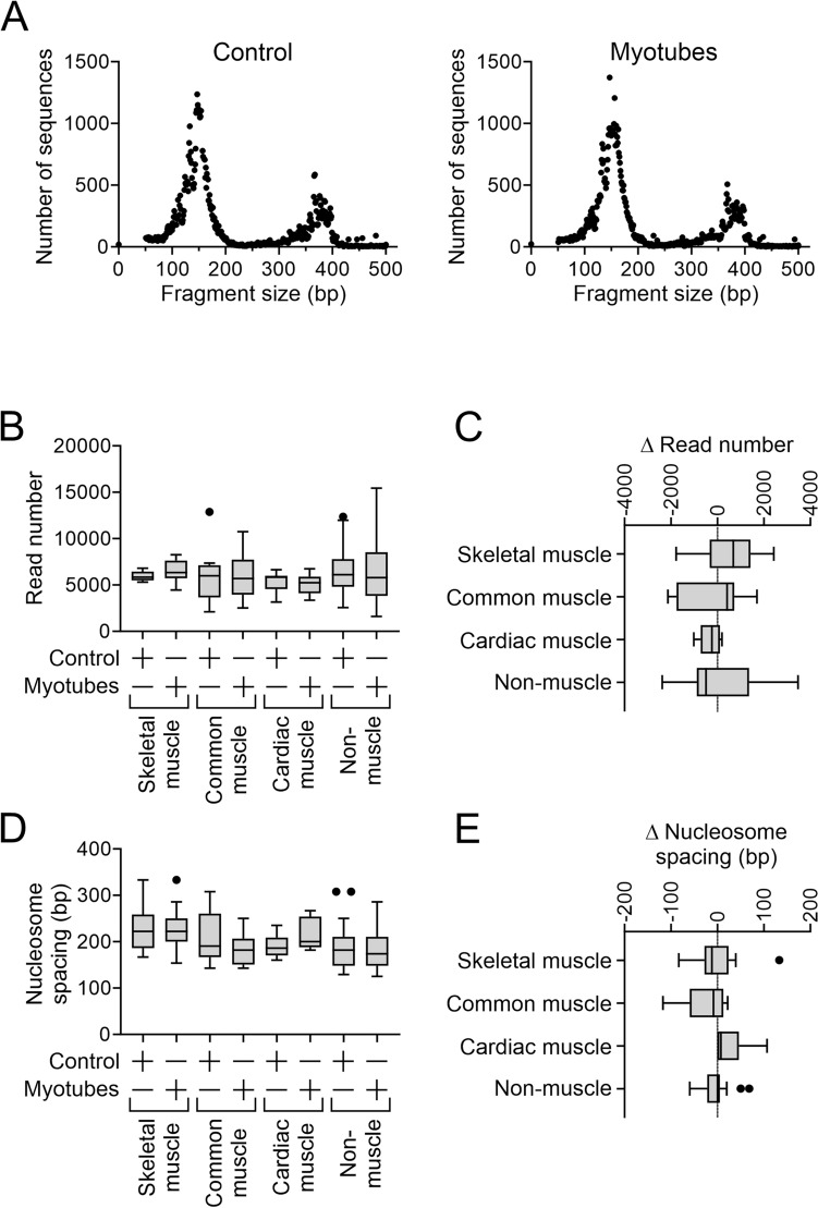
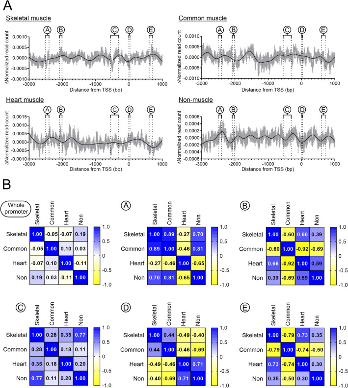
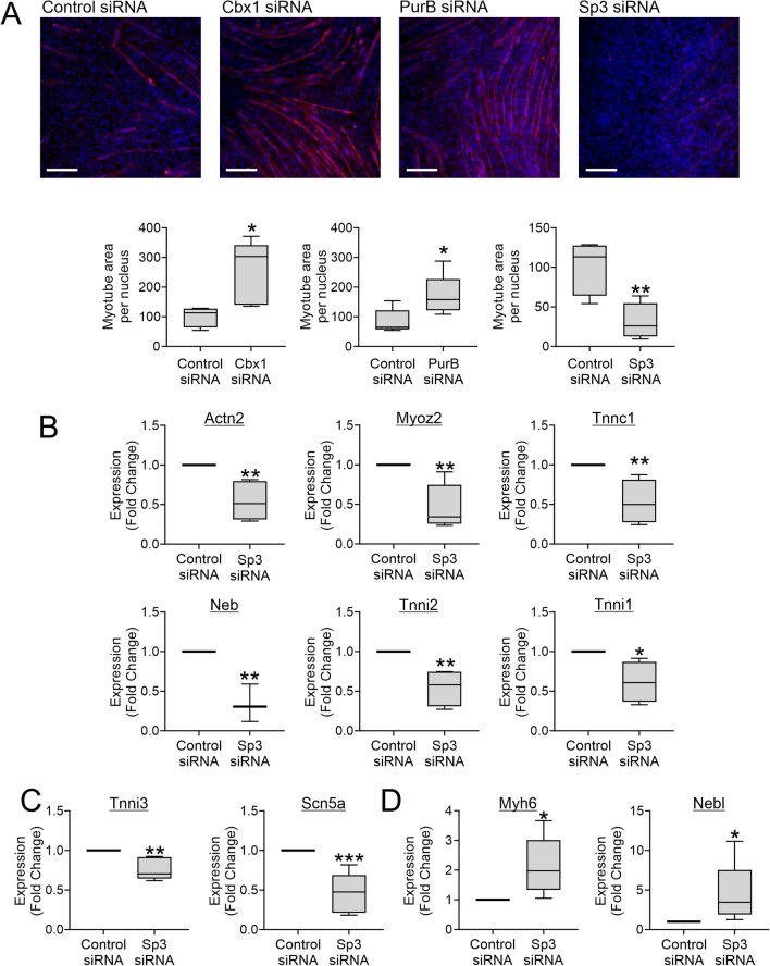
In a previous report, we demonstrated that Cbx1, PurB and Sp3 inhibited cardiac muscle differentiation by increasing nucleosome density around cardiac muscle gene promoters. Since cardiac and skeletal muscle express many of the same proteins, we asked if Cbx1, PurB and Sp3 similarly regulated skeletal muscle differentiation. In a C2C12 model of skeletal muscle differentiation, Cbx1 and PurB knockdown increased myotube formation. In contrast, Sp3 knockdown inhibited myotube formation, suggesting that Sp3 played opposing roles in cardiac muscle and skeletal muscle differentiation. Consistent with this finding, Sp3 knockdown also inhibited various muscle-specific genes. The Cbx1, PurB and Sp3 proteins are believed to influence gene-expression in part by altering nucleosome position. Importantly, we developed a statistical approach to determine if changes in nucleosome positioning were significant and applied it to understanding the architecture of muscle-specific genes. Through this novel statistical approach, we found that during myogenic differentiation, skeletal muscle-specific genes undergo a set of unique nucleosome changes which differ significantly from those shown in commonly expressed muscle genes. While Sp3 binding was associated with nucleosome loss, there appeared no correlation with the aforementioned nucleosome changes. In summary, we have identified a novel role for Sp3 in skeletal muscle differentiation and through the application of quantifiable MNase-seq have discovered unique fingerprints of nucleosome changes for various classes of muscle genes during myogenic differentiation.

Keywords: Cardiac muscle; ChIP-seq; MNase-seq; Skeletal muscle; Sp3.


Conflict of interest statement

The authors declare no competing interests.

Figures

Figure 1
Figure 1
Skeletal muscle differentiation is associated with dynamic re-positioning of nucleosomes. Chromatin was isolated from C2C12 cells immediately prior to the start of skeletal muscle differentiation (control) and seven days later when myotubes were evident. Once isolated, the chromatin was digested with MNase and the resultant digests were sequenced. (A) Sequence size distributions after MNase digestion. (B) Sequences were aligned to the mouse genome and the number of sequences (read count) which aligned to various promoters (defined as − 3kb to + 1kb with respect to the transcription start site (TSS) were determined at a 1bp resolution. Defined groups of muscle-specific and non-muscle genes were analyzed. The muscle-specific group were split further on the basis of their expression: (1) muscle-specific genes expressed solely in skeletal muscle; (2) muscle-specific genes expressed in both skeletal and cardiac muscle; and (3) muscle-specific genes expressed solely in cardiac muscle. These groups are called skeletal muscle-specific, common and heart muscle-specific respectively. The number of genes in each group ranged from 6 to 20. The graph shows the summed read number over the full length of the promoter. No statistical differences (ANOVA) were observed. (C) For each analyzed promoter, the change in read number (Δ Read count) following myogenic differentiation was calculated. N = 6–20 per group. No statistical differences (ANOVA) were observed. (D) For each analyzed promoter, the nucleosome number was determined. The graph shows average nucleosome spacing. Average nucleosome spacing was determined by dividing the promoter length (4kb) by the number of nucleosomes. N = 6–20 per group. No statistical differences (ANOVA) were observed. (E) For each analyzed promoter, the change in average nucleosome spacing (Δ Nucleosome spacing) following skeletal muscle differentiation was determined. The change in average nucleosome spacing was determined by subtracting average nucleosome spacing in control cells from the average nucleosome spacing observed in myotubes. N = 6–20 per group. No statistical differences (ANOVA) were observed.
Figure 2
Figure 2
Quantifying MNase-seq to determine changes in nucleosome positioning during skeletal muscle differentiation. (A) To compare nucleosome patterns within each group and across the four groups, the read counts were normalized. Normalization was carried out dividing the read count at each base-pair of the promoter by the sum of read counts across the promoter (a full description is provided in the methods) and was carried out to ensure that each promoter had equal weight. The graphs show the change in normalized read count for each group (skeletal muscle-specific, common muscle, heart muscle-specific, and non-muscle genes) following myogenic differentiation. The light grey bars show the mean and standard error a 1bp resolution. The black line represents the best-fit model of the data. Statistical analysis of the skeletal muscle-specific group identified five regions that were significantly different in control cells and myotubes. These regions are labelled A–E. (B) for regions A–E, correlations were determined between the four groups.
Figure 3
Figure 3
Cbx1, PurB and Sp3 play opposing roles on myotube formation. (AC) C2C12 cells were grown to confluence whereupon they underwent differentiation to generate myotubes. (A) RNA was isolated immediately prior to differentiation (control) and after 6 days of skeletal muscle differentiation. Expression of the muscle-specific genes Actn2 and Myh6 was determined by qPCR and expression levels relative to the housekeeping gene Gapdh are shown. N = 6. Independent T-test, **P < 0.01, ***P < 0.001. (B) Myotubes were visualized by immunostaining for the muscle-specific protein Actn2. Nuclei were counterstained with DAPI. Scale bar 200 microns. The image is representative of three independent experiments. (C) RNA was isolated immediately prior to differentiation (control) and after 8 days of skeletal muscle differentiation. Expression of Cbx1, PurB and Sp3 was determined by qPCR and expression levels relative to the housekeeping gene Gapdh are shown. N = 4. Independent T-test, *P < 0.05, **P < 0.01, ***P < 0.001. (D) C2C12 cells were transfected with siRNAs targeting Cbx1, PurB, or Sp3. A non-targeting siRNA was used as a control. Three days later, cells were analyzed for RNA levels. Expression of Cbx1, PurB and Sp3 was determined by qPCR and expression levels relative to the housekeeping gene Gapdh are shown. N = 4. Independent T-test, *P < 0.05, **P < 0.01, ***P < 0.001.
Figure 4
Figure 4
Sp3 plays opposing roles on muscle gene expression. (A), Three days prior to the start of skeletal muscle differentiation, C2C12 cells were transfected with siRNAs targeting Cbx1, PurB, or Sp3. A non-targeting siRNA was used as a control. Skeletal muscle differentiation was assessed by immunostaining cells with the muscle-specific protein Actn2. Images were taken at five random locations. Both myotube area and nuclei number were counted. Skeletal muscle differentiation is expressed as the myotube area per nucleus. Representative images (scale bar 200 microns) and quantification are shown from one of three independent experiments. Independent T-test, *P < 0.05, **P < 0.01. (B–D) C2C12 cells were underwent skeletal muscle differentiation for seven days. RNA was extracted and analyzed for the indicated muscle-specific mRNAs by qPCR. Gapdh was used as a housekeeping control. Expression is shown relative to cells transfected with non-targeting siRNA. (B) Skeletal muscle-specific and common muscle gene expression, C heart muscle-specific gene expression, (D) muscle genes up-regulated by Sp3 knockdown. N = 4–5. Independent T-test, *P < 0.05, **P < 0.01, ***P < 0.001.
Figure 5
Figure 5
Sp3 binding in muscle genes. (A) ChIP-seq was used to evaluate Sp3 binding in C2C12 cells immediately prior to differentiation (control) and after seven days of skeletal muscle differentiation (myotubes). The graph shows the number of muscle-specific and non-muscle genes with a validated Sp3 binding site. (B) For each Sp3 binding site, the change in nucleosome content following myogenic differentiation was calculated. The two bars represent Sp3 binding sites found only in non-differentiated and differentiated cells, respectively. (C) Sp3 binding sites (muscle and non-muscle) are shown with respect to the TSS and regions A–E identified in skeletal muscle-specific gene promoters. Sequences surrounding the binding peak are shown in Supplementary Table 1.

References

    1. Laumonier T, Menetrey J. Muscle injuries and strategies for improving their repair. J. Exp. Orthop. 2016;3:15. doi: 10.1186/s40634-016-0051-7. - DOI - PMC - PubMed
    1. Jiang CP, Wang L, Fu W. Emerging technologies and methods for musculoskeletal tissue repair and regeneration. Biomed. Res. Int. 2018;2018:8953079. doi: 10.1155/2018/8953079. - DOI - PMC - PubMed
    1. Baksh SS, Pratt RE, Gomez J, Dzau VJ, Hodgkinson CP. A novel Cbx1, PurB, and Sp3 complex mediates long-term silencing of tissue- and lineage-specific genes. J. Biol. Chem. 2022;298:102053. doi: 10.1016/j.jbc.2022.102053. - DOI - PMC - PubMed
    1. Baksh SS, Hu J, Pratt RE, Dzau VJ, Hodgkinson CP. Rig1 receptor plays a critical role in cardiac reprogramming via YY1 signaling. Am. J. Physiol. Cell Physiol. 2023;324:C843–C855. doi: 10.1152/ajpcell.00402.2022. - DOI - PMC - PubMed
    1. Lindskog C, et al. The human cardiac and skeletal muscle proteomes defined by transcriptomics and antibody-based profiling. BMC Genomics. 2015;16:475. doi: 10.1186/s12864-015-1686-y. - DOI - PMC - PubMed

Publication types