. 2024 Nov;34(6):e13263.
doi: 10.1111/bpa.13263. Epub 2024 Apr 25.

A role for vessel-associated extracellular matrix proteins in multiple sclerosis pathology

Affiliations

A role for vessel-associated extracellular matrix proteins in multiple sclerosis pathology

Marco Pisa et al. Brain Pathol. 2024 Nov.

Abstract

Multiple sclerosis (MS) is unsurpassed for its clinical and pathological hetherogeneity, but the biological determinants of this variability are unknown. HLA-DRB1*15, the main genetic risk factor for MS, influences the severity and distribution of MS pathology. This study set out to unravel the molecular determinants of the heterogeneity of MS pathology in relation to HLA-DRB1*15 status. Shotgun proteomics from a discovery cohort of MS spinal cord samples segregated by HLA-DRB*15 status revealed overexpression of the extracellular matrix (ECM) proteins, biglycan, decorin, and prolargin in HLA-DRB*15-positive cases, adding to established literature on a role of ECM proteins in MS pathology that has heretofore lacked systematic pathological validation. These findings informed a neuropathological characterisation of these proteins in a large autopsy cohort of 41 MS cases (18 HLA-DRB1*15-positive and 23 HLA-DRB1*15-negative), and seven non-neurological controls on motor cortical, cervical and lumbar spinal cord tissue. Biglycan and decorin demonstrate a striking perivascular expression pattern in controls that is reduced in MS (-36.5%, p = 0.036 and - 24.7%, p = 0.039; respectively) in lesional and non-lesional areas. A concomitant increase in diffuse parenchymal accumulation of biglycan and decorin is seen in MS (p = 0.015 and p = 0.001, respectively), particularly in HLA-DRB1*15-positive cases (p = 0.007 and p = 0.046, respectively). Prolargin shows a faint parenchymal pattern in controls that is markedly increased in MS cases where a perivascular deposition pattern is observed (motor cortex +97.5%, p = 0.001; cervical cord +49.1%, p = 0.016). Our findings point to ECM proteins and the vascular interface playing a central role in MS pathology within and outside the plaque area. As ECM proteins are known potent pro-inflammatory molecules, their parenchymal accumulation may contribute to disease severity. This study brings to light novel factors that may contribute to the heterogeneity of the topographical variation of MS pathology.

Keywords: ECM proteins; HLA‐DRB1*15; MS; pathology; topography; vessel.


Conflict of interest statement

The authors report no competing interests.

Figures

FIGURE 1
FIGURE 1
Functions of the ECM proteins decorin, biglycan and prolargin. Functions of the ECM proteins decorin, biglycan and prolargin. These three proteins are all part of the family of leucine‐rich proteoglycans. These proteins are expressed throughout the body. In the CNS these proteins can be found in the vascular basement membrane and in the interstitial matrix. Originally thought to be mainly important in bone growth, they have been found to be involved in a wide range of cellular functions. These include promoting autophagy (decorin), modulation of the immune system by interacting with toll like receptors and also cytokines such as tumour necrosis factor α, and in the formation of the ECM architecture [16, 26, 27, 28, 29, 30, 31, 48]. Created with BioRender.com.
FIGURE 2
FIGURE 2
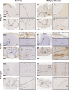
Immunoreactivity of biglycan, decorin, and prolargin in MS and controls. Biglycan staining in controls (A, C) and MS (B, D). In the motor cortex, biglycan shows minimal subcortical white matter deposition in controls (A) that contrasts with MS where an intense diffuse parenchymal staining pattern is observed (B). In the spinal cord, controls demonstrates a clear perivascular staining pattern (C, inset) that is markedly reduced in MS (D, inset) where a concomitant diffuse parenchymal staining is observed, particularly in white matter. Decorin staining in controls (E, G) and MS (F, H). In the motor cortex, decorin is homogeneously expressed in the meninges (E, top inset) and parenchymal perivascular spaces (E, bottom inset) of controls. In comparison, MS shows reduced decorin expression in the meninges (where it is limited to the adventitia of meningeal pial vessels) (F, top inset) and in parenchymal perivascular areas (F, bottom inset). In the spinal cord, controls show prominent perivascular and minimal parenchymal staining (G), which differs markedly in MS where reduced perivascular staining and increased diffuse parenchymal expression abutting Virchow Robin spaces (H) are seen. Prolargin staining in controls (I, K) and MS (J, L). In the motor cortex, controls show minimal prolargin staining (I), which contrasts significantly with MS where striking deposition is seen in the parenchyma abutting the Virchow Robin spaces, especially in the subpial and infragranular areas (J). In the spinal cord, controls have sparse staining (K) compared to MS case, which demonstrate a diffuse, faint parenchymal staining pattern in white matter with relative sparing of grey matter (L). The scale bar corresponds to 1 mm. GM, grey matter; WM, white matter.
FIGURE 3
FIGURE 3
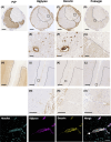
Immunoreactivity of biglycan, decorin, and prolargin along the neuraxis. Cervical spinal cord (A–G) and motor cortex (H–O) from a prototypical MS case immunolabelled for myelin (PLP) (A, H), biglycan (B, E, J, M), decorin (C, F, K, N), and prolargin (D, G, L, O). Well circumscribed areas of demyelination are seen in the spinal cord (A) and subpial cortex (H) (demarcated by dashed lines in A–D and H–L). In the spinal cord, biglycan (B) and decorin (C) show meningeal staining in the adventitia of pial vessels with an intense perivascular expression pattern within the Virchow Robin spaces of parenchymal vessels, particularly within lesional areas (E, F). Motor cortex shows reduced meningeal expression and minimal cortical parenchymal expression of biglycan (J) and decorin (K). Double immunofluorescence labelling of biglycan and decorin show co‐localisation of these proteins (bottom row images). Prolargin expression is diffuse throughout the spinal cord parenchyma, particularly in non‐lesional areas (D, G). In the motor cortex, prolargin demonstrates increased expression in the parenchyma abutting the Virchow Robin space (L, O), notably in subpial and infragranular cortical areas. Scale bars correspond to 1 mm in A–D and H–L, 50 μm in E–G and M–O.
FIGURE 4
FIGURE 4
Topographical distribution of vascular area in MS cases and controls. (A) Vascular area in non‐neurological controls (n = 3) is presented according to matter type (white and grey) and site (cortex, cervical and lumbar spinal cord). Total vascular area is higher in grey compared to white matter throughout the neuraxis, and in the primary motor cortex compared to cervical and lumbar levels of the spinal cord. (B) Vascular area in lesional and non‐lesional areas in MS (n = 39) and non‐neurological controls (n = 3) according to matter type. Vascular area is greater in grey matter compared to white matter in each area type with grey matter vascularity being reduced in MS tissue compared to controls. Bars illustrate mean ± SEM. Relevant corrected Wald pairwise comparisons are displayed: **p < 0.001; *p < 0.05.
FIGURE 5
FIGURE 5
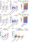
Topographical distribution of biglycan, decorin and prolargin expression in controls and MS. Biglycan (A–C), decorin (D–F), and prolargin (G, H) expression in controls and MS. (A) In controls, perivascular biglycan expression is increased in white compared to grey matter, particularly at cervical level. (B) In MS, perivascular biglycan expression is reduced compared to controls, regardless of spinal cord level or lesional status. In contrast, parenchymal biglycan expression (C) is increased in MS compared with controls, particularly in cases harbouring the HLA‐DRB1*15 allele. (D) In controls, perivascular decorin expression is increased in white compared with grey matter and in the lumbar compared with cervical cord levels. (E) Perivascular decorin expression is reduced in lesional and non‐lesional MS compared with controls at the lumbar cord level only. (F) In contrast, parenchymal decorin expression is increased in MS compared with controls, particularly in cases harboring the HLA‐DRB1*15 allele. (G) In controls, prolargin shows a gradient of expression along the neuraxis, being lowest in the cortex and highest in the lumbar cord. Within the cervical and lumbar cord, prolargin immunoreactivity is greater in white compared with grey matter. (H) Prolargin expression is increased in MS compared with controls in the cortex and cervical cord, particularly in non‐lesional white matter areas. Bars illustrate mean ± SEM. Relevant Wald pairwise comparisons are displayed: **p < 0.001; *p < 0.05. AI, area index.

References

    1. Popescu BFG, Pirko I, Lucchinetti CF. Pathology of multiple sclerosis: where do we stand? Contin Lifelong Learn Neurol. 2013;19(4):901–921. - PMC - PubMed
    1. Olsson T, Barcellos LF, Alfredsson L. Interactions between genetic, lifestyle and environmental risk factors for multiple sclerosis. Nat Rev Neurol. 2017;13(1):25–36. - PubMed
    1. Chung KK, Altmann D, Barkhof F, Miszkiel K, Brex PA, O'Riordan J, et al. A 30‐year clinical and magnetic resonance imaging observational study of multiple sclerosis and clinically isolated syndromes. Ann Neurol. 2020;87(1):63–74. - PMC - PubMed
    1. Brownlee WJ, Tur C, Manole A, Eshaghi A, Prados F, Miszkiel KA, et al. HLA‐DRB1*1501 influences long‐term disability progression and tissue damage on MRI in relapse‐onset multiple sclerosis. Mult Scler Houndmills Basingstoke Engl. 2023;29(3):333–342. - PubMed
    1. DeLuca GC, Alterman R, Martin JL, Mittal A, Blundell S, Bird S, et al. Casting light on multiple sclerosis heterogeneity: the role of HLA‐DRB1 on spinal cord pathology. Brain J Neurol. 2013;136(Pt 4):1025–1034. - PubMed

Publication types